
Synaptic Transmission
An action potential races down the axon and arrives at its destination - but it cannot simply jump across to the next cell. The gap between neurons, called the synapse, requires the electrical signal to be converted into a chemical one (or, in rare cases, passed directly through gap junctions). This conversion process is synaptic transmission, and it is where the nervous system gains its incredible flexibility. Synapses can be strengthened, weakened, or modulated by drugs, disease, and experience.
Understanding synaptic transmission is essential for MCAT questions spanning neuroscience, pharmacology, and behavioral science.
Anatomy of the Synapse
The synapse has three components:
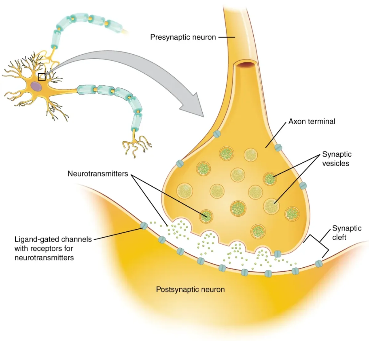
- Presynaptic terminal (axon terminal or synaptic bouton) - the end of the sending neuron’s axon. It contains mitochondria and synaptic vesicles filled with neurotransmitter molecules.
- Synaptic cleft - the narrow gap (~20 nm wide) between the two neurons. Neurotransmitters must diffuse across this space.
- Postsynaptic membrane - the receiving surface of the next neuron (or muscle cell, or gland cell). It is studded with receptors specific to the neurotransmitter being released.
Chemical Synapse: Step by Step
The sequence of events at a chemical synapse is one of the most testable processes on the MCAT. Know every step:
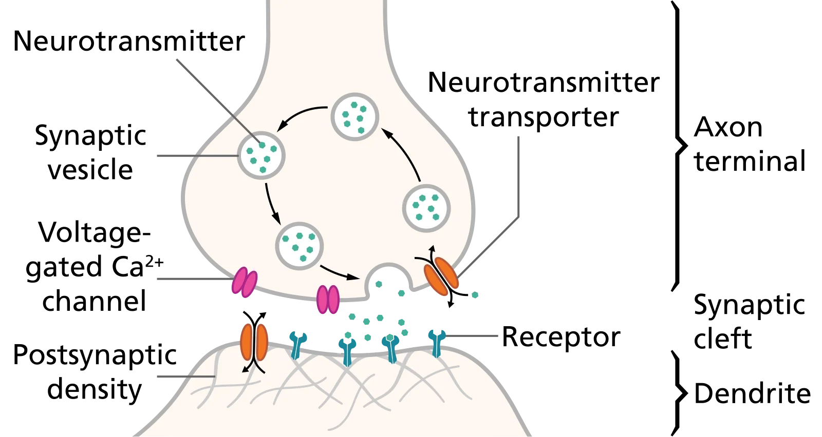
- Action potential arrives at the presynaptic terminal.
- Voltage-gated Ca2+ channels open. Depolarization of the terminal opens these channels, and Ca2+ floods into the presynaptic cell.
- Ca2+ triggers vesicle fusion. The rise in intracellular Ca2+ causes synaptic vesicles to fuse with the presynaptic membrane, releasing neurotransmitter into the cleft by exocytosis.
- Neurotransmitters diffuse across the synaptic cleft (~20 nm).
- Neurotransmitters bind postsynaptic receptors, opening or closing ion channels and changing the postsynaptic membrane potential.
- Signal termination occurs by one of three mechanisms: enzymatic degradation (e.g., acetylcholinesterase breaks down ACh), reuptake into the presynaptic terminal (e.g., serotonin transporter), or simple diffusion away from the cleft.
Electrical Synapses
Not all synapses use chemicals. Electrical synapses use gap junctions - protein channels (connexons) that directly connect the cytoplasm of two neurons. Ions flow directly from one cell to the next.
Electrical synapses are bidirectional and extremely fast because there is no chemical intermediary. However, they are less modulatable - you cannot easily fine-tune or inhibit them the way you can with chemical synapses.
Electrical synapses are found in cardiac muscle (allowing synchronized contraction), smooth muscle, and certain brain circuits where speed is critical.
EPSPs and IPSPs
When neurotransmitters bind postsynaptic receptors, the effect depends on which ion channels are opened or closed:
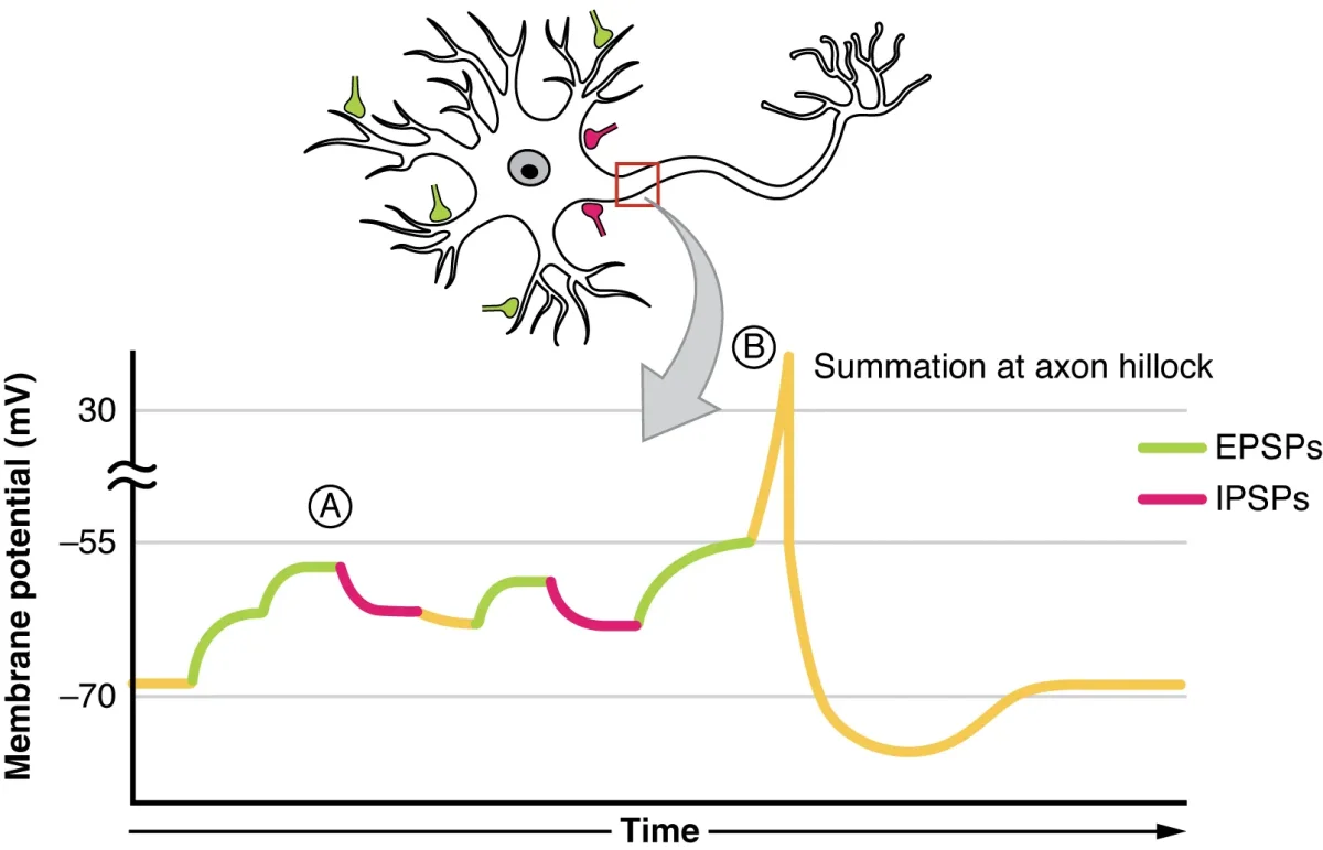
Excitatory postsynaptic potentials (EPSPs) result when the postsynaptic membrane is depolarized, bringing it closer to threshold. This typically happens when Na+ channels open and Na+ flows into the cell. EPSPs increase the probability that the postsynaptic neuron will fire an action potential.
Inhibitory postsynaptic potentials (IPSPs) result when the postsynaptic membrane is hyperpolarized, pushing it further from threshold. This happens when Cl- channels open (Cl- flows in) or K+ channels open (K+ flows out). IPSPs decrease the probability of firing.
A single EPSP is usually too small to reach threshold on its own. For the postsynaptic neuron to fire, multiple EPSPs must add together - a process called summation.
Summation: Adding Signals at the Axon Hillock
The axon hillock is the integration zone of the neuron. It receives all the EPSPs and IPSPs from the dendrites and cell body and determines whether their sum reaches threshold.
Temporal summation occurs when a single presynaptic neuron fires rapidly in succession. Each EPSP arrives before the previous one fades, and they stack on top of each other. Think of it as one person repeatedly pushing a swing - if they push fast enough, the swing goes higher each time.
Spatial summation occurs when multiple presynaptic neurons fire simultaneously, and their EPSPs (arriving at different locations on the dendrites) combine at the axon hillock. Think of it as several people pushing the same swing at the same time.
In reality, both temporal and spatial summation happen simultaneously. The axon hillock sums all excitatory and inhibitory inputs. If the net result reaches threshold, the neuron fires. If not, it stays quiet.
Ionotropic vs. Metabotropic Receptors
Postsynaptic receptors fall into two broad categories:
Ionotropic receptors (ligand-gated ion channels) are fast-acting. When the neurotransmitter binds, the receptor itself is the ion channel - it opens immediately, allowing ions to flow within milliseconds. Examples include nicotinic acetylcholine receptors and GABA-A receptors.
Metabotropic receptors (G-protein coupled receptors, or GPCRs) are slower but longer-lasting. Neurotransmitter binding activates an intracellular G-protein, which triggers a second messenger cascade (e.g., cAMP, IP3). This can open or close ion channels indirectly, modify gene expression, or produce other cellular effects. Examples include muscarinic acetylcholine receptors, dopamine receptors, and GABA-B receptors.
Major Neurotransmitters
| Neurotransmitter | Type | Key Functions |
|---|---|---|
| Acetylcholine (ACh) | Excitatory (at NMJ) | Neuromuscular junction, parasympathetic NS, memory |
| Dopamine | Modulatory | Reward, motivation, motor control |
| Serotonin (5-HT) | Modulatory | Mood, sleep, appetite |
| Norepinephrine (NE) | Excitatory | Alertness, attention, sympathetic NS |
| GABA | Inhibitory | Main inhibitory NT in the CNS |
| Glutamate | Excitatory | Main excitatory NT in the CNS |
| Glycine | Inhibitory | Major inhibitory NT in the spinal cord |
| Endorphins | Modulatory | Natural pain reduction, euphoria |
Signal Termination
Neurotransmitter signaling must be turned off quickly to allow precise control. Three mechanisms accomplish this:
-
Enzymatic degradation: Enzymes in the synaptic cleft break down the neurotransmitter. The classic example is acetylcholinesterase (AChE), which rapidly hydrolyzes ACh into acetate and choline.
-
Reuptake: Transporter proteins on the presynaptic membrane pump the neurotransmitter back into the presynaptic terminal for recycling. This is the primary termination mechanism for dopamine, serotonin, and norepinephrine.
-
Diffusion: The neurotransmitter simply drifts away from the synaptic cleft and is absorbed by surrounding glial cells or degraded elsewhere.