
The Brain
The brain is the most complex organ in the human body, containing roughly 86 billion neurons connected by trillions of synapses. For the MCAT, you do not need to memorize every gyrus and sulcus, but you absolutely need to know the major regions, what they do, and what happens when they are damaged. Brain anatomy shows up in biology, psychology, and even biochemistry passages.
This section covers the cerebrum, diencephalon, brainstem, cerebellum, limbic system, basal ganglia, meninges, and cerebrospinal fluid. It is one of the longest sections in this chapter for a reason - every one of these structures is fair game on test day.
The Cerebrum: Four Lobes
The cerebrum is the largest part of the brain, accounting for about 85% of total brain mass. Its surface is covered by the cerebral cortex, a thin layer of gray matter (neuron cell bodies) folded into ridges (gyri) and grooves (sulci) to maximize surface area. Beneath the cortex lies white matter (myelinated axons connecting different cortical regions).
The cerebrum is divided into two hemispheres (left and right) connected by the corpus callosum, a massive bundle of axons that allows the hemispheres to communicate. Each hemisphere is divided into four lobes.
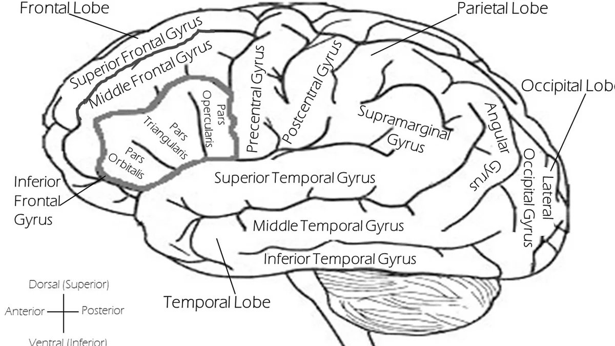
Frontal Lobe
The frontal lobe is the largest lobe, occupying the front third of each hemisphere. It handles some of the most “human” functions:
- Primary motor cortex (precentral gyrus): Initiates voluntary movement. It is organized as a motor homunculus - a map where different body parts are represented in proportion to how finely they are controlled (hands and face get disproportionately large areas).
- Prefrontal cortex: Executive functions - planning, judgment, personality, impulse control, working memory, and social behavior. Damage here produces dramatic personality changes.
Parietal Lobe
The parietal lobe sits behind the frontal lobe, separated by the central sulcus:
- Primary somatosensory cortex (postcentral gyrus): Processes touch, pressure, temperature, and pain from the body. It is also organized as a homunculus, with the most sensitive body parts (lips, fingertips) getting the most cortical real estate.
- Spatial awareness and integration: The parietal lobe integrates sensory information to create a sense of body position and spatial relationships. Damage to the right parietal lobe can cause hemispatial neglect - patients ignore the entire left side of their world.
Temporal Lobe
The temporal lobe sits below the lateral sulcus on each side of the brain:
- Primary auditory cortex: Processes sound.
- Wernicke’s area (usually left hemisphere): Language comprehension. Damage causes Wernicke’s aphasia - the patient speaks fluently but the words are nonsensical. They cannot understand spoken or written language (“Wernicke’s is Wordy but Wrong”).
- Hippocampus (medial temporal lobe): Critical for forming new long-term memories. Bilateral hippocampal damage causes anterograde amnesia - the inability to form new declarative memories (the case of patient H.M.).
Occipital Lobe
The occipital lobe occupies the back of the brain:
- Primary visual cortex: Processes visual information received from the eyes via the optic nerve and lateral geniculate nucleus of the thalamus.
- Damage to the occipital lobe causes cortical blindness - the eyes work fine, but the brain cannot process what they see.
The Diencephalon: Thalamus and Hypothalamus
The diencephalon sits deep within the brain, between the cerebrum and the brainstem.
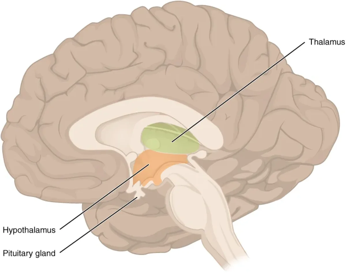
The thalamus is the brain’s sensory relay station. Nearly all sensory information passes through the thalamus before reaching the cerebral cortex. Vision goes through the lateral geniculate nucleus, hearing through the medial geniculate nucleus, and somatosensory information through the ventral posterior nucleus.
The one major exception: smell (olfaction) bypasses the thalamus entirely and projects directly to the olfactory cortex. This is a classic MCAT fact.
The hypothalamus is the master regulator of homeostasis. Despite being only about the size of an almond, it controls:
- Body temperature (thermoregulation)
- Hunger and thirst (feeding and satiety centers)
- Circadian rhythms (suprachiasmatic nucleus receives light input)
- Autonomic nervous system output (sympathetic and parasympathetic tone)
- Endocrine function - the hypothalamus controls the pituitary gland (the “master gland”) through releasing and inhibiting hormones. This hypothalamic-pituitary axis is one of the most tested endocrine concepts on the MCAT.
- Emotional responses (works with the limbic system)
The Brainstem: Midbrain, Pons, and Medulla Oblongata
The brainstem connects the cerebrum to the spinal cord and controls the most fundamental life-sustaining functions. It consists of three regions, from top to bottom:
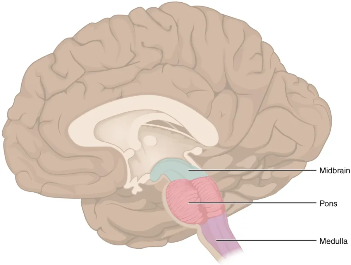
Midbrain (mesencephalon): Processes visual and auditory reflexes. Contains the superior colliculus (visual reflexes - e.g., turning your head toward a flash of light) and the inferior colliculus (auditory reflexes - e.g., orienting toward a sudden sound). Also contains the substantia nigra, which produces dopamine for the basal ganglia (degeneration here causes Parkinson’s disease).
Pons: Serves as a bridge (Latin pons = bridge) between the cerebellum and the cerebral cortex. It relays information between these structures and also plays a role in regulating breathing (works with the medulla’s respiratory centers). Several cranial nerve nuclei are located here.
Medulla oblongata: The most vital part of the brainstem. It controls autonomic functions that keep you alive:
- Cardiovascular center: Regulates heart rate and blood pressure
- Respiratory center: Controls the basic rhythm of breathing
- Other reflexes: Vomiting, coughing, sneezing, swallowing, hiccupping
Damage to the medulla is often fatal because these are the functions you cannot live without. The medulla is also where most descending motor tracts and ascending sensory tracts decussate (cross over to the opposite side), which is why the left brain controls the right body and vice versa.
Reticular formation: A network of neurons running through the entire length of the brainstem. It regulates arousal, consciousness, and the sleep-wake cycle. The reticular activating system (RAS) is the ascending portion that keeps the cortex alert. Damage to the RAS can cause coma.
The Cerebellum
The cerebellum (“little brain”) sits behind the brainstem at the back of the skull. It contains more neurons than the rest of the brain combined, despite being only about 10% of brain volume.
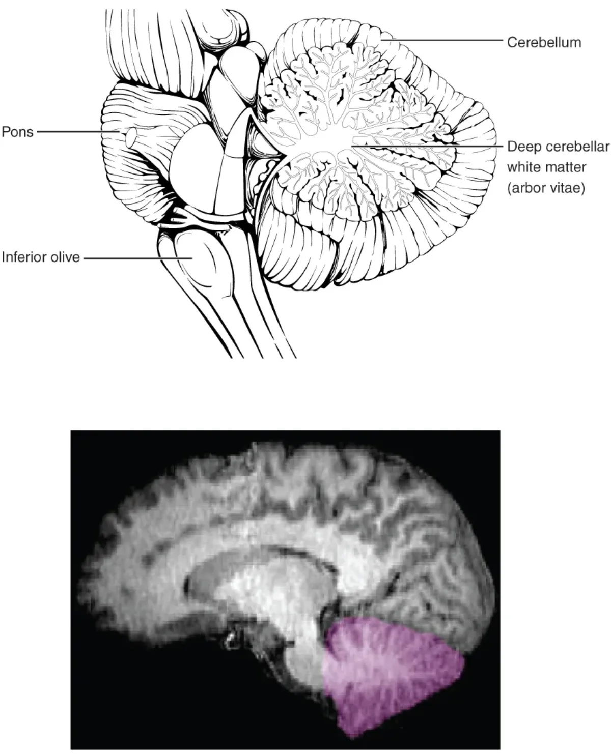
Key functions of the cerebellum:
- Motor coordination: Smooths and fine-tunes movements initiated by the motor cortex. Damage causes ataxia - jerky, uncoordinated movement.
- Balance and posture: Integrates input from the vestibular system to maintain equilibrium.
- Motor learning: Stores procedural memories like riding a bike, playing piano, or typing. Once learned, these tasks become “automatic” and run through cerebellar circuits.
A critical distinction: the cerebellum does not initiate movement. That is the job of the primary motor cortex (frontal lobe). The cerebellum compares intended movement (from the cortex) with actual movement (from proprioceptive feedback) and sends corrections in real time. Think of it as a GPS that constantly recalculates your route - it does not decide where you are going, but it makes sure you get there smoothly.
The Limbic System
The limbic system is a group of interconnected structures that sit deep within the brain, forming a ring around the top of the brainstem. It handles emotions, motivation, and memory formation.
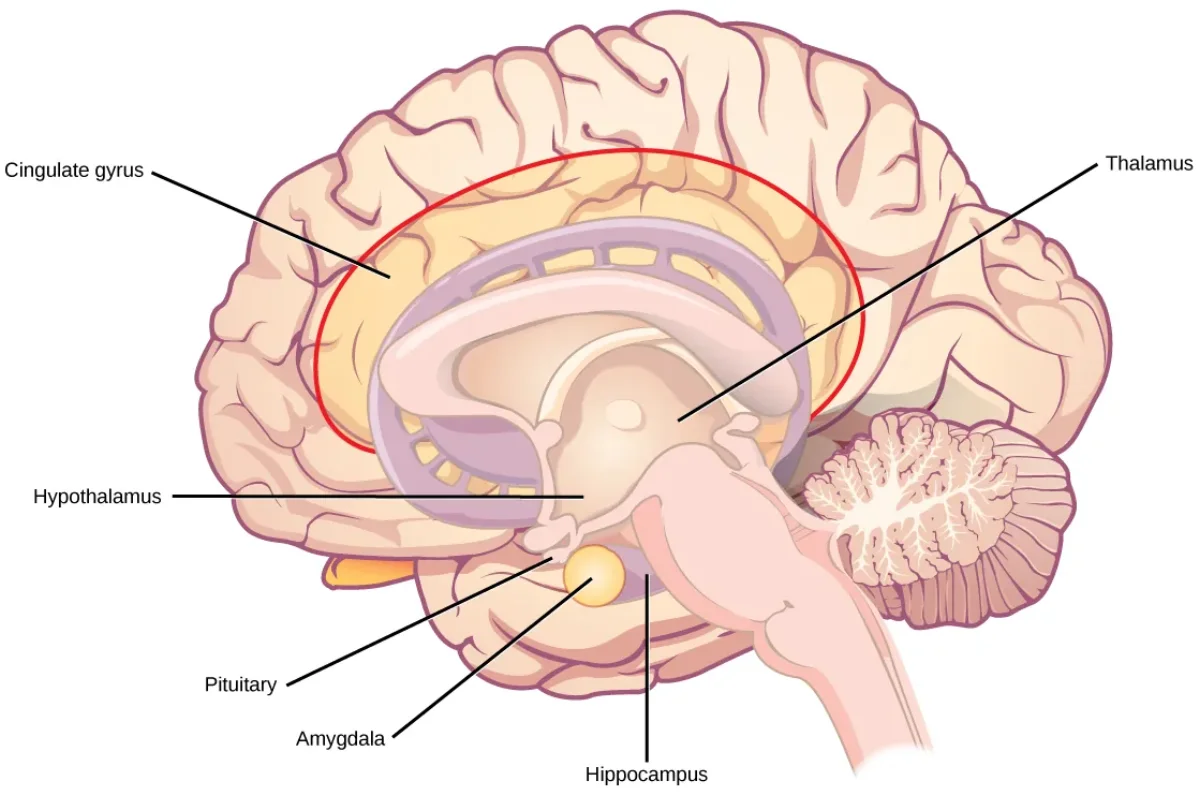
Key limbic structures:
- Hippocampus: Essential for converting short-term memories into long-term declarative memories (facts and events). It does not store memories permanently - it transfers them to the cortex over time. Bilateral damage causes anterograde amnesia.
- Amygdala: Processes fear, aggression, and emotional significance. It attaches emotional weight to memories (which is why emotionally charged events are remembered more vividly). Damage to the amygdala reduces fear responses.
- Cingulate gyrus: Links emotions to behavior and plays a role in attention and pain perception.
The limbic system works closely with the hypothalamus (emotional responses trigger autonomic and endocrine changes) and the prefrontal cortex (which modulates limbic output - this is how you can suppress an emotional impulse).
Basal Ganglia (Basal Nuclei)
The basal ganglia are a group of subcortical nuclei (caudate nucleus, putamen, globus pallidus, and associated structures) involved in motor planning and initiation.
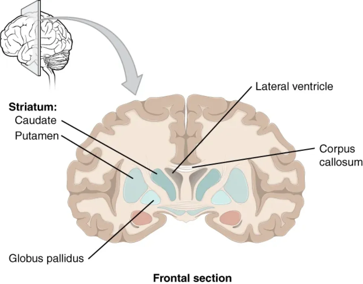
The basal ganglia form a loop with the motor cortex and thalamus. They help select which movements to perform and suppress unwanted movements. Disruption of this circuit - whether from loss of dopaminergic input or degeneration of the basal ganglia neurons themselves - produces characteristic movement disorders.
The Meninges
The brain and spinal cord are wrapped in three protective membranes called meninges, from outermost to innermost:
- Dura mater (“tough mother”): The thickest, outermost layer. It is a dense, fibrous membrane that adheres to the inner surface of the skull. Epidural bleeding (between the skull and dura) is typically caused by arterial rupture and is a neurosurgical emergency.
- Arachnoid mater (“spider mother”): A web-like middle layer. The space beneath it - the subarachnoid space - is filled with cerebrospinal fluid (CSF). Subarachnoid hemorrhage causes “the worst headache of my life.”
- Pia mater (“gentle mother”): The thinnest, innermost layer. It clings directly to the surface of the brain, following every gyrus and sulcus.
Cerebrospinal Fluid (CSF)
CSF is a clear fluid that bathes the brain and spinal cord. It serves three functions: cushioning (shock absorption), buoyancy (reduces the effective weight of the brain from ~1,400 g to ~50 g), and waste removal.
CSF is produced by the choroid plexus, a network of capillaries and ependymal cells lining the ventricles. It circulates through the four ventricles (two lateral ventricles, third ventricle, fourth ventricle), exits into the subarachnoid space, and is eventually reabsorbed into the venous blood via arachnoid granulations.
Blockage of CSF flow causes a dangerous buildup of fluid that increases intracranial pressure.
Putting It All Together: Brain Region Summary
| Structure | Key Functions | Clinical Connection |
|---|---|---|
| Frontal lobe | Motor initiation, planning, personality, Broca’s area (speech production) | Broca’s aphasia, personality changes |
| Parietal lobe | Somatosensory processing, spatial awareness | Hemispatial neglect |
| Temporal lobe | Auditory processing, Wernicke’s area (language comprehension), hippocampus | Wernicke’s aphasia, amnesia |
| Occipital lobe | Visual processing | Cortical blindness |
| Thalamus | Sensory relay (all senses except smell) | Sensory processing disruption |
| Hypothalamus | Homeostasis, pituitary control, ANS regulation | Endocrine dysfunction |
| Midbrain | Visual/auditory reflexes, substantia nigra | Parkinson’s disease |
| Pons | Bridge to cerebellum, breathing regulation | Breathing dysfunction |
| Medulla | Heart rate, breathing, blood pressure, vomiting | Fatal if damaged |
| Cerebellum | Motor coordination, balance, motor learning | Ataxia |
| Hippocampus | Memory formation (short-term to long-term) | Anterograde amnesia |
| Amygdala | Fear, emotion, emotional memory | Reduced fear response |
| Basal ganglia | Motor planning, suppressing unwanted movement | Parkinson’s, Huntington’s |