
Spinal Cord & Reflexes
The spinal cord is far more than a simple relay cable between the brain and body. It is an integration center in its own right, capable of processing sensory information and generating motor commands without any input from the brain. Every time you jerk your hand away from a hot surface before you consciously feel pain, your spinal cord has already handled the situation.
Understanding spinal cord anatomy and reflex arcs is a high-yield MCAT topic that connects neuron structure, signal transmission, and nervous system organization into a single testable pathway.
Spinal Cord Gross Anatomy
The spinal cord is a cylindrical structure roughly 45 cm long in adults. It begins at the foramen magnum, where it is continuous with the brainstem (medulla oblongata), and extends inferiorly to approximately the L1-L2 vertebral level. Below this point, the spinal cord tapers into the conus medullaris, and only a bundle of nerve roots called the cauda equina (“horse’s tail”) continues through the lower vertebral canal.
The spinal cord is protected by the vertebral column, three layers of meninges (dura mater, arachnoid mater, pia mater), and cerebrospinal fluid circulating in the subarachnoid space. Two notable enlargements exist: the cervical enlargement (C4-T1), which serves the upper limbs, and the lumbar enlargement (L1-S3), which serves the lower limbs.
A total of 31 pairs of spinal nerves emerge from the spinal cord: 8 cervical, 12 thoracic, 5 lumbar, 5 sacral, and 1 coccygeal. Each spinal nerve is a mixed nerve containing both sensory (afferent) and motor (efferent) fibers.
Cross-Sectional Anatomy: Grey and White Matter
When you look at a cross-section of the spinal cord, a distinctive butterfly-shaped (or “H”-shaped) region of grey matter sits in the center, surrounded by white matter on the outside. This arrangement is the opposite of the brain, where grey matter forms the outer cortex and white matter is internal.
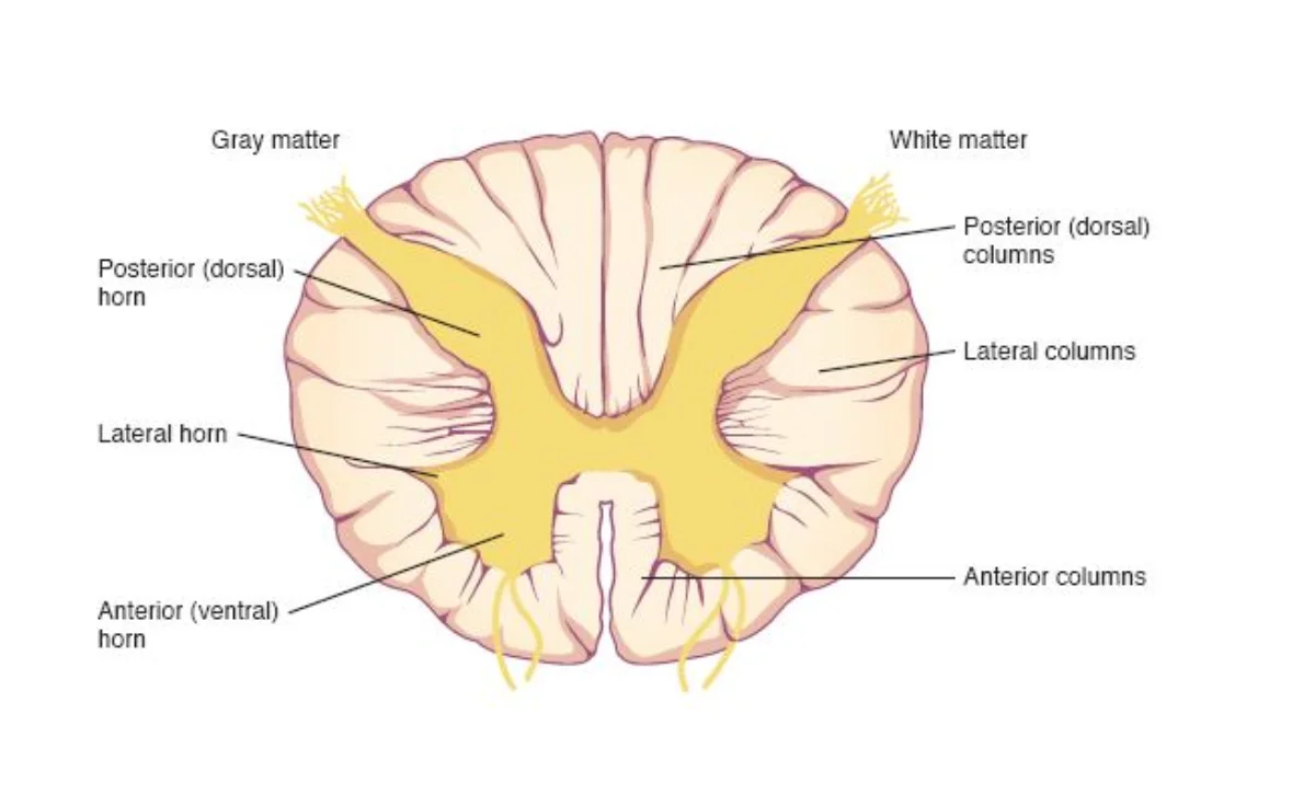
Grey matter contains neuron cell bodies, dendrites, unmyelinated axons, and interneurons. It is the site of synaptic integration. The grey matter is organized into horns:
- Dorsal horns (posterior): receive incoming sensory information from afferent neurons. Think “D” for Dorsal, “D” for Data coming in.
- Ventral horns (anterior): contain motor neuron cell bodies that send commands out to skeletal muscles. Think “V” for Ventral, “V” for Vacate (signals leaving).
- Lateral horns (present only at thoracic and upper lumbar levels, T1-L2): contain cell bodies of preganglionic sympathetic neurons.
White matter surrounds the grey matter and is composed of myelinated axon tracts. The myelin gives it its white appearance. These tracts are organized into three columns (funiculi) on each side: dorsal, lateral, and ventral columns.
Dorsal and Ventral Roots
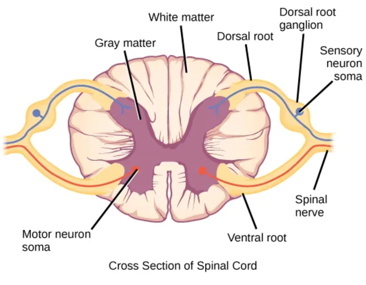
Each spinal nerve forms from the union of a dorsal root and a ventral root.
The dorsal root carries sensory (afferent) fibers into the spinal cord. Just before the dorsal root enters the cord, it swells into the dorsal root ganglion (DRG), which houses the cell bodies of sensory neurons. These are pseudounipolar neurons - their single process splits into a peripheral branch (extending to the receptor) and a central branch (entering the spinal cord).
The ventral root carries motor (efferent) fibers out of the spinal cord. The cell bodies of these motor neurons reside in the ventral horn of the grey matter.
The dorsal and ventral roots merge just lateral to the spinal cord to form a single mixed spinal nerve. This is why spinal nerves carry both sensory and motor information.
Ascending and Descending Tracts
The white matter tracts of the spinal cord serve as information highways connecting the spinal cord to the brain.
Ascending tracts carry sensory information upward from the body to the brain. Two major ascending pathways to know:
- Dorsal column-medial lemniscus pathway: transmits fine touch, vibration, and proprioception. Fibers ascend ipsilaterally (same side) in the dorsal columns and cross over in the medulla.
- Spinothalamic tract: transmits pain, temperature, and crude touch. Fibers cross over in the spinal cord itself and ascend contralaterally (opposite side).
Descending tracts carry motor commands downward from the brain to the body. The most important is:
- Corticospinal (pyramidal) tract: transmits voluntary motor commands from the motor cortex. Most fibers cross over in the medullary pyramids (decussation of pyramids), which is why the left brain controls the right body and vice versa.
| Tract | Direction | Information Carried | Where It Crosses |
|---|---|---|---|
| Dorsal column-medial lemniscus | Ascending | Fine touch, vibration, proprioception | Medulla |
| Spinothalamic | Ascending | Pain, temperature, crude touch | Spinal cord |
| Corticospinal | Descending | Voluntary motor commands | Medullary pyramids |
The Reflex Arc
A reflex is a rapid, involuntary, predictable motor response to a stimulus. Reflexes are mediated by a neural circuit called the reflex arc, which is the simplest functional unit of the nervous system.
Every reflex arc has five components, always in this order:
- Receptor - detects the stimulus (e.g., stretch receptor in muscle, pain receptor in skin)
- Sensory (afferent) neuron - transmits the signal from the receptor to the CNS via the dorsal root
- Integration center - processes the signal within the CNS (spinal cord for spinal reflexes); this may be a single synapse or involve interneurons
- Motor (efferent) neuron - carries the response command from the CNS to the effector via the ventral root
- Effector - the muscle or gland that carries out the response
Monosynaptic vs. Polysynaptic Reflexes
Monosynaptic reflexes involve only one synapse - a direct connection between the sensory neuron and the motor neuron with no interneuron in between. The classic example is the patellar (knee-jerk) reflex. When the patellar tendon is tapped, muscle spindles in the quadriceps detect the stretch. The sensory neuron synapses directly onto a motor neuron in the ventral horn, which fires and contracts the quadriceps, causing the leg to kick forward.
Polysynaptic reflexes involve one or more interneurons between the sensory and motor neurons. The withdrawal (flexor) reflex is the classic example. When you step on a sharp object, sensory neurons activate interneurons in the spinal cord, which then activate motor neurons to flex the injured limb away from the stimulus. At the same time, a crossed-extensor reflex activates extensors in the opposite leg to maintain balance.
Supraspinal Modulation of Reflexes
Spinal reflexes do not operate in isolation. Descending pathways from the brain continuously modulate the local reflex arc, either suppressing it (most of the time) or enhancing it when needed. This is why you can consciously override reflex urges, why reflex strength changes with attention or stress, and why upper motor neuron damage produces hyperreflexia (the “brakes” from above are lost, leaving the intact local arc unchecked).
The take-home: a reflex arc is the fastest possible response, but it is always running inside a larger loop that includes supraspinal feedback from the motor cortex, brainstem, and cerebellum.
Clinical Relevance: Upper vs. Lower Motor Neuron Lesions
Understanding the reflex arc helps explain clinical findings that appear on the MCAT. The key concept is that the reflex arc itself is a local spinal cord circuit. Whether a lesion is “above” or “below” this circuit determines the pattern of symptoms.