
Sensory & Motor Pathways
Every sensation you experience - the pressure of a handshake, the burn of a hot pan, the position of your limbs in space - begins with a sensory receptor converting a stimulus into an electrical signal. That signal must then travel along specific pathways to reach the brain, where it is consciously perceived. Likewise, every voluntary movement originates in the motor cortex and travels along descending pathways to reach the muscles. The MCAT tests both the receptor types and the major pathways, along with the twelve cranial nerves that serve as direct lines between the brain and specific structures.
Sensory Receptor Types by Stimulus
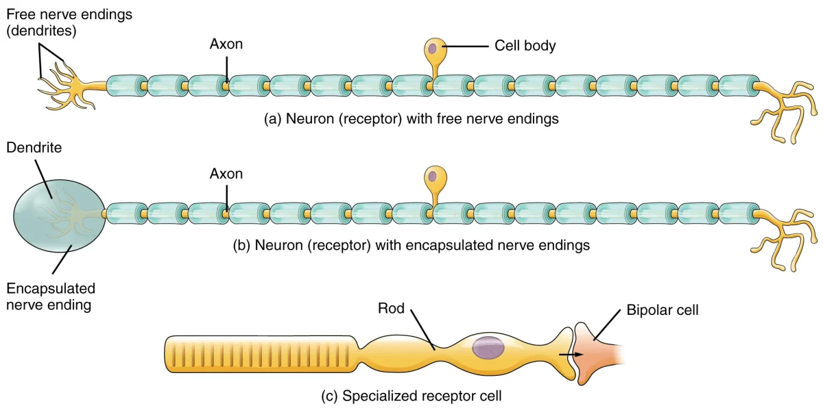
Sensory receptors are classified by the type of stimulus they detect. Each receptor type responds best to one specific form of energy, known as its adequate stimulus.
- Mechanoreceptors respond to mechanical forces - pressure, vibration, stretch, and touch. Examples include Meissner’s corpuscles (light touch), Pacinian corpuscles (deep pressure and vibration), Merkel’s discs (sustained pressure), and muscle spindles (stretch).
- Thermoreceptors respond to changes in temperature. Separate populations of free nerve endings detect warming versus cooling.
- Nociceptors respond to potentially damaging stimuli and generate the sensation of pain. They are free nerve endings activated by extreme heat, extreme cold, intense mechanical force, or inflammatory chemicals.
- Chemoreceptors respond to specific chemical molecules. Examples include olfactory receptors (smell), taste receptors (gustation), and peripheral chemoreceptors that monitor blood oxygen and pH.
- Photoreceptors respond to light. Rods and cones in the retina are the primary examples.
Sensory Receptor Classification by Location
Receptors can also be grouped by where in the body they are found:
- Exteroceptors are located at or near the body surface and detect external stimuli - touch, pressure, temperature, pain, and light. They keep you aware of the outside environment.
- Interoceptors (visceroceptors) are located within internal organs and blood vessels. They monitor conditions such as blood pressure, blood oxygen levels, and organ stretch. You are usually not consciously aware of these signals.
- Proprioceptors are located in muscles, tendons, joints, and the inner ear. They provide information about body position, movement, and equilibrium. Muscle spindles detect stretch, Golgi tendon organs detect tension, and joint kinesthetic receptors detect joint position.
Ascending (Sensory) Pathways
Sensory information from the body travels to the brain through two major ascending pathways. Both ultimately reach the somatosensory cortex in the parietal lobe, but they carry different types of information and cross the midline at different levels.
Dorsal column-medial lemniscus (DCML) pathway: Carries fine touch, vibration, two-point discrimination, and proprioception. First-order neurons ascend ipsilaterally in the dorsal columns of the spinal cord, synapse in the medulla, cross the midline there, and then ascend to the thalamus. Third-order neurons relay the signal from the thalamus to the somatosensory cortex.
Spinothalamic tract: Carries pain, temperature, and crude (non-discriminative) touch. First-order neurons synapse soon after entering the spinal cord. Second-order neurons cross the midline within the spinal cord and then ascend contralaterally to the thalamus. Third-order neurons relay to the somatosensory cortex.
| Feature | DCML Pathway | Spinothalamic Tract |
|---|---|---|
| Carries | Fine touch, vibration, proprioception | Pain, temperature, crude touch |
| Crosses midline at | Medulla | Spinal cord |
| Ascends on | Ipsilateral side (until medulla) | Contralateral side |
Descending (Motor) Pathways
Motor commands from the brain travel down to the spinal cord and out to muscles through descending pathways.
Corticospinal (pyramidal) tract: This is the primary pathway for voluntary movement. Upper motor neurons originate in the primary motor cortex (precentral gyrus) and descend through the brainstem. Approximately 90% of fibers cross the midline at the medullary pyramids (pyramidal decussation) and descend in the lateral corticospinal tract. They synapse on lower motor neurons in the ventral horn of the spinal cord, which then innervate skeletal muscles.
This crossing explains why the left motor cortex controls the right side of the body and vice versa.
Upper motor neurons (UMNs) reside entirely within the CNS - their cell bodies are in the motor cortex and their axons descend through the brainstem and spinal cord. Lower motor neurons (LMNs) have cell bodies in the ventral horn of the spinal cord (or brainstem motor nuclei for cranial nerves) and their axons exit the CNS to directly innervate skeletal muscles.
The Twelve Cranial Nerves
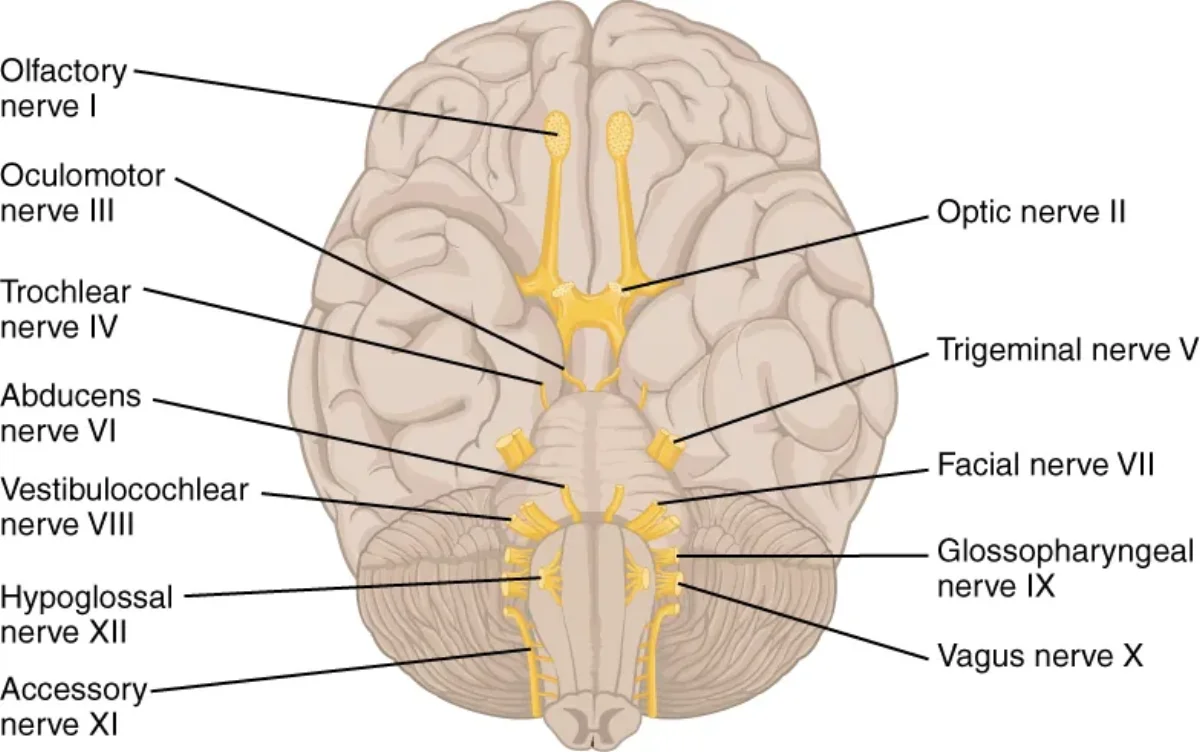
Unlike spinal nerves, which emerge from the spinal cord, cranial nerves emerge directly from the brain (mostly the brainstem). There are 12 pairs, numbered I through XII in order from anterior to posterior. Some are purely sensory, some are purely motor, and some carry both types of fibers.
| Number | Name | Type | Key Functions |
|---|---|---|---|
| I | Olfactory | Sensory | Smell |
| II | Optic | Sensory | Vision |
| III | Oculomotor | Motor | Most eye movements; pupil constriction; lens accommodation |
| IV | Trochlear | Motor | Superior oblique muscle (eye movement - looking down and inward) |
| V | Trigeminal | Both | Facial sensation (touch, pain, temperature); mastication (chewing) |
| VI | Abducens | Motor | Lateral rectus muscle (eye abduction) |
| VII | Facial | Both | Facial expression; taste (anterior of tongue); salivation; lacrimation |
| VIII | Vestibulocochlear | Sensory | Hearing (cochlear branch); balance/equilibrium (vestibular branch) |
| IX | Glossopharyngeal | Both | Taste (posterior of tongue); swallowing; monitors carotid body/sinus |
| X | Vagus | Both | Parasympathetic to thoracic and abdominal viscera; voice; swallowing; taste (epiglottis) |
| XI | Accessory (Spinal Accessory) | Motor | Trapezius and sternocleidomastoid muscles (head/shoulder movement) |
| XII | Hypoglossal | Motor | Tongue movement |
Somatotopic Organization
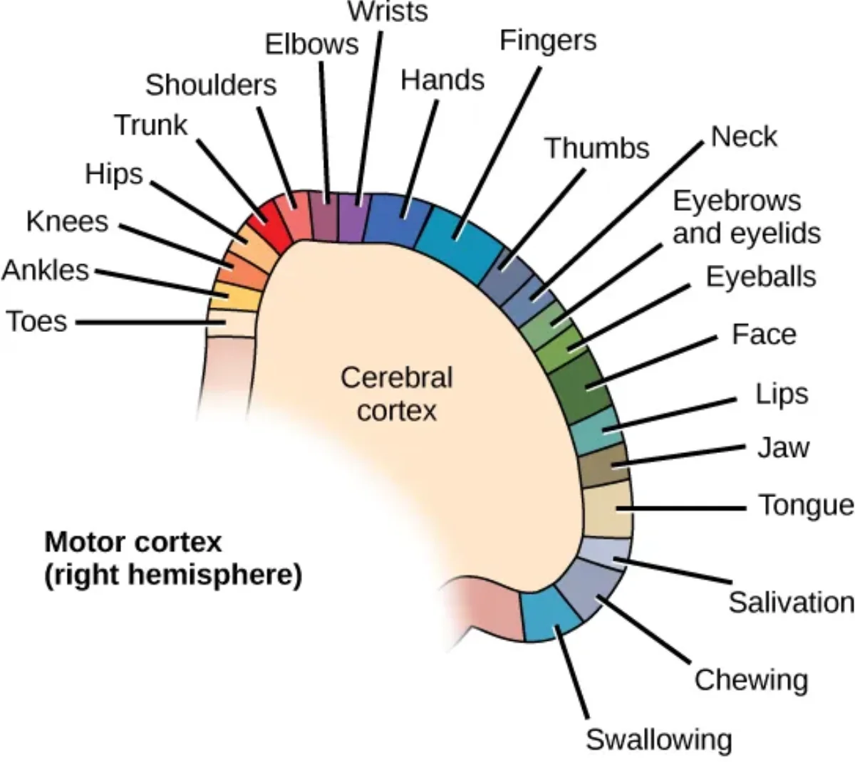
Both the somatosensory cortex (in the postcentral gyrus) and the motor cortex (in the precentral gyrus) are organized somatotopically, meaning that specific cortical regions correspond to specific body parts. This creates the homunculus - a distorted “little human” map where the size of each body region reflects the density of its innervation, not its physical size.
The hands, lips, and tongue occupy disproportionately large areas in both the sensory and motor homunculi because these regions require extremely fine sensory discrimination and precise motor control. In contrast, the trunk and legs occupy relatively small cortical areas.