
Heart Anatomy
The heart is a muscular organ roughly the size of your fist, located slightly left of center in the thoracic cavity between the lungs. Its sole job is deceptively simple: pump blood. But the engineering behind that job is anything but simple. The heart must simultaneously send deoxygenated blood to the lungs and oxygenated blood to the entire body, and it must do this without ever letting the two streams mix.
Four Chambers, Two Pumps
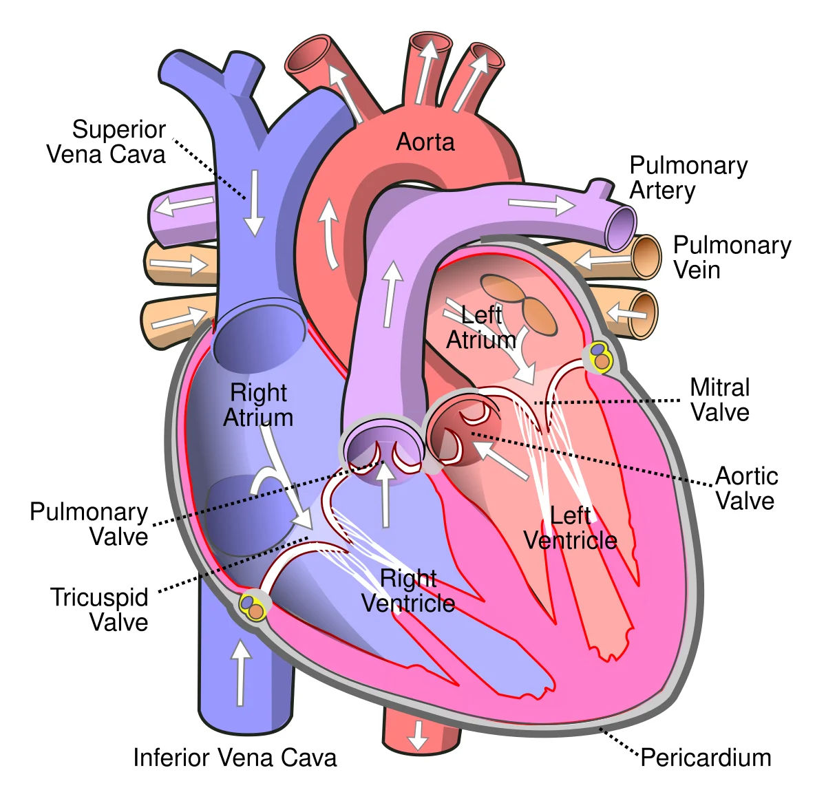
The heart has four chambers arranged as two side-by-side pumps.
The right atrium receives deoxygenated blood from the body via two large veins: the superior vena cava (draining the upper body) and the inferior vena cava (draining the lower body). The coronary sinus also empties deoxygenated blood from the heart muscle itself into the right atrium.
The right ventricle receives blood from the right atrium and pumps it to the lungs via the pulmonary trunk, which splits into the left and right pulmonary arteries. This is the only place in the body where arteries carry deoxygenated blood.
The left atrium receives oxygenated blood returning from the lungs via four pulmonary veins. These are the only veins in the body that carry oxygenated blood.
The left ventricle is the powerhouse. It receives blood from the left atrium and pumps it into the aorta, which distributes blood to every organ in the body. Because it must generate enough pressure to reach your toes, the left ventricular wall is approximately three times thicker than the right.
The Four Valves
Valves ensure blood flows in only one direction. They open passively when pressure pushes blood forward and snap shut when blood tries to flow backward.
There are two types of valves:
Atrioventricular (AV) valves sit between atria and ventricles:
- Tricuspid valve - right side, three leaflets
- Bicuspid (mitral) valve - left side, two leaflets
Semilunar valves sit at the exits of the ventricles:
- Pulmonary valve - exit of right ventricle, opens into pulmonary trunk
- Aortic valve - exit of left ventricle, opens into aorta
Heart Wall and Support Structures
The heart wall is composed predominantly of cardiac muscle (myocardium), which generates the force for contraction. The myocardium is much thicker in the left ventricle than the right, reflecting the greater workload of pumping blood through the systemic circulation.
The entire heart sits inside the pericardial sac (pericardium), a double-walled membrane filled with serous fluid that reduces friction as the heart beats.
The AV valves are anchored by chordae tendineae - tough cords connected to papillary muscles in the ventricular walls. These prevent the valve leaflets from being blown back into the atria during ventricular contraction.
The Three Circulations
Pulmonary circulation: Right ventricle → pulmonary arteries → lungs (gas exchange) → pulmonary veins → left atrium. This is a low-pressure circuit (~ mmHg).
Systemic circulation: Left ventricle → aorta → arteries → capillaries (nutrient/gas exchange) → veins → vena cavae → right atrium. This is a high-pressure circuit (~ mmHg).
Coronary circulation: The heart feeds itself first. The left and right coronary arteries branch off the aorta immediately above the aortic valve. After delivering oxygen to the myocardium, coronary veins drain into the coronary sinus, which empties into the right atrium.
Portal Systems
In most circulations, blood passes through one capillary bed before returning to the heart. In a portal system, blood passes through two capillary beds in series before returning. There are three portal systems to know:
| Portal System | Pathway | Purpose |
|---|---|---|
| Hepatic portal | Intestinal capillaries → hepatic portal vein → liver capillaries | Delivers absorbed nutrients to the liver for processing before entering general circulation |
| Hypophyseal portal | Hypothalamic capillaries → portal veins → anterior pituitary capillaries | Allows hypothalamic releasing hormones to reach the anterior pituitary directly |
| Renal portal | Glomerular capillaries → efferent arteriole → peritubular capillaries (vasa recta) | Allows reabsorption of filtered substances back into the blood |
Fetal Circulation
Before birth, the lungs are collapsed and non-functional. The fetus receives oxygen from the placenta, not from breathing. Three temporary shunts redirect blood away from the lungs:
| Shunt | Connects | Purpose | Closes after birth |
|---|---|---|---|
| Foramen ovale | Right atrium → left atrium | Bypasses right ventricle/lungs | Becomes fossa ovalis |
| Ductus arteriosus | Pulmonary artery → aorta | Diverts blood past the lungs | Becomes ligamentum arteriosum |
| Ductus venosus | Umbilical vein → inferior vena cava | Bypasses the liver | Becomes ligamentum venosum |
All three shunts close shortly after birth when the newborn takes its first breaths and pulmonary resistance drops dramatically.