
Blood Vessels
The circulatory system uses five types of blood vessels, each engineered for a specific job. Understanding their structural differences is the key to understanding why blood flows the way it does - and why it fails in disease.
The Three Layers of Blood Vessels
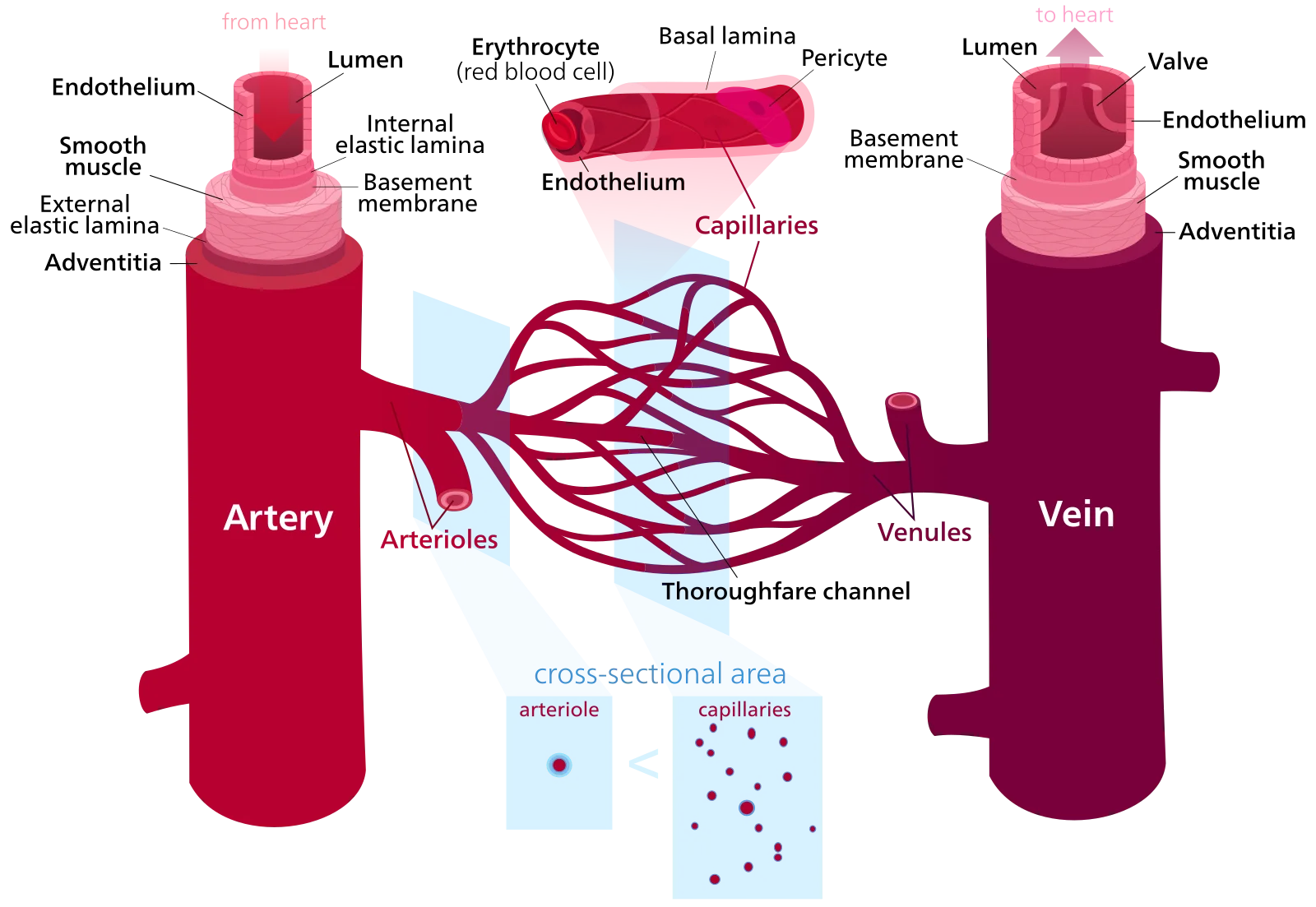
Most blood vessels share a common three-layer wall structure:
| Layer | Composition | Function |
|---|---|---|
| Tunica intima (innermost) | Single layer of endothelial cells | Smooth, non-thrombogenic surface for blood flow; regulates permeability |
| Tunica media (middle) | Smooth muscle + elastic fibers | Vasoconstriction/vasodilation; maintains vessel tone |
| Tunica externa (outermost) | Connective tissue (collagen) | Anchors vessel to surrounding tissue; structural support |
Arteries
Arteries carry blood away from the heart under high pressure. They have thick walls with abundant smooth muscle and elastic fibers in the tunica media.
Elastic (conducting) arteries - the largest arteries (aorta, pulmonary trunk, carotid arteries). Their walls are rich in elastic fibers that stretch during systole and recoil during diastole, smoothing out the pulsatile flow from the heart. This is why you still have blood flow during diastole - the elastic recoil of these arteries acts like a secondary pump.
Muscular (distributing) arteries - medium-sized arteries (brachial, femoral, renal arteries). More smooth muscle, less elastic tissue. They distribute blood to specific organs and can constrict or dilate to redirect flow.
Arterioles: The Resistance Vessels
Arterioles are the smallest arteries (diameter: 10-100 micrometers) and the primary regulators of blood pressure and blood flow distribution. Their smooth muscle walls can constrict (vasoconstriction) or relax (vasodilation) to change resistance dramatically.
A small change in arteriole radius produces a massive change in resistance. This relationship follows Poiseuille’s law: resistance is inversely proportional to the fourth power of the radius. Halve the radius and resistance increases 16-fold.
Capillaries: The Exchange Vessels
Capillaries are the thinnest vessels (diameter: 5-10 micrometers, just wide enough for a single red blood cell). Their walls are only one endothelial cell thick - no tunica media, no tunica externa. This thinness is essential for diffusion of gases, nutrients, and waste.
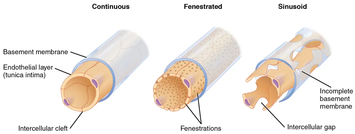
There are three types of capillaries:
| Type | Structure | Where Found | What Passes Through |
|---|---|---|---|
| Continuous | Tight junctions, no pores | Muscle, skin, lungs, brain (BBB) | Small molecules, water, ions only |
| Fenestrated | Small pores (60-80 nm) in endothelial cells | Kidneys, intestines, endocrine glands | Small proteins, filtered fluids |
| Sinusoidal | Large gaps (30-40 micrometers) | Liver, spleen, bone marrow | RBCs, WBCs, large proteins - almost everything |
Velocity and Cross-Sectional Area
Blood flows fastest in the aorta and slowest in the capillaries. This seems counterintuitive - the capillaries are tiny, so shouldn’t flow speed up? No, because while each individual capillary is narrow, the total cross-sectional area of all capillaries combined is enormous (about 600 times the area of the aorta).
By the continuity equation (A1v1 = A2v2), when total cross-sectional area increases, velocity must decrease. This slow flow in capillaries is essential - it gives time for gas and nutrient exchange.
Veins: The Capacitance Vessels
Veins return blood to the heart under low pressure. Their walls are thinner and more compliant (stretchable) than arteries. Veins hold about 60-65% of total blood volume at any time, which is why they are called capacitance vessels.
Because venous pressure is so low (~5-10 mmHg), veins use several mechanisms to push blood back to the heart:
- Venous valves - one-way flaps that prevent backflow, especially in the legs where blood must travel against gravity
- Skeletal muscle pump - contraction of surrounding muscles squeezes veins, pushing blood upward
- Respiratory pump - during inhalation, decreased thoracic pressure creates a pressure gradient that pulls venous blood toward the heart
Vessel Comparison Summary
| Feature | Arteries | Arterioles | Capillaries | Venules | Veins |
|---|---|---|---|---|---|
| Wall thickness | Thickest | Thick | Thinnest (1 cell) | Thin | Moderate |
| Lumen diameter | Large | Small | Tiny (5-10 um) | Small | Large |
| Blood pressure | Highest | High | Low | Very low | Lowest |
| Flow velocity | Fast | Moderate | Slowest | Slow | Moderate |
| Special features | Elastic recoil | Resistance control | Gas exchange | WBC migration | Valves, high volume |