Think of your respiratory system as a highway system. Air enters through the on-ramp (nose or mouth), travels down an increasingly narrow series of roads (pharynx, larynx, trachea, bronchi, bronchioles), and finally arrives at the parking lot where actual business happens (alveoli). Everything before the alveoli is just getting air to the right place - the conducting zone. The alveoli are the only place where gas exchange actually occurs - the respiratory zone.
This distinction matters for the MCAT: the conducting zone warms, humidifies, and filters air but does zero gas exchange. The respiratory zone is where oxygen enters your blood and CO2 leaves it.
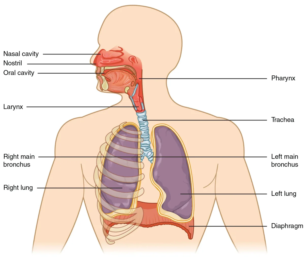
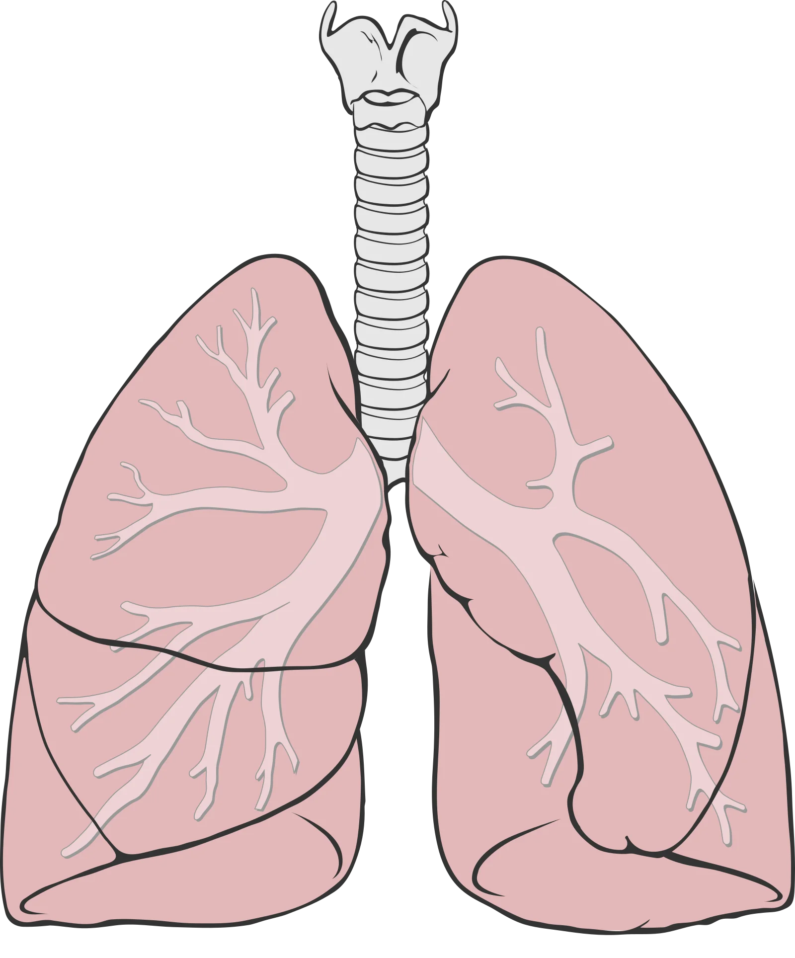
The respiratory system from the nasal cavity to the alveoli, showing the upper and lower respiratory tracts. Credit: OpenStax Anatomy & Physiology 2e, CC BY 4.0
The Upper Respiratory Tract
The upper respiratory tract includes the nasal cavity, pharynx, and larynx. These structures sit above the trachea and serve as the entry point and first line of defense for incoming air.
The Nasal Cavity
Air enters through the nostrils and passes into the nasal cavity, which is divided by the nasal septum. Three bony projections called nasal conchae (or turbinates) protrude into each side of the cavity. These conchae create turbulent airflow, forcing air to swirl and make maximum contact with the mucosal lining.
The nasal cavity performs three critical functions simultaneously:
Warming - A rich blood supply in the mucosal lining heats incoming air to body temperature
Humidifying - Mucus and serous secretions add moisture, protecting the delicate lower airway tissues
Filtering - Nasal hairs (vibrissae) trap large particles, while mucus produced by goblet cells captures smaller particles and pathogens
The nasal cavity also contains the olfactory epithelium in its superior region, which is responsible for the sense of smell.
The Pharynx
The pharynx is a shared passageway for both air and food. It is divided into three regions:
Region
Location
Function
Nasopharynx
Behind the nasal cavity
Air passage only; contains pharyngeal tonsils (adenoids)
Oropharynx
Behind the oral cavity
Shared air and food passage
Laryngopharynx
Above the larynx
Shared passage; diverges into larynx (air) and esophagus (food)
The pharynx is where the respiratory and digestive pathways cross. This crossover is why you can choke - food can accidentally enter the airway instead of the esophagus.
The Larynx
The larynx (voice box) sits at the top of the trachea and serves two functions: sound production and airway protection.
The epiglottis is a flap of elastic cartilage that covers the opening of the larynx during swallowing, preventing food from entering the airway. When you swallow, the larynx elevates and the epiglottis folds down like a trap door over the glottis (the opening between the vocal cords).
The vocal cords (vocal folds) stretch across the larynx. Air passing between them causes vibration, producing sound. Tension in the cords controls pitch - tighter cords produce higher-pitched sounds.
The Lower Respiratory Tract
Below the larynx, the lower respiratory tract begins with the trachea and extends through the bronchial tree to the alveoli.
The Trachea
The trachea (windpipe) is a tube roughly 10-12 cm long that connects the larynx to the bronchi. It is reinforced by 16-20 C-shaped cartilage rings that keep it open during pressure changes. These rings are C-shaped (not complete circles) because the open side faces the esophagus posteriorly, allowing the esophagus to expand when you swallow food.
The trachea is lined with pseudostratified ciliated columnar epithelium containing goblet cells. This is the beginning of the mucociliary escalator - a defense system where mucus traps particles and cilia beat in coordinated waves to push the mucus upward toward the pharynx, where it is swallowed or expelled.
The Mucociliary Escalator
The mucociliary escalator deserves special attention because it is one of the respiratory system’s most important innate defense mechanisms. Here is how it works:
Goblet cells secrete sticky mucus that lines the airways
Cilia on epithelial cells beat in coordinated waves (about 10-20 beats per second)
The beating cilia propel the mucus - along with any trapped bacteria, dust, or debris - upward toward the pharynx
At the pharynx, the mucus is swallowed (and destroyed by stomach acid) or coughed out
Smoking paralyzes cilia and increases mucus production simultaneously. This is why smokers develop a chronic “smoker’s cough” - without functioning cilia, coughing becomes the only way to clear mucus from the airways.
Other Respiratory Defenses
The mucociliary escalator is not the only defense mechanism. The respiratory system also uses:
Lysozyme - an enzyme found in nasal secretions, tears, and saliva that attacks the peptidoglycan cell walls of gram-positive bacteria
IgA antibodies - mucosal surfaces are coated with secretory IgA, which binds pathogens and prevents them from attaching to epithelial cells
Alveolar macrophages (dust cells) - immune cells that patrol the alveolar surfaces, engulfing bacteria and particles that make it past the upper defenses
Mast cells - immune cells in the lungs with antibodies on their surface. When triggered, they release inflammatory chemicals. Mast cells are also responsible for allergic reactions in the airways (like asthma)
Why are the cartilage rings in the trachea C-shaped rather than complete circles?
Click to reveal answer
The open side of the C faces the esophagus posteriorly. This allows the esophagus to expand into the tracheal space when a food bolus is swallowed. Complete rings would prevent this expansion and interfere with swallowing.
What is the mucociliary escalator, and what happens to it in smokers?
Click to reveal answer
The mucociliary escalator is the defense system in which goblet cells produce mucus to trap particles, and cilia beat to push that mucus upward to the pharynx. Smoking paralyzes cilia while increasing mucus production, forcing the person to rely on coughing to clear the airways - hence the chronic "smoker's cough."
Thermoregulation
The respiratory system plays an important role in temperature regulation. As air passes through the nasal cavity and upper airways, the rich capillary beds in the mucosal lining warm incoming air to body temperature and humidify it to near 100% humidity. This protects the delicate lower airways from cold, dry air damage.
During exhalation, heat and water vapor are carried out of the body with the exhaled air. This represents a source of evaporative heat loss. When body temperature rises (during exercise or fever), increased respiratory rate accelerates this heat loss. In some animals, panting is a primary thermoregulation mechanism - rapid shallow breathing maximizes evaporative cooling from the respiratory surfaces.
Conducting Zone vs. Respiratory Zone
This is a critical distinction for the MCAT:
Feature
Conducting Zone
Respiratory Zone
Structures
Nose to terminal bronchioles
Respiratory bronchioles, alveolar ducts, alveoli
Gas exchange?
No
Yes
Cartilage support?
Yes (decreases distally)
No
Smooth muscle?
Yes (increases in bronchioles)
Minimal
Epithelium
Pseudostratified ciliated columnar
Simple squamous
Also called
”Anatomical dead space"
"Gas exchange surface”
The conducting zone is called anatomical dead space because air sitting in these passages is not participating in gas exchange. Typical anatomical dead space volume is about 150 mL. This means that of a normal 500 mL tidal breath, only about 350 mL actually reaches the alveoli for gas exchange.
A patient takes a 500 mL tidal breath. If anatomical dead space is 150 mL, how much air actually participates in gas exchange?
Click to reveal answer
350 mL. Only air reaching the alveoli participates in gas exchange. Dead space volume (150 mL) fills the conducting airways but does not contact gas exchange surfaces. Alveolar ventilation = tidal volume - dead space = 500 - 150 = 350 mL per breath.
If the trachea is the trunk of a tree, the bronchi are the main branches, the bronchioles are the smaller twigs, and the alveoli are the leaves - tiny, numerous, and where the real work happens. Your lungs contain about 300 million alveoli, creating a total gas exchange surface area roughly the size of a tennis court (about 70 square meters). All of that surface area is packed inside your chest because the alveoli are incredibly small - each one is only about 0.2 mm in diameter.
Interactive 3D Bronchi and Alveoli. Zoom in from the bronchial tree down to the alveolar sacs where gas exchange occurs.Credit: zames1992 via Sketchfab, CC BY
The Bronchial Tree
At the carina (the ridge where the trachea divides), the trachea splits into the right and left main (primary) bronchi. Each main bronchus enters its respective lung at the hilum - the medial surface where blood vessels, nerves, and lymphatics also enter.
The branching pattern is:
Main (primary) bronchi - one per lung; the right is wider, shorter, and more vertical than the left
Lobar (secondary) bronchi - one per lobe (3 right, 2 left)
Bronchioles - no cartilage, walls are smooth muscle
Terminal bronchioles - the smallest conducting airways (end of the conducting zone)
Respiratory bronchioles - have some alveoli budding from their walls (beginning of the respiratory zone)
Alveolar ducts and alveolar sacs - clusters of alveoli where gas exchange occurs
Key Structural Changes Along the Bronchial Tree
As you move from the trachea toward the alveoli, several important structural changes occur:
Feature
Trachea/Bronchi
Bronchioles
Alveoli
Cartilage
C-rings/plates (structural support)
None
None
Smooth muscle
Minimal
Abundant (controls airflow)
Minimal
Epithelium
Pseudostratified ciliated columnar
Simple ciliated columnar/cuboidal
Simple squamous
Diameter
Large
Small (< 1 mm)
Tiny (~0.2 mm)
Function
Conduct air
Conduct air, regulate airflow
Gas exchange
Two trends to remember: cartilage decreases while smooth muscle increases as airways get smaller. Large airways need cartilage to stay open, but small airways need smooth muscle to regulate airflow distribution. This is why asthma (bronchiolar constriction) affects exhalation so dramatically: the small airways have no cartilage to keep them propped open against the squeeze.
The Lungs
The two lungs are not identical:
Right lung: 3 lobes (superior, middle, inferior), separated by the horizontal and oblique fissures
Left lung: 2 lobes (superior, inferior), separated by the oblique fissure; has a cardiac notch - an indentation where the heart sits
The Pleural Membranes
Each lung is enclosed in a double-layered membrane called the pleura:
Visceral pleura - the inner layer, directly adherent to the lung surface. It dips into the fissures between lobes.
Parietal pleura - the outer layer, lining the inside of the chest wall and the superior surface of the diaphragm.
Pleural cavity - the potential space between the two layers, containing a thin film of pleural fluid.
The pleural fluid serves two functions: it lubricates the pleural surfaces so the lungs can slide smoothly during breathing, and it creates surface tension that keeps the lungs adhered to the chest wall (like two wet glass slides stuck together).
Intrapleural Pressure
The pressure within the pleural cavity (intrapleural pressure) is normally negative - about -4 mmHg at rest relative to atmospheric pressure. This negative pressure is critical because it keeps the lungs inflated.
Why is it negative? The lungs have elastic recoil that constantly tries to collapse them inward, while the chest wall has its own elastic tendency to spring outward. These two opposing forces pull the pleural layers slightly apart, creating a suction effect (negative pressure) in the pleural space.
Pneumothorax
A pneumothorax occurs when air enters the pleural space, breaking the negative pressure seal. Without negative intrapleural pressure, the elastic recoil of the lung is no longer opposed, and the lung collapses.
A pneumothorax can result from chest trauma or spontaneous rupture of a small air pocket on the lung surface. The key concept is understanding why loss of negative intrapleural pressure leads to lung collapse.
The Alveoli
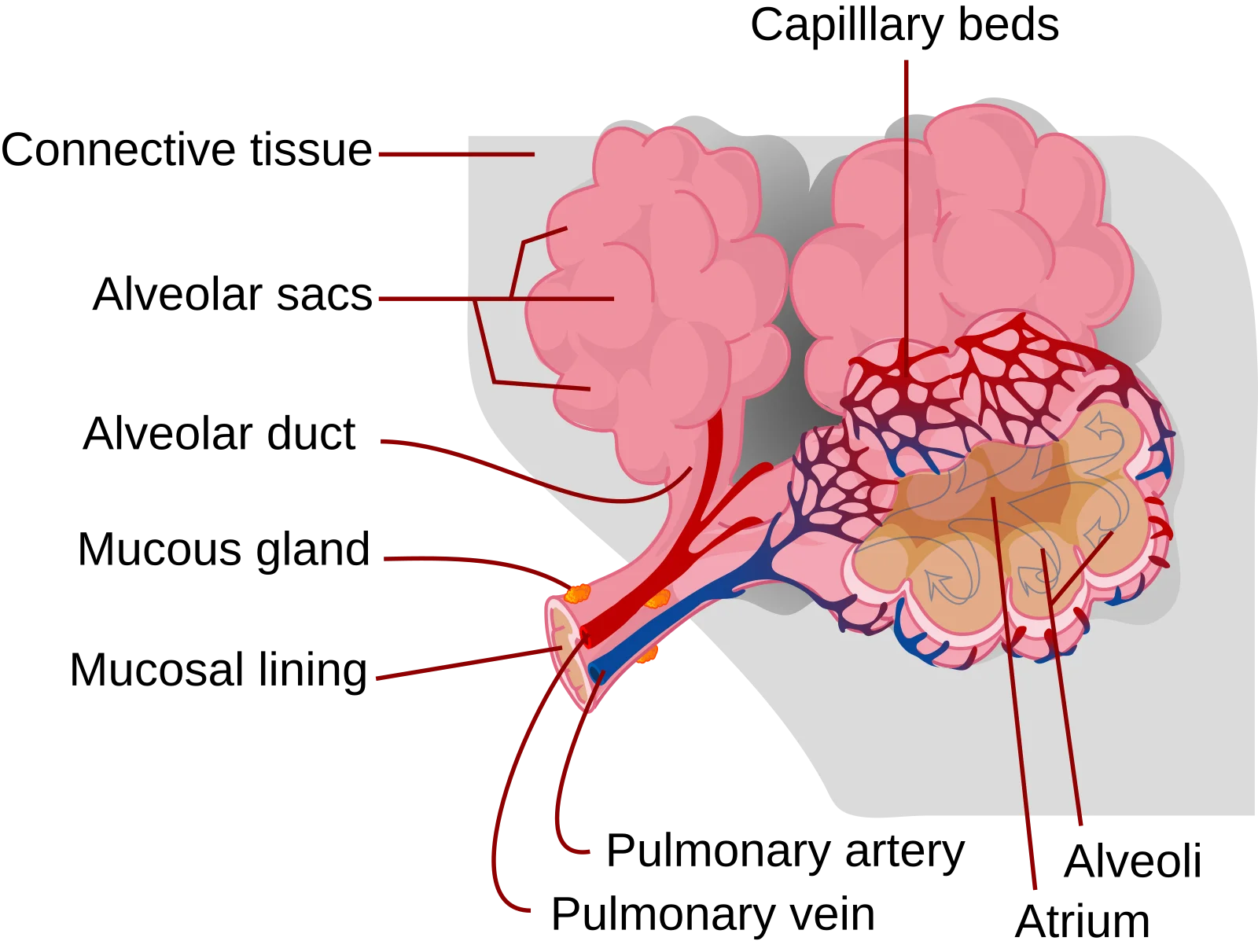
The alveoli are the functional units of the lung - tiny, thin-walled sacs where all gas exchange occurs. Each alveolus is wrapped in a dense network of capillaries, and the barrier between alveolar air and capillary blood is extraordinarily thin - only about 0.5 micrometers.
This barrier, called the respiratory membrane, consists of:
A thin layer of fluid lining the alveolus (with surfactant)
The alveolar epithelium (Type I cells)
A fused basement membrane
The capillary endothelium
Structure of an alveolus. Each alveolus is surrounded by capillaries for gas exchange across the thin respiratory membrane. Credit: LadyofHats / Wikimedia Commons, Public Domain
Two types of cells make up the alveolar epithelium:
Cell Type
Structure
Function
Type I pneumocytes (alveolar cells)
Very thin, flat squamous cells; cover ~95% of alveolar surface
Gas exchange - their thinness minimizes diffusion distance
Type II pneumocytes (alveolar cells)
Cuboidal cells; cover ~5% of surface but are more numerous
Secrete surfactant; can divide to regenerate Type I cells
Alveolar macrophages (dust cells) patrol the alveolar surfaces, engulfing bacteria, dust, and other particles that make it past the mucociliary escalator. They are the last line of defense before the gas exchange surface.
Surfactant
Surfactant is a phospholipid-rich substance (primarily dipalmitoylphosphatidylcholine, or DPPC) secreted by Type II pneumocytes. It reduces surface tension within the alveoli, preventing them from collapsing during exhalation.
Why is this necessary? The alveoli are essentially tiny wet bubbles. Surface tension in a wet sphere naturally tries to collapse it (think of a soap bubble popping). Without surfactant, the smallest alveoli would collapse first (because LaPlace’s law tells us that smaller spheres have higher collapsing pressure for the same surface tension), and each breath would require enormous effort to re-inflate them.
What do Type I and Type II pneumocytes do, and which is more numerous?
Click to reveal answer
Type I pneumocytes are thin, flat cells that cover ~95% of the alveolar surface and are specialized for gas exchange. Type II pneumocytes are cuboidal cells that secrete surfactant and can regenerate Type I cells. Type II cells are actually more numerous by cell count, but Type I cells cover far more surface area due to their flattened shape.
Why would a pneumothorax cause a lung to collapse?
Click to reveal answer
A pneumothorax introduces air into the pleural space, eliminating the negative intrapleural pressure. Normally, negative intrapleural pressure opposes the elastic recoil of the lung, keeping it inflated. When air enters the pleural space and pressure equalizes to atmospheric, nothing opposes elastic recoil, so the lung collapses inward.
How does surfactant prevent alveolar collapse, and what law explains why small alveoli are most vulnerable?
Click to reveal answer
Surfactant reduces surface tension in alveoli. LaPlace's law (P = 2T/r) shows that smaller alveoli have higher collapsing pressure for a given surface tension. Without surfactant, small alveoli would collapse into larger ones. Surfactant disproportionately reduces surface tension in smaller alveoli (where it is more concentrated), equalizing pressure and preventing collapse.
Here is a question that trips up many students: do you actively suck air into your lungs? No. You create a pressure difference, and the atmosphere pushes air in for you. Breathing is entirely about pressure gradients, and the physics behind it is Boyle’s Law - one of the simplest gas laws you will encounter on the MCAT.
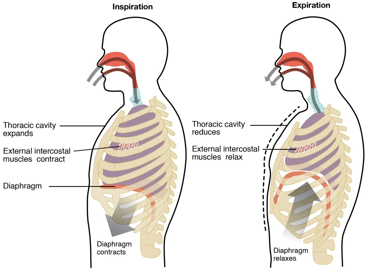
Mechanics of breathing: during inspiration, the diaphragm contracts and flattens, expanding the thoracic cavity and drawing air in. During expiration, the diaphragm relaxes and the elastic recoil of the lungs pushes air out. Credit: OpenStax Anatomy & Physiology 2e, CC BY 4.0The diaphragm and rib cage during breathing. Note the bucket-handle motion of the ribs during inspiration (external intercostals contract, lifting ribs up and out). Credit: Lumen Learning / OpenStax Anatomy & Physiology, CC BY 4.0
Inspiration (Inhalation)
Inspiration is an active process. It requires muscle contraction and energy expenditure. Here is what happens in sequence:
Step 1: Muscle contraction. The diaphragm (the dome-shaped muscle separating the thoracic and abdominal cavities) contracts and flattens downward, increasing the vertical dimension of the thoracic cavity. Simultaneously, the external intercostal muscles contract, pulling the ribs upward and outward (think of a bucket handle lifting), increasing the anteroposterior and lateral dimensions.
Step 2: Volume increases. The thoracic cavity expands in all three dimensions.
Step 3: Pressure drops. Per Boyle’s Law, as volume increases, intrapulmonary pressure (pressure inside the lungs) drops below atmospheric pressure. This creates a negative pressure gradient.
Step 4: Air rushes in. Air always flows from high pressure to low pressure. Since atmospheric pressure (760 mmHg at sea level) is now higher than intrapulmonary pressure, air flows into the lungs until pressures equalize.
Expiration (Exhalation)
Quiet expiration is a passive process. It requires no muscle contraction:
Step 1: Muscles relax. The diaphragm relaxes and domes upward. The external intercostal muscles relax, and the ribs move down and inward due to gravity and elastic recoil of the chest wall.
Step 2: Volume decreases. The thoracic cavity shrinks.
Step 3: Pressure rises. Per Boyle’s Law, decreased volume means increased intrapulmonary pressure, which now exceeds atmospheric pressure.
Step 4: Air is pushed out. Air flows from the higher-pressure lungs to the lower-pressure atmosphere.
The key insight: quiet expiration is driven entirely by elastic recoil of the lungs and chest wall. The lungs are stretched during inspiration like a rubber band, and they snap back passively. No energy required.
Forced Expiration
During exercise, coughing, or any situation requiring rapid, forceful exhalation, forced expiration becomes an active process:
Internal intercostal muscles contract, pulling the ribs downward and inward
Abdominal muscles (rectus abdominis, obliques) contract, pushing the abdominal organs upward against the diaphragm
These muscles actively compress the thoracic cavity, rapidly decreasing volume and increasing pressure far above atmospheric pressure, forcing air out quickly.
Pressure Changes During Breathing
Understanding the three pressures involved in breathing is essential:
Pressure
Definition
During Inspiration
During Expiration
Atmospheric (Patm)
Pressure of outside air
760 mmHg (constant)
760 mmHg (constant)
Intrapulmonary (Palv)
Pressure inside the alveoli
Drops below 760 mmHg
Rises above 760 mmHg
Intrapleural (Pip)
Pressure in the pleural space
Drops further negative (~-6 mmHg)
Less negative (~-4 mmHg)
The critical relationship: intrapleural pressure is always more negative than intrapulmonary pressure during normal breathing. This keeps the lungs inflated. If intrapleural pressure ever equals atmospheric pressure (as in a pneumothorax), the lung collapses.
The Role of the Diaphragm
The diaphragm is the primary muscle of respiration. It is innervated by the phrenic nerve, which arises from cervical spinal cord levels C3, C4, and C5.
Compliance and Elastance
Two opposing properties determine how easily the lungs move:
Compliance = how easily the lungs stretch. High compliance = inflate easily. Emphysema destroys alveolar walls and elastic tissue, so compliance goes up.
Elastance = how readily the lungs snap back. High elastance = strong recoil. Pulmonary fibrosis deposits stiff scar tissue, so elastance goes up.
The two trade off: emphysema is easy to inflate but hard to deflate (air trapping). Fibrosis is hard to inflate but easy to deflate (restrictive pattern). Surfactant raises compliance by lowering surface tension, so its absence (preterm RDS) makes the alveoli hard to inflate.
During heavy exercise or respiratory distress, accessory muscles (neck muscles for inspiration, abdominals for forced expiration) join in to help the primary respiratory muscles.
Is quiet expiration active or passive, and what drives it?
Click to reveal answer
Quiet expiration is passive. It is driven by the elastic recoil of the lungs and chest wall, not by muscle contraction. The stretched lung tissue snaps back like a rubber band, decreasing thoracic volume, increasing intrapulmonary pressure above atmospheric, and pushing air out. Muscle contraction is only needed for forced expiration.
A patient with a C4 spinal cord injury can still breathe independently, but a patient with a C2 injury cannot. Why?
Click to reveal answer
The phrenic nerve originates from C3-C5. A C4 injury preserves some phrenic nerve function, allowing partial diaphragm contraction. A C2 injury is above all phrenic nerve roots, completely paralyzing the diaphragm. Without diaphragm function, the patient cannot generate the negative pressure needed for inspiration and requires mechanical ventilation.
Imagine your lungs as a water bottle. During quiet breathing, you only sip from the top (tidal volume). But you could chug much more if you tried (inspiratory reserve). After a normal sip, you could force out more water by squeezing (expiratory reserve). And no matter how hard you squeeze, some water always stays at the bottom, trapped in the crevices (residual volume). Understanding these volumes - and how they combine into capacities - is essential for MCAT questions about pulmonary function.
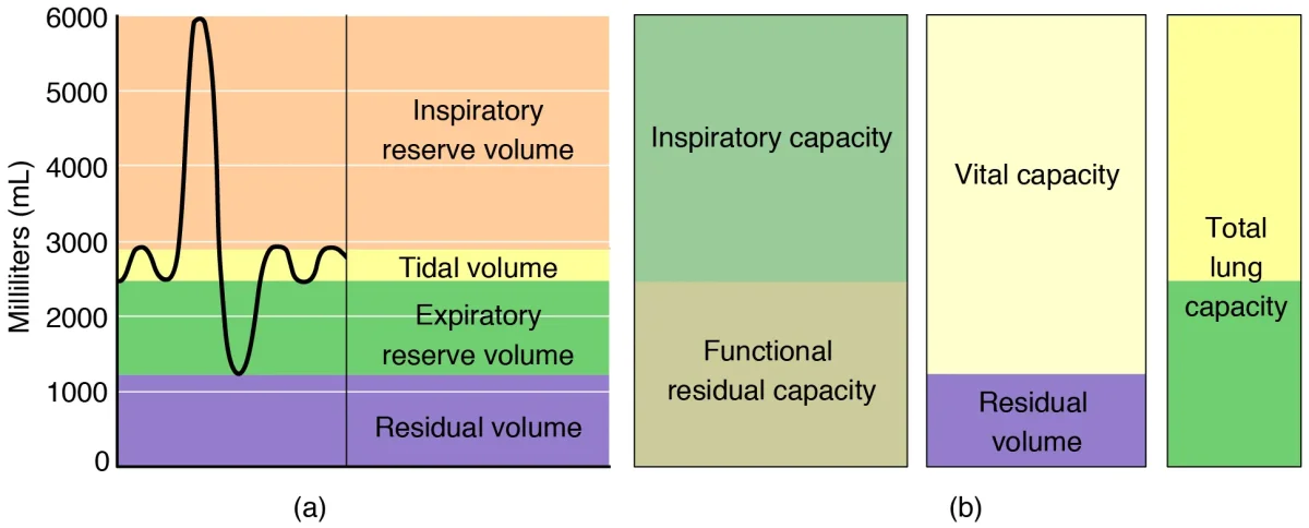
A spirometry tracing showing the standard lung volumes and capacities. Tidal volume (TV) is the normal breathing range; inspiratory and expiratory reserves extend above and below it. Residual volume remains after maximal exhalation. Credit: OpenStax Anatomy & Physiology 2e, CC BY 4.0
The Four Lung Volumes
There are four non-overlapping lung volumes that cannot be subdivided further:
Volume
Abbreviation
Definition
Typical Value
Tidal Volume
TV
Air moved in or out during a normal, quiet breath
~500 mL
Inspiratory Reserve Volume
IRV
Extra air you can inhale beyond a normal tidal breath (with maximal effort)
~3,000 mL
Expiratory Reserve Volume
ERV
Extra air you can exhale beyond a normal tidal exhalation (with effort)
~1,200 mL
Residual Volume
RV
Air that remains in the lungs even after maximal forced exhalation
~1,200 mL
The residual volume exists because the airways and alveoli do not completely collapse during exhalation. RV prevents alveolar collapse and maintains gas exchange even between breaths.
Lung Capacities
Lung capacities are combinations of two or more volumes. There are four:
Capacity
Formula
Definition
Typical Value
Inspiratory Capacity (IC)
TV + IRV
Maximum air you can inhale from the end of a normal exhalation
~3,500 mL
Functional Residual Capacity (FRC)
ERV + RV
Air remaining in the lungs after a normal exhalation
~2,400 mL
Vital Capacity (VC)
TV + IRV + ERV
Maximum air you can move in one breath (deepest inhale to hardest exhale)
~4,700 mL
Total Lung Capacity (TLC)
TV + IRV + ERV + RV
Total air the lungs can hold at maximum inflation
~5,900 mL
Spirometry
Spirometry is the clinical test used to measure lung volumes and capacities. The patient breathes into a device that records volume changes over time, producing a spirogram.
Important limitation: spirometry cannot measure residual volume directly. Since RV is the air you cannot blow out, it stays in the lungs during the test. This means spirometry also cannot directly measure FRC or TLC (both include RV).
To measure RV, clinicians use indirect methods such as gas dilution techniques. The key point for the MCAT: spirometry alone cannot give you RV.
Minute Ventilation and Alveolar Ventilation
Minute ventilation is the total volume of air moved per minute:
But minute ventilation overestimates how much air is actually useful, because it includes dead space air. Alveolar ventilation corrects for this:
Why Breathing Pattern Matters
Consider two patients who both have a minute ventilation of 6 L/min:
Patient
TV
RR
Minute Ventilation
Alveolar Ventilation
A
500 mL
12/min
6,000 mL/min
(500-150) x 12 = 4,200 mL/min
B
200 mL
30/min
6,000 mL/min
(200-150) x 30 = 1,500 mL/min
Patient B has the same minute ventilation but far less alveolar ventilation. Shallow, rapid breathing is inefficient because a larger proportion of each breath is wasted filling dead space. This is why deep, slow breaths are more effective than shallow, fast ones.
Obstructive vs. Restrictive Lung Disease
Pulmonary function tests are used to distinguish two broad categories of lung disease:
Feature
Obstructive Disease
Restrictive Disease
Problem
Airflow out is blocked
Lungs cannot fully expand
Examples
Asthma, COPD (emphysema, chronic bronchitis)
Pulmonary fibrosis, scoliosis, obesity
FEV1 (air exhaled in 1 sec)
Decreased
Decreased
FVC (total air exhaled forcefully)
Normal or slightly decreased
Decreased
FEV1/FVC ratio
Decreased (< 0.7)
Normal or increased (> 0.7)
TLC
Normal or increased (air trapping)
Decreased
RV
Increased (can’t blow it all out)
Decreased
Compliance
Increased (emphysema)
Decreased (stiff lungs)
Lung anatomy overview. Focus on: the branching airway from trachea to bronchi to bronchioles, and how obstructive diseases (emphysema, asthma) vs. restrictive diseases affect airflow and volumes differently. Credit: Patrick J. Lynch, CC BY 2.5
Which lung volumes and capacities cannot be measured by spirometry alone?
Click to reveal answer
Residual volume (RV), functional residual capacity (FRC), and total lung capacity (TLC). Spirometry measures air flow in and out, but RV is the air that remains after maximal exhalation - it never leaves the lungs. Since FRC = ERV + RV and TLC = VC + RV, both include RV and cannot be determined by spirometry alone.
A patient has a FEV1/FVC ratio of 0.55. Is this obstructive or restrictive disease, and why?
Click to reveal answer
Obstructive disease. A normal FEV1/FVC ratio is about 0.80 (80% of air is exhaled in the first second). A ratio of 0.55 means the patient can only blow out 55% in the first second - exhalation is prolonged due to airway obstruction. In restrictive disease, both FEV1 and FVC decrease proportionally, so the ratio stays normal or even increases.
Gas exchange is the entire reason the respiratory system exists. Every tube, every muscle, every membrane in the previous sections is designed to get oxygen from the atmosphere into your blood and carbon dioxide from your blood into the atmosphere. The mechanism is simple: diffusion down partial pressure gradients. No pumps, no active transport, no ATP. Just molecules moving from where there are more of them to where there are fewer.
Dalton’s Law of Partial Pressures
The total pressure of a gas mixture equals the sum of the individual partial pressures of each gas. This is the same Dalton’s law from general chemistry - here we apply it to respiration.
The composition of dry atmospheric air:
Gas
Percentage
Partial Pressure (at 760 mmHg)
Nitrogen (N2)
78%
593 mmHg
Oxygen (O2)
21%
160 mmHg
Argon (Ar)
0.9%
7 mmHg
Carbon dioxide (CO2)
0.04%
0.3 mmHg
Alveolar Gas Composition
The air inside alveoli is not the same as atmospheric air. It differs because:
Water vapor is added (airways humidify incoming air to 100% humidity), which dilutes all other gas partial pressures
Oxygen is continuously being absorbed into the blood
Carbon dioxide is continuously being added from the blood
Gas
Atmospheric Air
Alveolar Air
PO2
160 mmHg
~104 mmHg
PCO2
0.3 mmHg
~40 mmHg
PH2O
Variable
47 mmHg (at 37°C)
The water vapor pressure at body temperature (37°C) is always 47 mmHg. This must be subtracted when calculating alveolar gas pressures.
Henry’s Law
Henry’s Law governs how much gas dissolves in a liquid (like blood):
Fick’s Law of Diffusion
Fick’s Law describes the rate at which a gas diffuses across a membrane like the respiratory membrane:
How Fick’s Law Applies to Disease
Every factor in Fick’s Law can be disrupted by disease:
Factor
Normal
Disease State
Effect
Surface area (A)
~70 m²
Emphysema (alveolar walls destroyed)
Decreased diffusion
Membrane thickness (T)
~0.5 µm
Pulmonary fibrosis, edema (thickened barrier)
Decreased diffusion
Pressure gradient (ΔP)
60 mmHg for O2
High altitude (low atmospheric PO2)
Decreased diffusion
Diffusion coefficient (D)
Normal
Not usually altered by disease
N/A
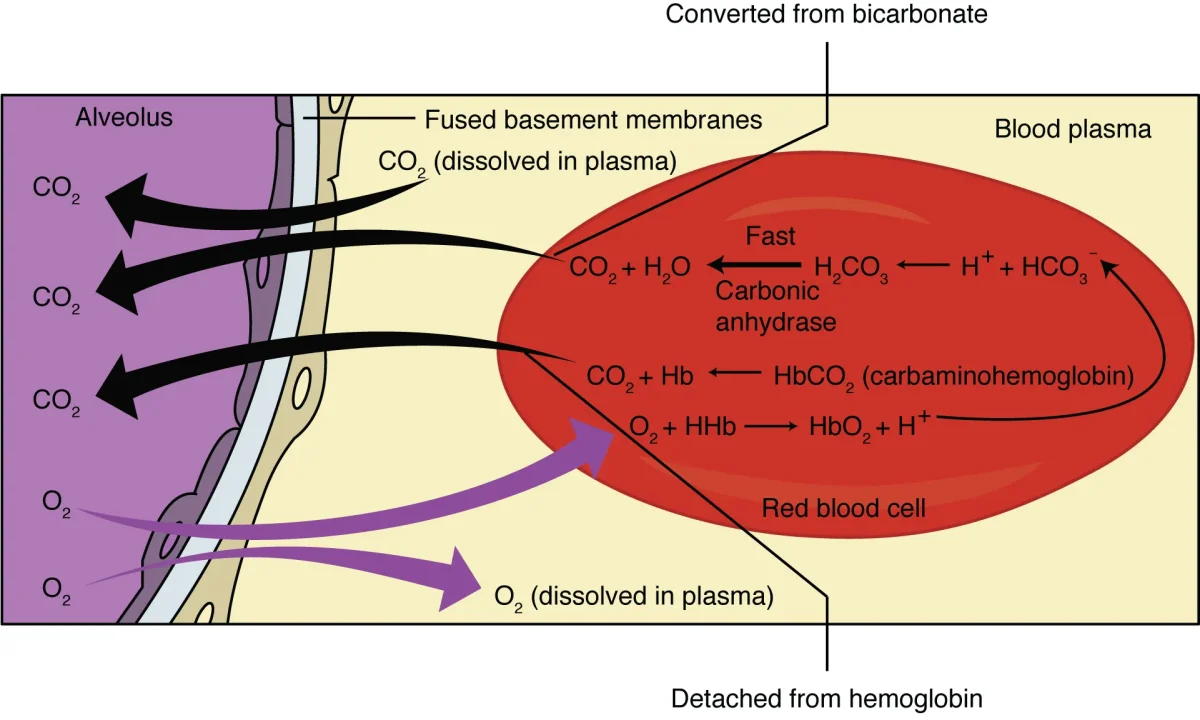
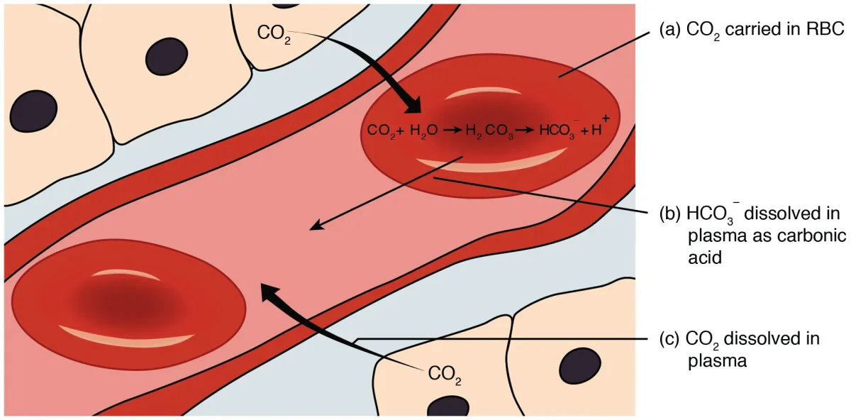
Gas exchange at the capillary level. CO2 enters the RBC and is converted to bicarbonate (HCO3-) by carbonic anhydrase, while O2 dissolves in plasma and binds hemoglobin. Credit: OpenStax Anatomy & Physiology 2e, CC BY 4.0
The Partial Pressure Gradients Driving Gas Exchange
Gas exchange happens at two locations: the lungs and the tissues.
At the lungs (external respiration):
Gas
Alveolar Air
Pulmonary Capillary Blood (arriving)
Direction
O2
104 mmHg
40 mmHg
Into blood
CO2
40 mmHg
46 mmHg
Into alveolus
Oxygen diffuses from the alveolus (PO2 = 104) into the blood (PO2 = 40) because of the 64 mmHg gradient. CO2 diffuses from the blood (PCO2 = 46) into the alveolus (PCO2 = 40) because of the 6 mmHg gradient. Blood leaving the lungs has PO2 of ~100 mmHg and PCO2 of ~40 mmHg.
At the tissues (internal respiration):
Gas
Systemic Capillary Blood
Tissue Cells
Direction
O2
100 mmHg
~40 mmHg
Into tissues
CO2
40 mmHg
~46 mmHg
Into blood
The process reverses: oxygen diffuses from blood into tissues, and CO2 diffuses from tissues into blood.
Normal vs. Impaired Gas Exchange
Under normal conditions, blood flowing through the pulmonary capillary has enough time to fully equilibrate with alveolar air - oxygen transfer is very efficient. In diseases that thicken the membrane (fibrosis, edema) or reduce the surface area (emphysema), diffusion slows and blood may not fully equilibrate before leaving the capillary, resulting in hypoxemia.
Why does CO2 diffuse faster than O2 across the respiratory membrane despite having a much smaller partial pressure gradient?
Click to reveal answer
CO2 is approximately 20 times more soluble than O2 in the aqueous respiratory membrane. According to Fick's Law, diffusion rate depends on the diffusion coefficient (which includes solubility) multiplied by the pressure gradient. CO2's much higher solubility more than compensates for its smaller pressure gradient (6 mmHg vs. 60 mmHg for O2).
According to Fick's Law, which two disease processes would impair gas exchange, and how?
Click to reveal answer
Emphysema reduces surface area (A) and pulmonary fibrosis/edema increases membrane thickness (T). Fick's Law: Rate = (A x D x ΔP) / T. Decreasing A (emphysema destroys alveolar walls) or increasing T (fibrosis deposits scar tissue, edema adds fluid to the membrane) both reduce diffusion rate. Both cause hypoxemia before hypercapnia because CO2 diffuses 20x faster than O2.
Oxygen is not very soluble in blood. If you relied on dissolved oxygen alone, your blood could carry only about 3 mL of O2 per liter - nowhere near enough to meet your body’s demand of roughly 250 mL of O2 per minute. The solution? A molecular shuttle bus called hemoglobin, which increases oxygen-carrying capacity by nearly 70-fold.
Hemoglobin Structure
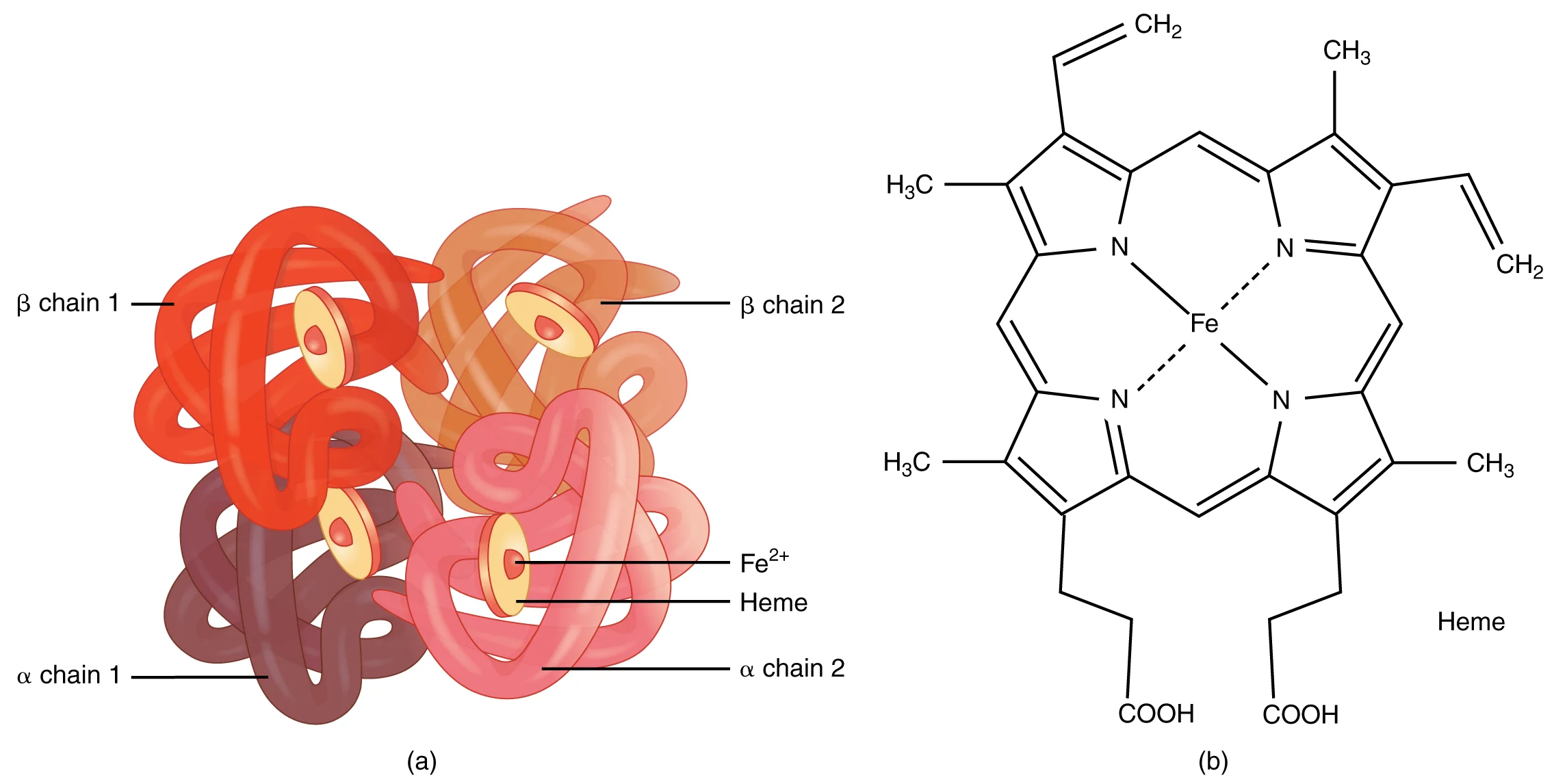
Hemoglobin is a quaternary protein consisting of 4 polypeptide subunits:
Each subunit contains a heme group - a porphyrin ring with a central iron atom (Fe2+) that can bind one O2 molecule.
Cooperative Binding and the T/R State Model
Hemoglobin exists in two conformational states:
State
Name
Oxygen Affinity
When?
T-state
Tense
Low affinity
Deoxyhemoglobin (no O2 bound)
R-state
Relaxed
High affinity
Oxyhemoglobin (O2 bound)
When the first O2 molecule binds to a heme group, it triggers a conformational change that shifts the entire molecule from T-state to R-state, making it easier for the next O2 molecules to bind. This is positive cooperativity - each O2 binding event facilitates the next.
The reverse is also true: when the first O2 is released in the tissues, the remaining O2 molecules are released more easily. This creates a very efficient loading/unloading system.
Hemoglobin is a quaternary protein with four subunits (2 alpha, 2 beta). Each subunit contains a heme group with an iron atom (Fe2+) that can bind one O2 molecule, for a total carrying capacity of four O2 per hemoglobin. Credit: OpenStax College / Wikimedia Commons, CC BY 3.0
The Oxygen-Hemoglobin Dissociation Curve
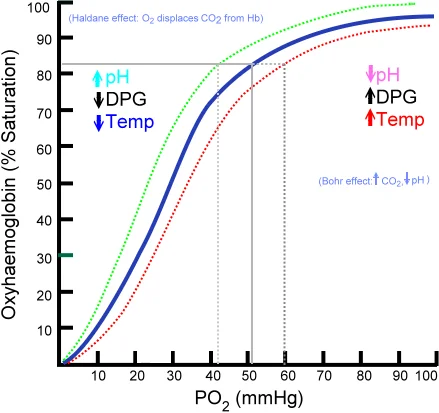
The O2-Hb dissociation curve plots hemoglobin saturation (%) against partial pressure of oxygen (PO2). Because of cooperative binding, the curve is sigmoidal (S-shaped), not linear.
Key points on the curve:
PO2
Saturation
Location
Meaning
~100 mmHg
~98%
Lungs
Hemoglobin is nearly fully loaded
~40 mmHg
~75%
Resting tissues
About 25% of O2 has been unloaded
~20 mmHg
~35%
Exercising tissues
About 65% of O2 has been unloaded
The steep middle portion of the curve (PO2 ~20-60 mmHg) is where small changes in PO2 cause large changes in saturation. This is the tissue range, where hemoglobin efficiently unloads oxygen in response to small drops in PO2.
The flat upper portion (PO2 > 70 mmHg) means that hemoglobin stays nearly saturated even if PO2 drops somewhat. This is a safety buffer - even at altitude, saturation remains high until PO2 drops significantly.
The oxygen-hemoglobin dissociation curve with shift factors. The sigmoidal shape reflects cooperative binding. Right shift (decreased pH, increased temp/CO22,3-BPG) promotes O2 release in active tissues. Left shift (increased pH, decreased temp/CO22,3-BPG) promotes O2 loading in the lungs. Credit: Wikimedia Commons, Public Domain
Right Shift - Decreased Affinity (Releases O2 More Easily)
A right shift means hemoglobin releases oxygen more readily at any given PO2. This is beneficial in the tissues where O2 is needed.
Factors causing a right shift:
Increased CO2 (hypercapnia)
Decreased pH / increased H+ (acidosis)
Increased temperature
Increased 2,3-BPG (2,3-bisphosphoglycerate)
All of these conditions are found in actively metabolizing tissues - muscles during exercise are hot, acidic, and producing CO2. The right shift ensures that hemoglobin dumps oxygen exactly where it is needed most.
Left Shift - Increased Affinity (Holds O2 Tighter)
A left shift means hemoglobin binds oxygen more tightly and is less willing to release it. This is beneficial in the lungs where O2 is being picked up.
Factors causing a left shift:
Decreased CO2
Increased pH (alkalosis)
Decreased temperature
Decreased 2,3-BPG
Fetal hemoglobin (HbF)
Carbon monoxide (CO) binding
The Bohr Effect
The Bohr effect specifically describes how CO2 and H+ concentration affect hemoglobin’s oxygen affinity:
In the tissues: high CO2 and low pH cause a right shift, promoting O2 release
In the lungs: low CO2 and high pH cause a left shift, promoting O2 binding
This creates an elegant delivery system: hemoglobin automatically releases more oxygen to tissues that are metabolically active (producing CO2 and acid) and picks up more oxygen in the lungs (where CO2 is being exhaled).
The Haldane Effect
The Haldane effect is the mirror image of the Bohr effect: it describes how oxygenation of hemoglobin affects its ability to carry CO2.
In the tissues: deoxygenated hemoglobin binds CO2 more readily (forming carbaminohemoglobin)
In the lungs: oxygenated hemoglobin releases CO2 more readily
The Bohr and Haldane effects work together: the Bohr effect promotes O2 delivery to tissues, and the Haldane effect promotes CO2 pickup from tissues and CO2 release in the lungs.
2,3-BPG (2,3-Bisphosphoglycerate)
2,3-BPG is a molecule produced by red blood cells during glycolysis. It binds to the central cavity of the deoxyhemoglobin tetramer, stabilizing the T-state and reducing oxygen affinity (right shift).
2,3-BPG levels increase in response to chronic hypoxia - for example, at high altitude or in anemia. This is an adaptive response that helps deliver more oxygen to tissues when overall oxygen availability is reduced.
Fetal Hemoglobin (HbF)
Fetal hemoglobin has gamma chains instead of beta chains. The gamma chains do not bind 2,3-BPG as well, so HbF has a higher oxygen affinity than adult HbA (left-shifted curve).
This is essential for fetal survival: at the placenta, fetal blood must “steal” oxygen from maternal hemoglobin. HbF’s higher affinity allows it to grab O2 even at the relatively low PO2 of the placenta.
Myoglobin
Myoglobin is a single-subunit oxygen-binding protein found in muscle tissue. Unlike hemoglobin’s 4 subunits, myoglobin has only one heme group and therefore shows no cooperativity.
Its dissociation curve is hyperbolic (not sigmoidal), and it has a much higher affinity for oxygen than hemoglobin. Myoglobin does not release oxygen until PO2 drops very low.
Function: myoglobin acts as an oxygen reservoir in muscle cells. During intense exercise, when capillary PO2 drops dramatically, myoglobin releases its stored O2 to sustain aerobic metabolism.
Feature
Hemoglobin
Myoglobin
Subunits
4 (quaternary)
1 (tertiary)
Heme groups
4
1
Curve shape
Sigmoidal
Hyperbolic
Cooperativity
Yes
No
O2 affinity
Lower (releases easily)
Higher (stores O2)
Location
Red blood cells
Muscle cells
Function
O2 transport
O2 storage
Carbon Monoxide Poisoning
Carbon monoxide (CO) binds hemoglobin with approximately 200-250 times greater affinity than oxygen. Even small amounts of CO in inhaled air can occupy a large fraction of heme binding sites, forming carboxyhemoglobin (COHb).
CO poisoning is devastating for two reasons:
Reduced carrying capacity - CO occupies heme sites that would otherwise carry O2
Left shift - CO binding causes the remaining heme groups to hold onto O2 more tightly (increased affinity), so even the O2 that is loaded cannot be delivered to tissues
Why is the O2-hemoglobin dissociation curve sigmoidal rather than hyperbolic?
Click to reveal answer
Because of cooperative binding. Hemoglobin has 4 subunits that interact. Binding of the first O2 triggers a conformational change (T-state to R-state) that increases the affinity of the remaining subunits for O2. This produces the S-shaped curve: slow initial binding, rapid middle section, and plateau at high PO2. Myoglobin, with only 1 subunit, shows no cooperativity and has a hyperbolic curve.
An exercising muscle has increased temperature, CO2, H+, and 2,3-BPG. How does this affect the O2-Hb curve, and why is this beneficial?
Click to reveal answer
All four factors shift the curve to the right (decreased O2 affinity). This is beneficial because a right shift means hemoglobin releases oxygen more readily at any given PO2. Exercising muscles need more oxygen, and these metabolic byproducts signal hemoglobin to "dump oxygen here." It is a brilliant self-regulating delivery system - the tissues that work hardest get the most oxygen.
Why does fetal hemoglobin (HbF) have a higher oxygen affinity than adult hemoglobin (HbA)?
Click to reveal answer
HbF has gamma chains instead of beta chains, and gamma chains bind 2,3-BPG poorly. Since 2,3-BPG normally stabilizes the T-state (low-affinity state) of hemoglobin, reduced 2,3-BPG binding means HbF stays in the R-state (high affinity). This left-shifted curve allows fetal blood to extract oxygen from maternal blood at the placenta.
If hemoglobin is the bus that carries oxygen, what about the return trip? Carbon dioxide - the waste product of aerobic metabolism - needs to get from the tissues back to the lungs for exhalation. But CO2 does not just hitch a ride on hemoglobin the way oxygen does. Instead, CO2 travels by three different routes, and the dominant one involves a clever chemical conversion that also happens to be your blood’s primary pH buffering system.
The Three Mechanisms of CO2 Transport
Mechanism
Percentage
How It Works
Dissolved CO2
~7-10%
CO2 dissolves directly in plasma (follows Henry’s Law)
Bicarbonate ion (HCO3-)
~70%
CO2 is converted to HCO3- inside RBCs by carbonic anhydrase
Carbaminohemoglobin
~20-23%
CO2 binds to the amino groups on hemoglobin (not the heme group)
The Bicarbonate Buffer System
The bicarbonate pathway is the most important CO2 transport mechanism and the body’s primary blood pH buffer. Here is the complete reaction:
The three methods of CO2 transport: dissolved in plasma (~7-10%), as bicarbonate (~70%), and as carbaminohemoglobin (~20-23%). Note the chloride shift maintaining electrical neutrality as HCO3- exits the RBC. Credit: OpenStax Anatomy & Physiology 2e, CC BY 4.0
The Process in the Tissues
CO2 diffuses from metabolizing tissue cells into the blood and enters red blood cells
Carbonic anhydrase (inside RBCs) catalyzes: CO2 + H2O → H2CO3
H+ binds to hemoglobin (buffered - this is the Bohr effect mechanism)
HCO3- is transported out of the RBC into the plasma
The Chloride Shift
When HCO3- exits the RBC into the plasma, it would create a charge imbalance (too much negative charge leaving). To maintain electrical neutrality, chloride ions (Cl-) move into the RBC from the plasma via an antiport protein. This 1:1 exchange - one HCO3- out, one Cl- in - is called the chloride shift.
The Process in the Lungs
Everything reverses:
In the pulmonary capillaries, the high PO2 environment promotes oxygen binding to hemoglobin
As hemoglobin binds O2 (Haldane effect), it releases H+ and CO2
Cl- exits the RBC, HCO3- re-enters (reverse chloride shift)
Inside the RBC: H+ + HCO3- → H2CO3 → CO2 + H2O (carbonic anhydrase runs in reverse)
CO2 diffuses into the alveolus and is exhaled
Blood pH and the Bicarbonate Buffer
Normal arterial blood pH is 7.35-7.45. The bicarbonate buffer system maintains this narrow range because the two sides of the equilibrium can be independently regulated:
The lungs control CO2 levels (respiratory component)
The kidneys control HCO3- levels (metabolic component)
Acid-Base Disturbances
There are four primary acid-base disturbances. Each alters the 20:1 ratio differently:
Disturbance
Primary Change
pH
Compensation
Respiratory acidosis
Increased CO2 (hypoventilation)
Decreased (< 7.35)
Kidneys retain HCO3-
Respiratory alkalosis
Decreased CO2 (hyperventilation)
Increased (> 7.45)
Kidneys excrete HCO3-
Metabolic acidosis
Decreased HCO3- or excess acid
Decreased (< 7.35)
Lungs hyperventilate (blow off CO2)
Metabolic alkalosis
Increased HCO3- or loss of acid
Increased (> 7.45)
Lungs hypoventilate (retain CO2)
The body always compensates by adjusting the opposite system: respiratory problems get renal compensation, and metabolic problems get respiratory compensation. Compensation returns the ratio toward 20:1 but rarely fully normalizes pH.
What is the most common form of CO2 transport in the blood, and what enzyme is required?
Click to reveal answer
Bicarbonate (HCO3-) accounts for ~70% of CO2 transport. The enzyme carbonic anhydrase (found inside red blood cells) catalyzes the conversion of CO2 + H2O to H2CO3, which then dissociates to H+ + HCO3-. The HCO3- exits the RBC via the chloride shift (exchanged for Cl-) and travels in plasma to the lungs, where the process reverses.
A patient is hyperventilating due to anxiety. What acid-base disturbance is this, and how would the kidneys compensate?
Click to reveal answer
Respiratory alkalosis. Hyperventilation blows off excess CO2, shifting the equilibrium: less CO2 means less H2CO3 means fewer H+ ions, so pH rises above 7.45. The kidneys compensate over hours to days by excreting more HCO3- (reducing the numerator in the Henderson-Hasselbalch equation) to bring the HCO3-/CO2 ratio back toward 20:1.
You breathe about 20,000 times a day without thinking about it. You can also hold your breath voluntarily - but only for so long before an overwhelming urge forces you to gasp. And during exercise, your breathing rate and depth increase automatically to match your metabolic demand. All of this happens through a sophisticated control system involving brainstem respiratory centers, chemical sensors in your blood vessels, and feedback loops that adjust ventilation breath by breath.
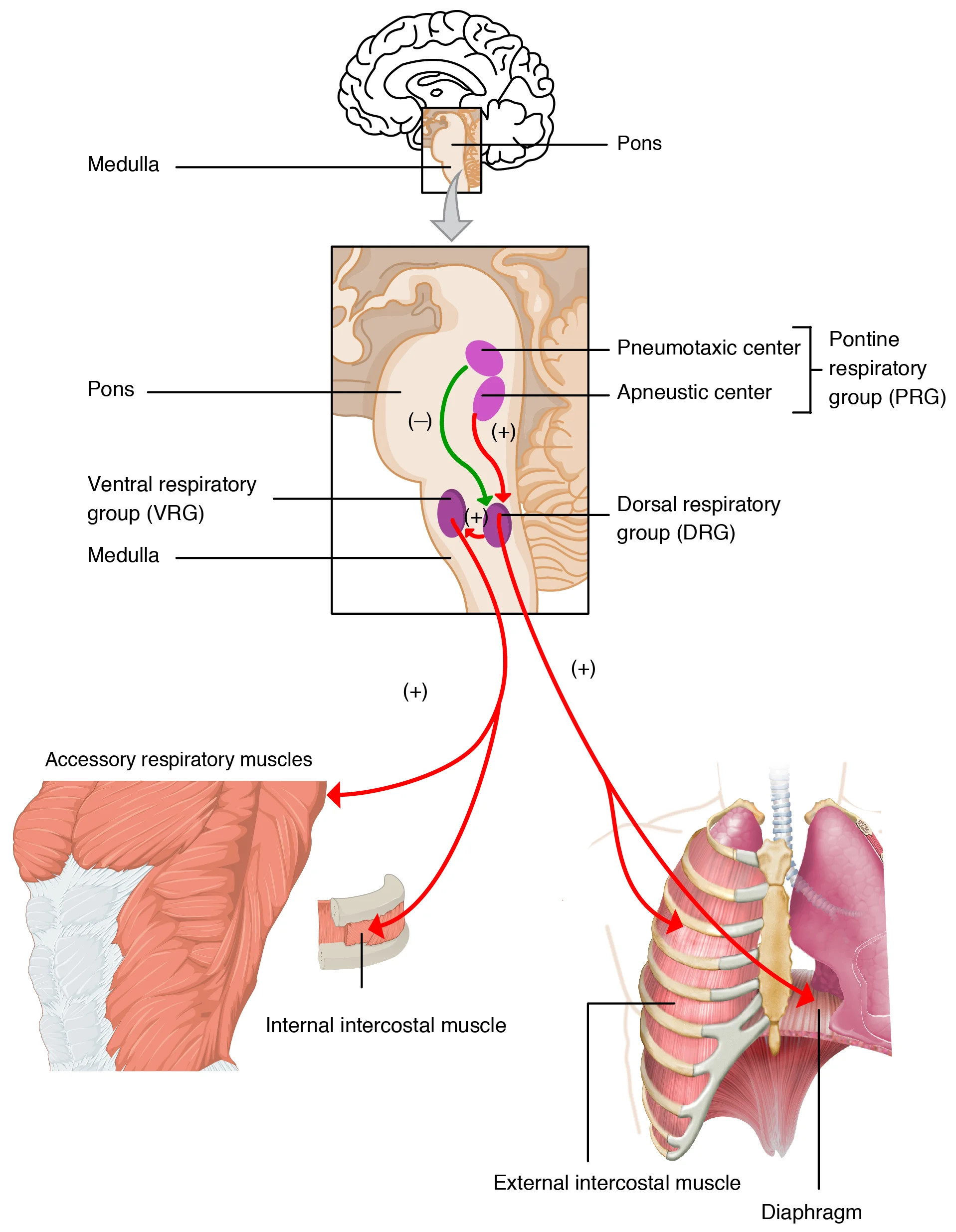
Brainstem respiratory centers. Focus on: the medulla sets the basic rhythm (DRG = inspiration, VRG = forced breathing), the pons fine-tunes it (pneumotaxic center limits inspiration). Central chemoreceptors detect CO2/H+; peripheral chemoreceptors detect low O2. Credit: OpenStax College, CC BY 3.0
The Brainstem Respiratory Centers
Breathing rhythm is generated by neurons in the medulla oblongata and modulated by the pons. There are four key centers:
Center
Location
Function
Dorsal Respiratory Group (DRG)
Medulla
Sets the basic rhythm of inspiration; fires rhythmically to trigger the phrenic nerve and diaphragm contraction
Ventral Respiratory Group (VRG)
Medulla
Active during forced breathing; controls both inspiration and active expiration; stimulates accessory muscles
Pneumotaxic Center
Upper pons
Limits the duration of inspiration by inhibiting the DRG; increases respiratory rate by shortening breath duration
Apneustic Center
Lower pons
Promotes prolonged inspiration by stimulating the DRG; normally inhibited by the pneumotaxic center
The Normal Breathing Cycle
During quiet breathing:
The DRG fires rhythmic signals via the phrenic nerve (to the diaphragm) and intercostal nerves (to the external intercostal muscles)
These signals last about 2 seconds, causing inspiration
The DRG then stops firing for about 3 seconds, allowing passive expiration through elastic recoil
The pneumotaxic center modulates this cycle by controlling when the DRG shuts off
During exercise or forced breathing, the VRG activates and recruits accessory muscles for both active inspiration and active expiration.
Chemical Control of Breathing
The most important regulators of breathing are chemical sensors that detect changes in blood gas levels and pH. There are two types:
Central Chemoreceptors
Located on the ventral surface of the medulla, central chemoreceptors are the primary drive to breathe. They detect changes in H+ concentration in the cerebrospinal fluid (CSF).
Here is the key mechanism: CO2 (but not H+ or HCO3-) can freely cross the blood-brain barrier. Once in the CSF, CO2 combines with water (via carbonic anhydrase) to form H2CO3, which dissociates into H+ and HCO3-. The central chemoreceptors detect this H+ increase and stimulate the respiratory centers to increase ventilation.
So while the central chemoreceptors technically sense H+, they are effectively monitoring blood CO2 levels because CO2 is the source of the H+.
Peripheral Chemoreceptors
Located in the carotid bodies (at the bifurcation of the common carotid arteries) and the aortic bodies (in the aortic arch), peripheral chemoreceptors detect:
PO2 - the primary O2 sensor; responds to critically low oxygen (PO2 < 60 mmHg)
PCO2 - responds faster than central chemoreceptors but plays a secondary role
The carotid bodies send signals via the glossopharyngeal nerve (CN IX).
The aortic bodies send signals via the vagus nerve (CN X).
CO2 is the Primary Drive to Breathe
This is one of the most important concepts in respiratory physiology: under normal conditions, CO2 (not O2) is the primary stimulus for breathing.
Even small increases in PCO2 (from 40 to 44 mmHg) cause large increases in ventilation. In contrast, PO2 must drop dramatically (below 60 mmHg) before it significantly stimulates breathing. This makes sense evolutionarily - CO2 is constantly produced by metabolism and needs to be continuously eliminated. Oxygen levels in the atmosphere are usually adequate, so fine-tuning breathing based on CO2 is more useful.
Why Hyperventilation is Dangerous
Hyperventilation blows off CO2, reducing PCO2 below normal. This removes the primary drive to breathe. The key concept: without CO2 buildup to trigger breathing, a person can lose consciousness from hypoxia before feeling any urge to breathe.
Oxygen as a Backup Drive: The Hypoxic Drive
If central chemoreceptors become desensitized to chronically elevated CO2, a person may switch to using low O2 as their primary breathing stimulus - this is the hypoxic drive.
Other Inputs to Breathing
Beyond chemical sensors, several other factors influence breathing:
Hering-Breuer reflex: stretch receptors in the lungs detect overinflation and send signals via the vagus nerve to inhibit further inspiration (prevents over-inflation)
Irritant receptors: detect smoke, dust, or chemical irritants in the airways, triggering coughing or bronchoconstriction
Cortical input: you can voluntarily override automatic breathing (holding breath, singing, speaking) - but CO2 buildup eventually overrides voluntary control
Limbic system: emotions (fear, excitement) can alter breathing patterns
Temperature: fever increases respiratory rate; hypothermia decreases it
What is the primary chemical stimulus for breathing under normal conditions, and where is it detected?
Click to reveal answer
CO2 is the primary stimulus, detected by central chemoreceptors on the ventral medulla. CO2 crosses the blood-brain barrier, is converted to H+ by carbonic anhydrase in the CSF, and H+ stimulates the central chemoreceptors. Even small rises in PCO2 strongly stimulate ventilation. O2 is a secondary stimulus detected by peripheral chemoreceptors, and it only becomes significant when PO2 drops below 60 mmHg.
Why is giving high-flow oxygen dangerous for a patient with severe COPD and chronic CO2 retention?
Click to reveal answer
COPD patients with chronic CO2 retention rely on hypoxic drive. Their central chemoreceptors have adapted to high CO2 and no longer respond to it. Low O2 (detected by peripheral chemoreceptors) becomes their main breathing stimulus. High-flow oxygen raises PO2, removing this stimulus. With neither CO2 nor O2 driving breathing, the patient may develop respiratory depression and hypoventilate.
Imagine a factory with workers and raw materials. For maximum efficiency, you need the right number of workers at each station AND the right amount of raw materials delivered to them. Too many workers with no materials? Wasted labor. Materials pouring in but no workers? Wasted resources. In the lungs, ventilation is the “materials delivery” (air) and perfusion is the “workers” (blood flow). The ratio between them - the V/Q ratio - determines how efficiently each region of the lung exchanges gas.
The V/Q Ratio
The ventilation-perfusion ratio (V/Q) compares the amount of air reaching the alveoli (ventilation, V) to the amount of blood flow past those alveoli (perfusion, Q).
V/Q Value
Meaning
Consequence
V/Q = 1.0
Perfect match
Ideal gas exchange
V/Q > 1.0
More ventilation than perfusion
Ventilated but not perfused = “dead space”
V/Q < 1.0
More perfusion than ventilation
Perfused but not ventilated = “shunt”
V/Q = infinity
Ventilation with zero perfusion
Complete dead space (e.g., pulmonary embolism blocking blood flow)
V/Q = 0
Perfusion with zero ventilation
Complete shunt (e.g., airway obstruction)
The overall V/Q ratio for the entire lung is approximately 0.8 (alveolar ventilation ~4 L/min, cardiac output ~5 L/min).
Dead Space vs. Shunt
Dead space (high V/Q): alveoli are ventilated but not perfused. Air reaches the alveoli, but there is no blood flow to pick up the oxygen. The air is “wasted.” This happens when blood flow is blocked - for example, a pulmonary embolism blocks perfusion to a region while ventilation continues.
There are two types of dead space:
Anatomical dead space: the conducting airways (~150 mL) - these never participate in gas exchange
Alveolar dead space: ventilated alveoli with no blood flow
Physiological dead space = anatomical + alveolar dead space (total wasted ventilation)
Shunt (low V/Q): alveoli are perfused but not ventilated. Blood flows past alveoli that have no fresh air in them, so the blood passes through without picking up oxygen. This results in deoxygenated blood mixing with oxygenated blood. Examples include airway obstruction (mucus plug, tumor), atelectasis (collapsed alveolus), and pneumonia (alveolus filled with fluid).
Gravity and the V/Q Ratio
In an upright person, gravity creates a gradient in both ventilation and perfusion from the apex (top) to the base (bottom) of the lung:
Region
Ventilation
Perfusion
V/Q Ratio
Apex (top)
Lower
Much lower (gravity pulls blood down)
Higher (V/Q > 1)
Base (bottom)
Higher
Much higher
Lower (V/Q < 1)
Both ventilation and perfusion are greater at the base due to gravity, but perfusion increases more steeply than ventilation as you move from apex to base. This means:
The apex is relatively over-ventilated compared to its blood flow (higher V/Q - closer to dead space)
The base is relatively over-perfused compared to its ventilation (lower V/Q - closer to shunt)
Hypoxic Pulmonary Vasoconstriction
The lungs have a unique response to low oxygen that is the opposite of what systemic blood vessels do:
Systemic blood vessels: low O2 causes vasodilation (to bring more blood to hypoxic tissue)
This response is called hypoxic pulmonary vasoconstriction (HPV). It is a brilliant matching mechanism: if an alveolus is not receiving enough air (low O2 in that region), the pulmonary arterioles serving that alveolus constrict, redirecting blood to better-ventilated regions. This optimizes V/Q matching.
V/Q Mismatch in Disease
Any condition that disrupts the match between airflow and blood flow impairs gas exchange and leads to hypoxemia.
How does the pulmonary vasculature respond to local hypoxia, and how does this differ from systemic vasculature?
Click to reveal answer
Pulmonary vessels constrict in response to low O2 (hypoxic pulmonary vasoconstriction), which is the opposite of systemic vessels, which dilate. This unique response diverts blood away from poorly ventilated alveoli and toward well-ventilated ones, optimizing V/Q matching. Systemic vasodilation in hypoxia increases blood flow to hypoxic tissues. The lung's opposite response ensures blood only goes where air is available for gas exchange.
A patient has a pulmonary embolism blocking blood flow to the right lower lobe. What happens to the V/Q ratio in that region?
Click to reveal answer
The V/Q ratio approaches infinity (dead space). Ventilation continues normally (air reaches the alveoli), but perfusion is blocked by the embolus (no blood flow). Without blood flow to pick up O2, the ventilation is wasted. This is "alveolar dead space" - ventilated but not perfused. The patient becomes hypoxemic because the affected region contributes nothing to gas exchange.
Everything you have learned about respiratory physiology assumes you are near sea level, breathing normal atmospheric air at 760 mmHg. But what happens when those conditions change? At high altitude, atmospheric pressure drops and oxygen becomes scarce. Underwater, pressure increases and gases dissolve into your tissues in dangerous quantities. Both environments push the respiratory system to its limits and reveal the elegance - and vulnerabilities - of human physiology.
High Altitude Physiology
At high altitude, atmospheric pressure decreases. Since the fraction of oxygen in air remains 21%, the partial pressure of oxygen (PO2) decreases proportionally.
Altitude
Atmospheric Pressure
PO2 (in air)
Approximate Alveolar PO2
Sea level
760 mmHg
160 mmHg
104 mmHg
3,000 m (~10,000 ft)
523 mmHg
110 mmHg
~67 mmHg
5,500 m (~18,000 ft)
379 mmHg
80 mmHg
~45 mmHg
8,848 m (Everest summit)
253 mmHg
53 mmHg
~28 mmHg
Acute Responses to High Altitude
When you first arrive at high altitude, your body responds immediately:
Hyperventilation - peripheral chemoreceptors detect low PO2 and stimulate increased ventilation. This is the most important acute response.
Increased heart rate - sympathetic activation increases cardiac output to deliver more blood per minute.
The respiratory alkalosis from hyperventilation is actually a problem: the elevated pH shifts the O2-Hb curve to the LEFT, making hemoglobin hold onto oxygen more tightly. This partially counteracts the benefit of hyperventilation.
Acclimatization (Chronic Responses)
Over days to weeks at altitude, the body acclimatizes through several mechanisms:
Renal compensation (days): The kidneys excrete excess HCO3- to compensate for the respiratory alkalosis, bringing pH back toward normal. This allows the O2-Hb curve to shift back toward the right.
Increased 2,3-BPG (hours to days): Red blood cells increase production of 2,3-BPG, which binds to hemoglobin and shifts the curve to the RIGHT. This facilitates oxygen delivery to tissues despite the lower arterial PO2.
Increased EPO and red blood cell production (days to weeks): The kidneys detect hypoxia and release erythropoietin (EPO), which stimulates the bone marrow to produce more red blood cells (polycythemia). More RBCs means more hemoglobin and greater total oxygen-carrying capacity.
Increased capillary density (weeks to months): Tissues develop more capillaries (angiogenesis) to improve oxygen extraction.
Increased mitochondrial density: Muscle cells increase their mitochondrial content to more efficiently utilize available oxygen.
Acute Mountain Sickness (AMS)
Rapid ascent to altitudes above 2,500 m without acclimatization can cause acute mountain sickness, characterized by headache, nausea, fatigue, and dizziness.
Diving Physiology
Underwater, the opposite problem occurs: pressure increases. For every 10 meters (33 feet) of depth, pressure increases by 1 atmosphere (760 mmHg).
Depth
Total Pressure
Gas Partial Pressures
Surface
1 atm (760 mmHg)
PN2 = 593 mmHg, PO2 = 160 mmHg
10 m
2 atm (1520 mmHg)
PN2 = 1186 mmHg, PO2 = 320 mmHg
30 m
4 atm (3040 mmHg)
PN2 = 2372 mmHg, PO2 = 640 mmHg
According to Henry’s Law (C = k x Pgas), the amount of gas dissolved in a liquid increases proportionally with the partial pressure of that gas. At increased depth, more nitrogen and oxygen dissolve in blood and tissues.
Nitrogen Narcosis
At great depths, the high partial pressure of nitrogen causes nitrogen narcosis - impaired judgment and disorientation similar to alcohol intoxication. The effect reverses upon ascent.
Decompression Sickness (The Bends)
If a diver ascends too quickly, the dissolved nitrogen in their tissues comes out of solution as gas bubbles (like opening a carbonated soda - rapidly reducing pressure causes dissolved gas to form bubbles). These nitrogen bubbles can lodge in joints, blood vessels, and tissues, causing:
Joint and muscle pain (the most common symptom - hence “the bends”)
Skin rash
Neurological symptoms (paralysis, confusion)
Pulmonary embolism (if bubbles reach the lungs)
Prevention: ascend slowly, allowing nitrogen to gradually diffuse out of tissues and be exhaled. The concept to understand is Henry’s Law in reverse - reducing pressure decreases solubility.
Oxygen Toxicity
While O2 is essential, too much is toxic. At very high partial pressures, excess oxygen generates reactive oxygen species (ROS) that damage cells. This is why oxygen concentration must be carefully controlled in clinical settings.
Summary: Altitude vs. Diving
Feature
High Altitude
Diving
Pressure change
Decreased atmospheric pressure
Increased hydrostatic pressure
O2 issue
Hypoxia (low PO2)
Potential hyperoxia (O2 toxicity at depth)
N2 issue
Not significant
Nitrogen narcosis, decompression sickness
Primary gas law
Dalton’s law (PO2 = fraction x Ptotal)
Henry’s law (gas solubility increases with pressure)
Key adaptation
Increased ventilation, EPO, 2,3-BPG
Controlled ascent, gas mixtures
Danger
Acute mountain sickness
Nitrogen narcosis, the bends, O2 toxicity
A mountain climber at 5,500 m has been acclimatizing for two weeks. Name three physiological changes that have occurred.
Click to reveal answer
(1) Increased 2,3-BPG - shifts the O2-Hb curve right, facilitating O2 release to tissues. (2) Increased EPO secretion leading to polycythemia - more red blood cells increase oxygen-carrying capacity. (3) Renal excretion of HCO3- - compensates for respiratory alkalosis from chronic hyperventilation, normalizing pH. Additional changes: increased capillary density and mitochondrial content in tissues.
A scuba diver ascends rapidly from 30 meters. Using Henry's Law, explain why gas bubbles form in the blood.
Click to reveal answer
Henry's Law: C = k x Pgas. At 30 meters (4 atm), the high partial pressure of nitrogen caused large amounts of N2 to dissolve in blood and tissues. Upon rapid ascent, the pressure drops suddenly. The dissolved N2 concentration now exceeds what can stay in solution at the lower pressure, so the excess nitrogen comes out of solution as gas bubbles. These bubbles can lodge in joints, blood vessels, and tissues, causing decompression sickness.