
Breathing Regulation
You breathe about 20,000 times a day without thinking about it. You can also hold your breath voluntarily - but only for so long before an overwhelming urge forces you to gasp. And during exercise, your breathing rate and depth increase automatically to match your metabolic demand. All of this happens through a sophisticated control system involving brainstem respiratory centers, chemical sensors in your blood vessels, and feedback loops that adjust ventilation breath by breath.
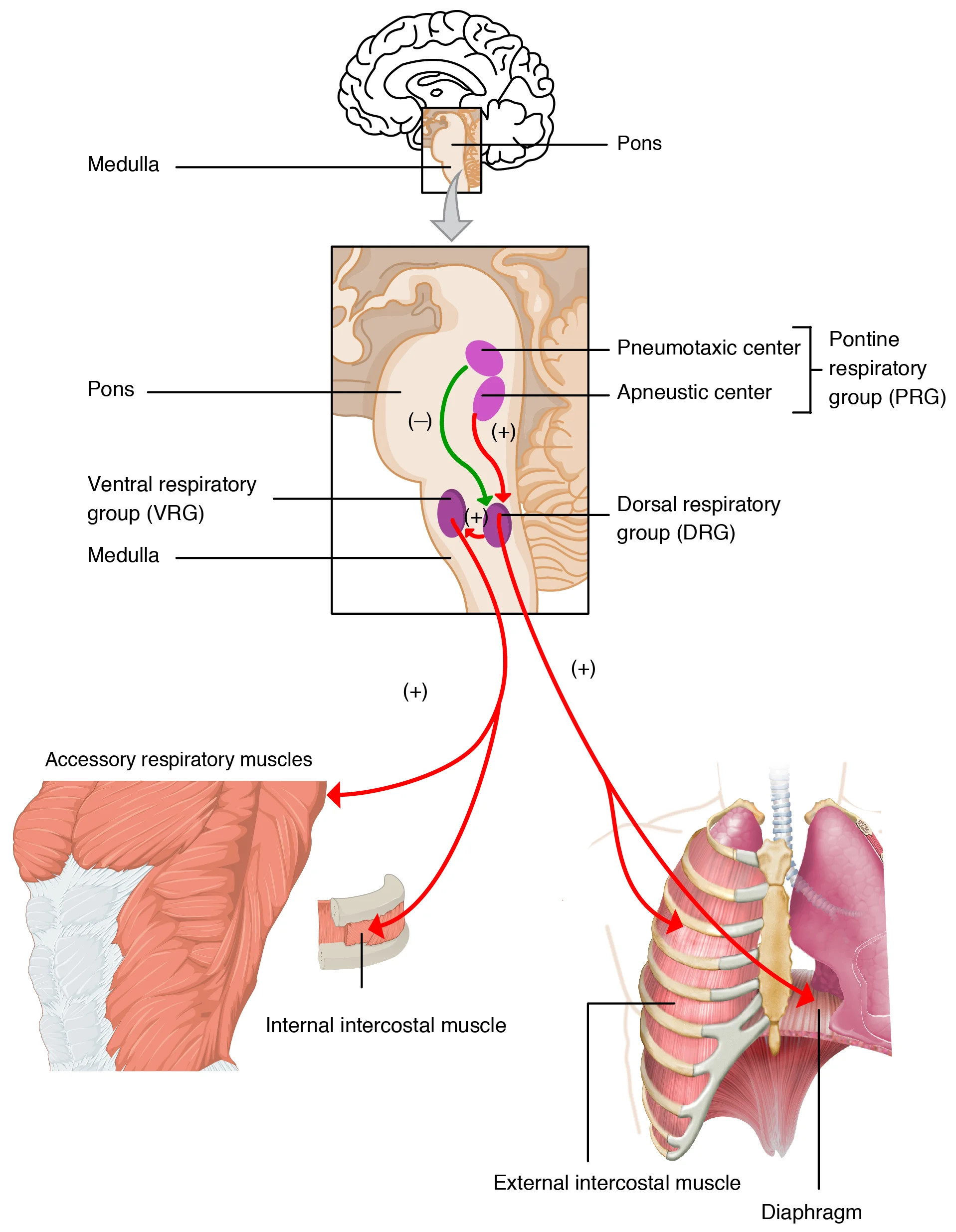
The Brainstem Respiratory Centers
Breathing rhythm is generated by neurons in the medulla oblongata and modulated by the pons. There are four key centers:
| Center | Location | Function |
|---|---|---|
| Dorsal Respiratory Group (DRG) | Medulla | Sets the basic rhythm of inspiration; fires rhythmically to trigger the phrenic nerve and diaphragm contraction |
| Ventral Respiratory Group (VRG) | Medulla | Active during forced breathing; controls both inspiration and active expiration; stimulates accessory muscles |
| Pneumotaxic Center | Upper pons | Limits the duration of inspiration by inhibiting the DRG; increases respiratory rate by shortening breath duration |
| Apneustic Center | Lower pons | Promotes prolonged inspiration by stimulating the DRG; normally inhibited by the pneumotaxic center |
The Normal Breathing Cycle
During quiet breathing:
- The DRG fires rhythmic signals via the phrenic nerve (to the diaphragm) and intercostal nerves (to the external intercostal muscles)
- These signals last about 2 seconds, causing inspiration
- The DRG then stops firing for about 3 seconds, allowing passive expiration through elastic recoil
- The pneumotaxic center modulates this cycle by controlling when the DRG shuts off
During exercise or forced breathing, the VRG activates and recruits accessory muscles for both active inspiration and active expiration.
Chemical Control of Breathing
The most important regulators of breathing are chemical sensors that detect changes in blood gas levels and pH. There are two types:
Central Chemoreceptors
Located on the ventral surface of the medulla, central chemoreceptors are the primary drive to breathe. They detect changes in H+ concentration in the cerebrospinal fluid (CSF).
Here is the key mechanism: CO2 (but not H+ or HCO3-) can freely cross the blood-brain barrier. Once in the CSF, CO2 combines with water (via carbonic anhydrase) to form H2CO3, which dissociates into H+ and HCO3-. The central chemoreceptors detect this H+ increase and stimulate the respiratory centers to increase ventilation.
So while the central chemoreceptors technically sense H+, they are effectively monitoring blood CO2 levels because CO2 is the source of the H+.
Peripheral Chemoreceptors
Located in the carotid bodies (at the bifurcation of the common carotid arteries) and the aortic bodies (in the aortic arch), peripheral chemoreceptors detect:
- - the primary O2 sensor; responds to critically low oxygen ( < 60 mmHg)
- - responds faster than central chemoreceptors but plays a secondary role
- pH - detects metabolic acidosis directly (since H+ cannot cross the BBB)
The carotid bodies send signals via the glossopharyngeal nerve (CN IX).
The aortic bodies send signals via the vagus nerve (CN X).
CO2 is the Primary Drive to Breathe
This is one of the most important concepts in respiratory physiology: under normal conditions, CO2 (not O2) is the primary stimulus for breathing.
Even small increases in (from 40 to 44 mmHg) cause large increases in ventilation. In contrast, must drop dramatically (below 60 mmHg) before it significantly stimulates breathing. This makes sense evolutionarily - CO2 is constantly produced by metabolism and needs to be continuously eliminated. Oxygen levels in the atmosphere are usually adequate, so fine-tuning breathing based on CO2 is more useful.
Why Hyperventilation is Dangerous
Hyperventilation blows off CO2, reducing below normal. This removes the primary drive to breathe. The key concept: without CO2 buildup to trigger breathing, a person can lose consciousness from hypoxia before feeling any urge to breathe.
Oxygen as a Backup Drive: The Hypoxic Drive
If central chemoreceptors become desensitized to chronically elevated CO2, a person may switch to using low O2 as their primary breathing stimulus - this is the hypoxic drive.
Other Inputs to Breathing
Beyond chemical sensors, several other factors influence breathing:
- Hering-Breuer reflex: stretch receptors in the lungs detect overinflation and send signals via the vagus nerve to inhibit further inspiration (prevents over-inflation)
- Irritant receptors: detect smoke, dust, or chemical irritants in the airways, triggering coughing or bronchoconstriction
- Cortical input: you can voluntarily override automatic breathing (holding breath, singing, speaking) - but CO2 buildup eventually overrides voluntary control
- Limbic system: emotions (fear, excitement) can alter breathing patterns
- Temperature: fever increases respiratory rate; hypothermia decreases it