
Respiratory Anatomy
Think of your respiratory system as a highway system. Air enters through the on-ramp (nose or mouth), travels down an increasingly narrow series of roads (pharynx, larynx, trachea, bronchi, bronchioles), and finally arrives at the parking lot where actual business happens (alveoli). Everything before the alveoli is just getting air to the right place - the conducting zone. The alveoli are the only place where gas exchange actually occurs - the respiratory zone.
This distinction matters for the MCAT: the conducting zone warms, humidifies, and filters air but does zero gas exchange. The respiratory zone is where oxygen enters your blood and CO2 leaves it.
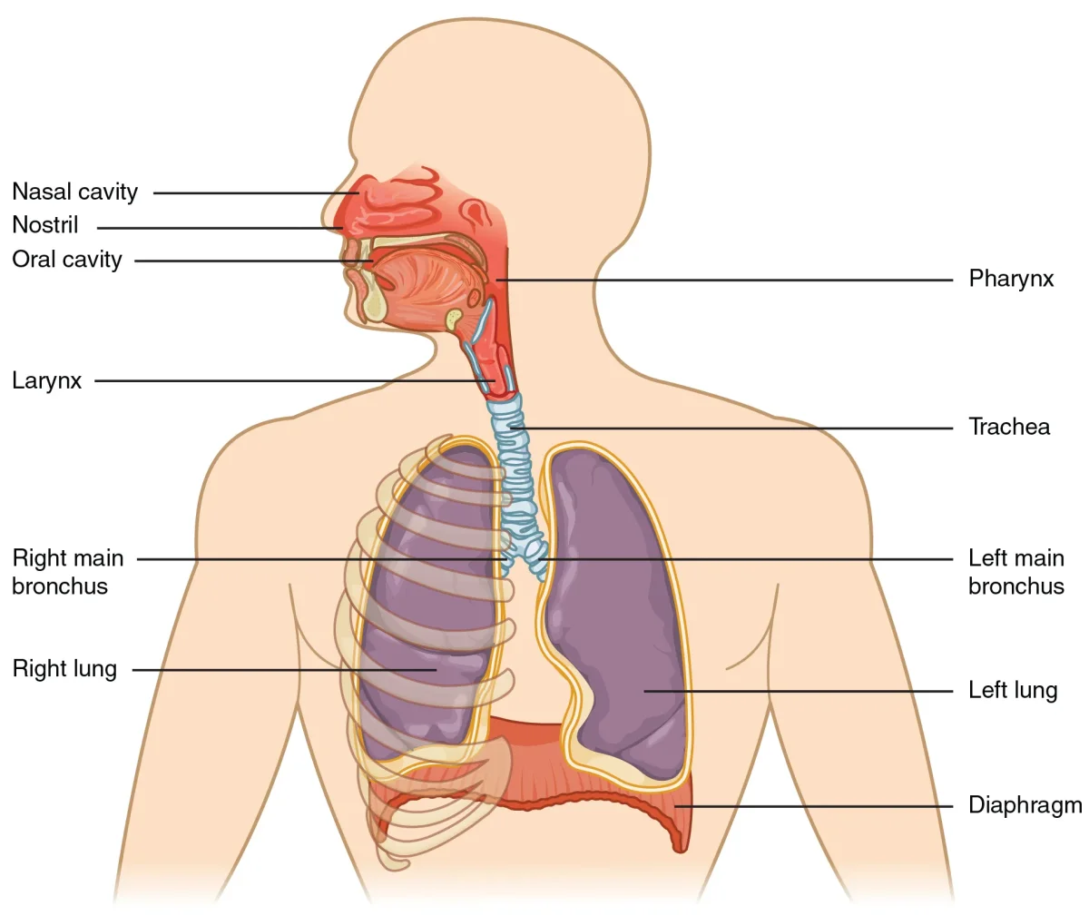
The Upper Respiratory Tract
The upper respiratory tract includes the nasal cavity, pharynx, and larynx. These structures sit above the trachea and serve as the entry point and first line of defense for incoming air.
The Nasal Cavity
Air enters through the nostrils and passes into the nasal cavity, which is divided by the nasal septum. Three bony projections called nasal conchae (or turbinates) protrude into each side of the cavity. These conchae create turbulent airflow, forcing air to swirl and make maximum contact with the mucosal lining.
The nasal cavity performs three critical functions simultaneously:
- Warming - A rich blood supply in the mucosal lining heats incoming air to body temperature
- Humidifying - Mucus and serous secretions add moisture, protecting the delicate lower airway tissues
- Filtering - Nasal hairs (vibrissae) trap large particles, while mucus produced by goblet cells captures smaller particles and pathogens
The nasal cavity also contains the olfactory epithelium in its superior region, which is responsible for the sense of smell.
The Pharynx
The pharynx is a shared passageway for both air and food. It is divided into three regions:
| Region | Location | Function |
|---|---|---|
| Nasopharynx | Behind the nasal cavity | Air passage only; contains pharyngeal tonsils (adenoids) |
| Oropharynx | Behind the oral cavity | Shared air and food passage |
| Laryngopharynx | Above the larynx | Shared passage; diverges into larynx (air) and esophagus (food) |
The pharynx is where the respiratory and digestive pathways cross. This crossover is why you can choke - food can accidentally enter the airway instead of the esophagus.
The Larynx
The larynx (voice box) sits at the top of the trachea and serves two functions: sound production and airway protection.
The epiglottis is a flap of elastic cartilage that covers the opening of the larynx during swallowing, preventing food from entering the airway. When you swallow, the larynx elevates and the epiglottis folds down like a trap door over the glottis (the opening between the vocal cords).
The vocal cords (vocal folds) stretch across the larynx. Air passing between them causes vibration, producing sound. Tension in the cords controls pitch - tighter cords produce higher-pitched sounds.
The Lower Respiratory Tract
Below the larynx, the lower respiratory tract begins with the trachea and extends through the bronchial tree to the alveoli.
The Trachea
The trachea (windpipe) is a tube roughly 10-12 cm long that connects the larynx to the bronchi. It is reinforced by 16-20 C-shaped cartilage rings that keep it open during pressure changes. These rings are C-shaped (not complete circles) because the open side faces the esophagus posteriorly, allowing the esophagus to expand when you swallow food.
The trachea is lined with pseudostratified ciliated columnar epithelium containing goblet cells. This is the beginning of the mucociliary escalator - a defense system where mucus traps particles and cilia beat in coordinated waves to push the mucus upward toward the pharynx, where it is swallowed or expelled.
The Mucociliary Escalator
The mucociliary escalator deserves special attention because it is one of the respiratory system’s most important innate defense mechanisms. Here is how it works:
- Goblet cells secrete sticky mucus that lines the airways
- Cilia on epithelial cells beat in coordinated waves (about 10-20 beats per second)
- The beating cilia propel the mucus - along with any trapped bacteria, dust, or debris - upward toward the pharynx
- At the pharynx, the mucus is swallowed (and destroyed by stomach acid) or coughed out
Smoking paralyzes cilia and increases mucus production simultaneously. This is why smokers develop a chronic “smoker’s cough” - without functioning cilia, coughing becomes the only way to clear mucus from the airways.
Other Respiratory Defenses
The mucociliary escalator is not the only defense mechanism. The respiratory system also uses:
- Lysozyme - an enzyme found in nasal secretions, tears, and saliva that attacks the peptidoglycan cell walls of gram-positive bacteria
- IgA antibodies - mucosal surfaces are coated with secretory IgA, which binds pathogens and prevents them from attaching to epithelial cells
- Alveolar macrophages (dust cells) - immune cells that patrol the alveolar surfaces, engulfing bacteria and particles that make it past the upper defenses
- Mast cells - immune cells in the lungs with antibodies on their surface. When triggered, they release inflammatory chemicals. Mast cells are also responsible for allergic reactions in the airways (like asthma)
Thermoregulation
The respiratory system plays an important role in temperature regulation. As air passes through the nasal cavity and upper airways, the rich capillary beds in the mucosal lining warm incoming air to body temperature and humidify it to near 100% humidity. This protects the delicate lower airways from cold, dry air damage.
During exhalation, heat and water vapor are carried out of the body with the exhaled air. This represents a source of evaporative heat loss. When body temperature rises (during exercise or fever), increased respiratory rate accelerates this heat loss. In some animals, panting is a primary thermoregulation mechanism - rapid shallow breathing maximizes evaporative cooling from the respiratory surfaces.
Conducting Zone vs. Respiratory Zone
This is a critical distinction for the MCAT:
| Feature | Conducting Zone | Respiratory Zone |
|---|---|---|
| Structures | Nose to terminal bronchioles | Respiratory bronchioles, alveolar ducts, alveoli |
| Gas exchange? | No | Yes |
| Cartilage support? | Yes (decreases distally) | No |
| Smooth muscle? | Yes (increases in bronchioles) | Minimal |
| Epithelium | Pseudostratified ciliated columnar | Simple squamous |
| Also called | ”Anatomical dead space" | "Gas exchange surface” |
The conducting zone is called anatomical dead space because air sitting in these passages is not participating in gas exchange. Typical anatomical dead space volume is about 150 mL. This means that of a normal 500 mL tidal breath, only about 350 mL actually reaches the alveoli for gas exchange.