
Lung Structure
If the trachea is the trunk of a tree, the bronchi are the main branches, the bronchioles are the smaller twigs, and the alveoli are the leaves - tiny, numerous, and where the real work happens. Your lungs contain about 300 million alveoli, creating a total gas exchange surface area roughly the size of a tennis court (about 70 square meters). All of that surface area is packed inside your chest because the alveoli are incredibly small - each one is only about 0.2 mm in diameter.
The Bronchial Tree
At the carina (the ridge where the trachea divides), the trachea splits into the right and left main (primary) bronchi. Each main bronchus enters its respective lung at the hilum - the medial surface where blood vessels, nerves, and lymphatics also enter.
The branching pattern is:
- Main (primary) bronchi - one per lung; the right is wider, shorter, and more vertical than the left
- Lobar (secondary) bronchi - one per lobe (3 right, 2 left)
- Bronchioles - no cartilage, walls are smooth muscle
- Terminal bronchioles - the smallest conducting airways (end of the conducting zone)
- Respiratory bronchioles - have some alveoli budding from their walls (beginning of the respiratory zone)
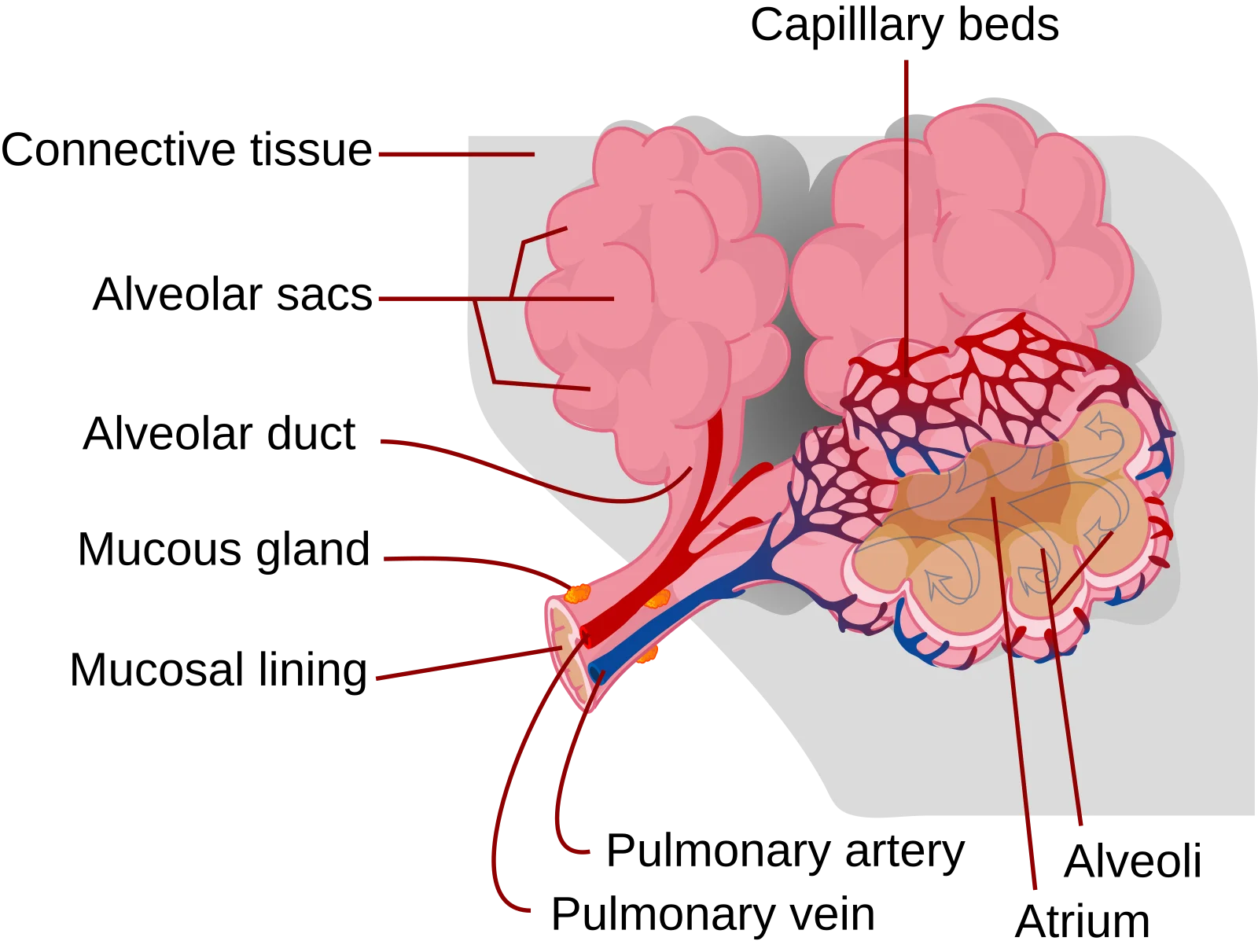
- Alveolar ducts and alveolar sacs - clusters of alveoli where gas exchange occurs
Key Structural Changes Along the Bronchial Tree
As you move from the trachea toward the alveoli, several important structural changes occur:
| Feature | Trachea/Bronchi | Bronchioles | Alveoli |
|---|---|---|---|
| Cartilage | C-rings/plates (structural support) | None | None |
| Smooth muscle | Minimal | Abundant (controls airflow) | Minimal |
| Epithelium | Pseudostratified ciliated columnar | Simple ciliated columnar/cuboidal | Simple squamous |
| Diameter | Large | Small (< 1 mm) | Tiny (~0.2 mm) |
| Function | Conduct air | Conduct air, regulate airflow | Gas exchange |
Two trends to remember: cartilage decreases while smooth muscle increases as airways get smaller. Large airways need cartilage to stay open, but small airways need smooth muscle to regulate airflow distribution. This is why asthma (bronchiolar constriction) affects exhalation so dramatically: the small airways have no cartilage to keep them propped open against the squeeze.
The Lungs
The two lungs are not identical:
- Right lung: 3 lobes (superior, middle, inferior), separated by the horizontal and oblique fissures
- Left lung: 2 lobes (superior, inferior), separated by the oblique fissure; has a cardiac notch - an indentation where the heart sits
The Pleural Membranes
Each lung is enclosed in a double-layered membrane called the pleura:
- Visceral pleura - the inner layer, directly adherent to the lung surface. It dips into the fissures between lobes.
- Parietal pleura - the outer layer, lining the inside of the chest wall and the superior surface of the diaphragm.
- Pleural cavity - the potential space between the two layers, containing a thin film of pleural fluid.
The pleural fluid serves two functions: it lubricates the pleural surfaces so the lungs can slide smoothly during breathing, and it creates surface tension that keeps the lungs adhered to the chest wall (like two wet glass slides stuck together).
Intrapleural Pressure
The pressure within the pleural cavity (intrapleural pressure) is normally negative - about -4 mmHg at rest relative to atmospheric pressure. This negative pressure is critical because it keeps the lungs inflated.
Why is it negative? The lungs have elastic recoil that constantly tries to collapse them inward, while the chest wall has its own elastic tendency to spring outward. These two opposing forces pull the pleural layers slightly apart, creating a suction effect (negative pressure) in the pleural space.
Pneumothorax
A pneumothorax occurs when air enters the pleural space, breaking the negative pressure seal. Without negative intrapleural pressure, the elastic recoil of the lung is no longer opposed, and the lung collapses.
A pneumothorax can result from chest trauma or spontaneous rupture of a small air pocket on the lung surface. The key concept is understanding why loss of negative intrapleural pressure leads to lung collapse.
The Alveoli
The alveoli are the functional units of the lung - tiny, thin-walled sacs where all gas exchange occurs. Each alveolus is wrapped in a dense network of capillaries, and the barrier between alveolar air and capillary blood is extraordinarily thin - only about 0.5 micrometers.
This barrier, called the respiratory membrane, consists of:
- A thin layer of fluid lining the alveolus (with surfactant)
- The alveolar epithelium (Type I cells)
- A fused basement membrane
- The capillary endothelium
Two types of cells make up the alveolar epithelium:
| Cell Type | Structure | Function |
|---|---|---|
| Type I pneumocytes (alveolar cells) | Very thin, flat squamous cells; cover ~95% of alveolar surface | Gas exchange - their thinness minimizes diffusion distance |
| Type II pneumocytes (alveolar cells) | Cuboidal cells; cover ~5% of surface but are more numerous | Secrete surfactant; can divide to regenerate Type I cells |
Alveolar macrophages (dust cells) patrol the alveolar surfaces, engulfing bacteria, dust, and other particles that make it past the mucociliary escalator. They are the last line of defense before the gas exchange surface.
Surfactant
Surfactant is a phospholipid-rich substance (primarily dipalmitoylphosphatidylcholine, or DPPC) secreted by Type II pneumocytes. It reduces surface tension within the alveoli, preventing them from collapsing during exhalation.
Why is this necessary? The alveoli are essentially tiny wet bubbles. Surface tension in a wet sphere naturally tries to collapse it (think of a soap bubble popping). Without surfactant, the smallest alveoli would collapse first (because LaPlace’s law tells us that smaller spheres have higher collapsing pressure for the same surface tension), and each breath would require enormous effort to re-inflate them.