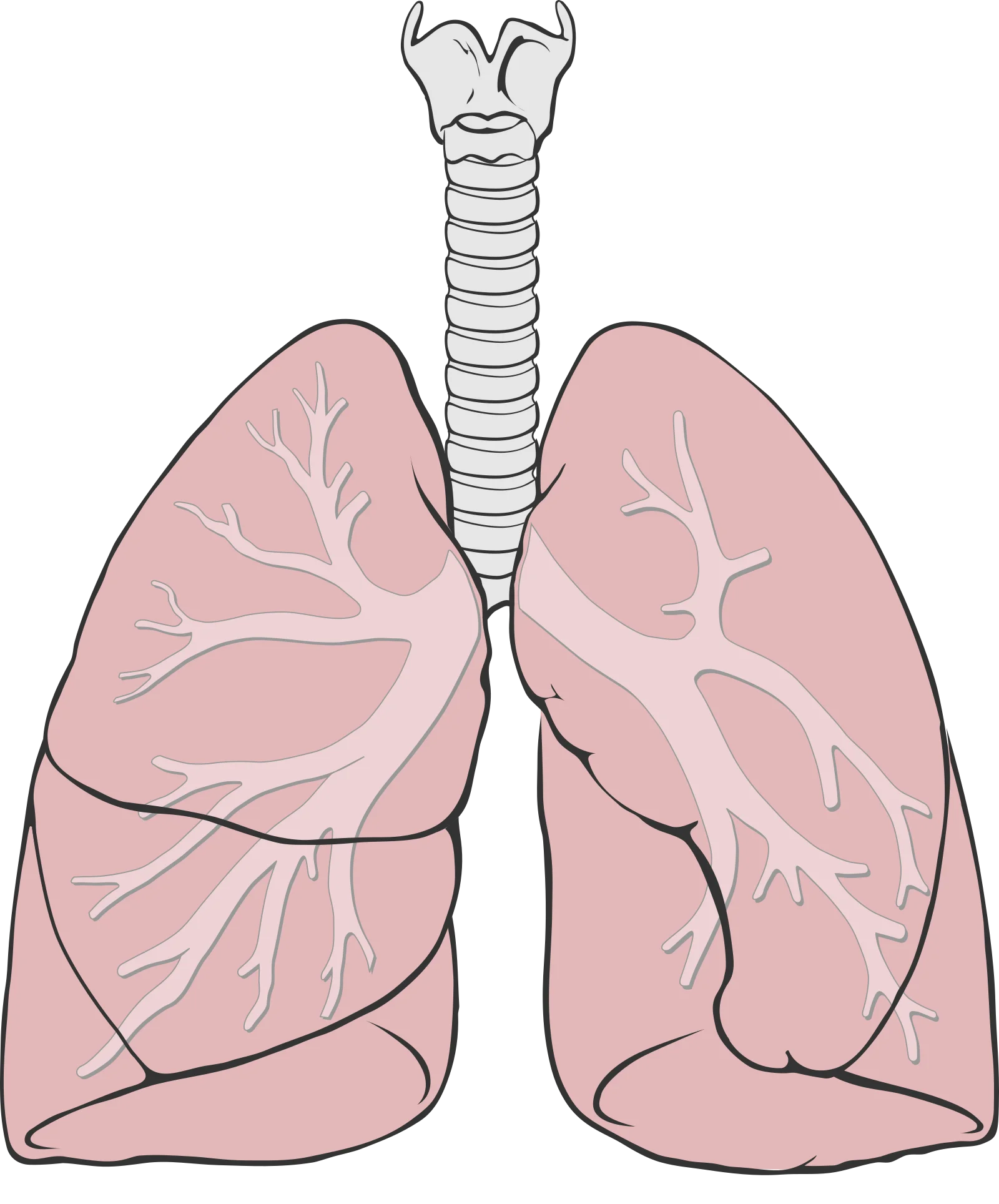
Lung Volumes
Imagine your lungs as a water bottle. During quiet breathing, you only sip from the top (tidal volume). But you could chug much more if you tried (inspiratory reserve). After a normal sip, you could force out more water by squeezing (expiratory reserve). And no matter how hard you squeeze, some water always stays at the bottom, trapped in the crevices (residual volume). Understanding these volumes - and how they combine into capacities - is essential for MCAT questions about pulmonary function.
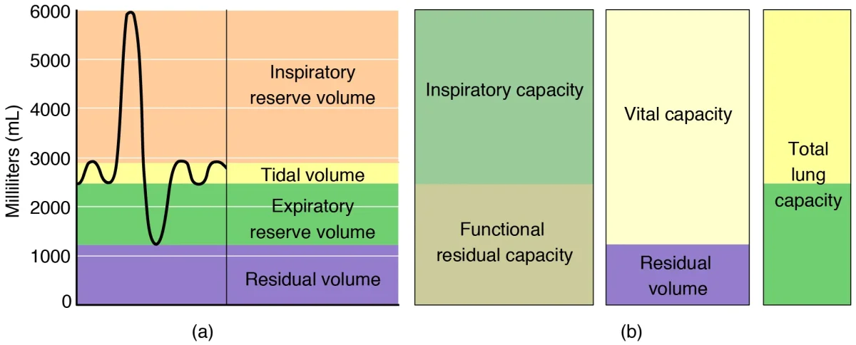
The Four Lung Volumes
There are four non-overlapping lung volumes that cannot be subdivided further:
| Volume | Abbreviation | Definition | Typical Value |
|---|---|---|---|
| Tidal Volume | TV | Air moved in or out during a normal, quiet breath | ~500 mL |
| Inspiratory Reserve Volume | IRV | Extra air you can inhale beyond a normal tidal breath (with maximal effort) | ~3,000 mL |
| Expiratory Reserve Volume | ERV | Extra air you can exhale beyond a normal tidal exhalation (with effort) | ~1,200 mL |
| Residual Volume | RV | Air that remains in the lungs even after maximal forced exhalation | ~1,200 mL |
The residual volume exists because the airways and alveoli do not completely collapse during exhalation. RV prevents alveolar collapse and maintains gas exchange even between breaths.
Lung Capacities
Lung capacities are combinations of two or more volumes. There are four:
| Capacity | Formula | Definition | Typical Value |
|---|---|---|---|
| Inspiratory Capacity (IC) | TV + IRV | Maximum air you can inhale from the end of a normal exhalation | ~3,500 mL |
| Functional Residual Capacity (FRC) | ERV + RV | Air remaining in the lungs after a normal exhalation | ~2,400 mL |
| Vital Capacity (VC) | TV + IRV + ERV | Maximum air you can move in one breath (deepest inhale to hardest exhale) | ~4,700 mL |
| Total Lung Capacity (TLC) | TV + IRV + ERV + RV | Total air the lungs can hold at maximum inflation | ~5,900 mL |
Spirometry
Spirometry is the clinical test used to measure lung volumes and capacities. The patient breathes into a device that records volume changes over time, producing a spirogram.
Important limitation: spirometry cannot measure residual volume directly. Since RV is the air you cannot blow out, it stays in the lungs during the test. This means spirometry also cannot directly measure FRC or TLC (both include RV).
To measure RV, clinicians use indirect methods such as gas dilution techniques. The key point for the MCAT: spirometry alone cannot give you RV.
Minute Ventilation and Alveolar Ventilation
Minute ventilation is the total volume of air moved per minute:
But minute ventilation overestimates how much air is actually useful, because it includes dead space air. Alveolar ventilation corrects for this:
Why Breathing Pattern Matters
Consider two patients who both have a minute ventilation of 6 L/min:
| Patient | TV | RR | Minute Ventilation | Alveolar Ventilation |
|---|---|---|---|---|
| A | 500 mL | 12/min | 6,000 mL/min | (500-150) x 12 = 4,200 mL/min |
| B | 200 mL | 30/min | 6,000 mL/min | (200-150) x 30 = 1,500 mL/min |
Patient B has the same minute ventilation but far less alveolar ventilation. Shallow, rapid breathing is inefficient because a larger proportion of each breath is wasted filling dead space. This is why deep, slow breaths are more effective than shallow, fast ones.
Obstructive vs. Restrictive Lung Disease
Pulmonary function tests are used to distinguish two broad categories of lung disease:
| Feature | Obstructive Disease | Restrictive Disease |
|---|---|---|
| Problem | Airflow out is blocked | Lungs cannot fully expand |
| Examples | Asthma, COPD (emphysema, chronic bronchitis) | Pulmonary fibrosis, scoliosis, obesity |
| FEV1 (air exhaled in 1 sec) | Decreased | Decreased |
| FVC (total air exhaled forcefully) | Normal or slightly decreased | Decreased |
| FEV1/FVC ratio | Decreased (< 0.7) | Normal or increased (> 0.7) |
| TLC | Normal or increased (air trapping) | Decreased |
| RV | Increased (can’t blow it all out) | Decreased |
| Compliance | Increased (emphysema) | Decreased (stiff lungs) |