
Skeletal Muscle Structure
Before we can understand how muscles contract, we need to understand what they are made of. Skeletal muscle has an elegant, repeating architecture that is organized like a set of Russian nesting dolls - each level is a smaller version of the one above it.
The Muscle Hierarchy
From largest to smallest:
| Level | What It Is | Wrapped In |
|---|---|---|
| Muscle | The whole organ (e.g., biceps brachii) | Epimysium (dense connective tissue) |
| Fascicle | A bundle of muscle fibers | Perimysium |
| Muscle fiber | A single multinucleated cell | Endomysium |
| Myofibril | A contractile thread running the length of the fiber | Sarcolemma encloses all myofibrils |
| Sarcomere | The functional unit of contraction | Bounded by Z lines |
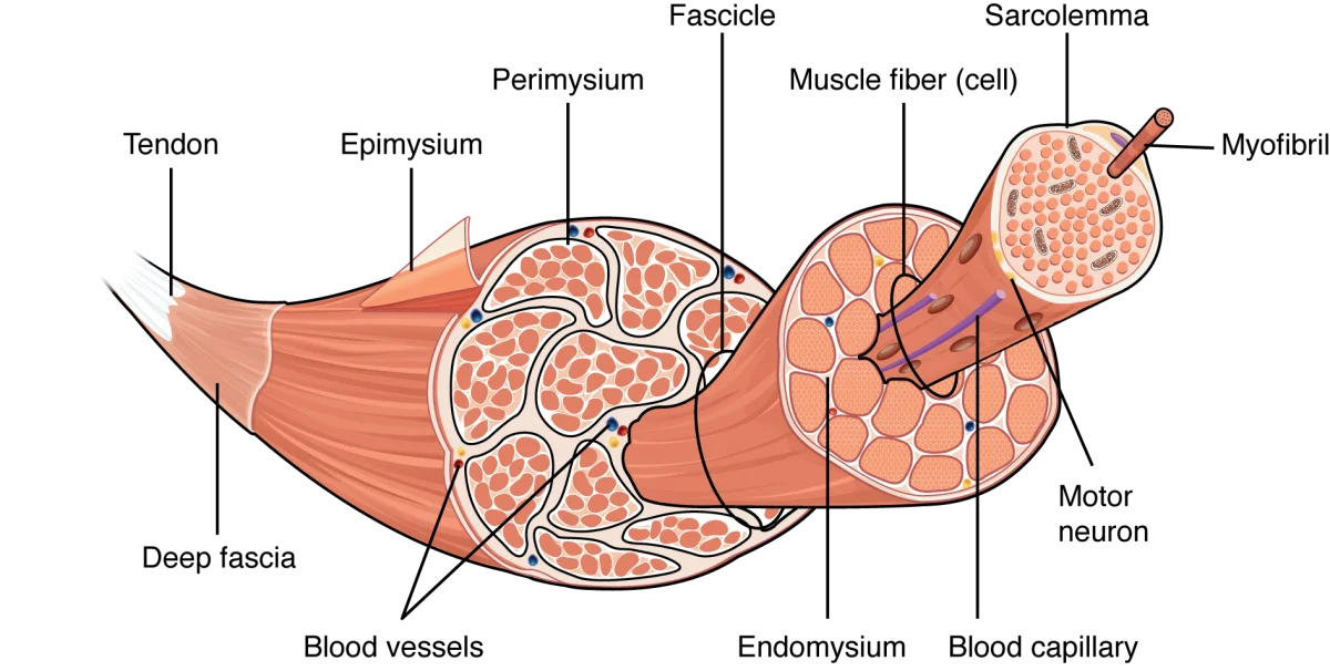
Key Features of a Muscle Fiber (Cell)
A single skeletal muscle fiber is one of the largest and most unusual cells in the body:
- Multinucleated - each fiber contains dozens to hundreds of nuclei, pushed to the periphery of the cell. This is because muscle fibers form by the fusion of many precursor cells (myoblasts) during development.
- Sarcolemma - the cell membrane of a muscle fiber
- Sarcoplasm - the cytoplasm of a muscle fiber
- Sarcoplasmic reticulum (SR) - a specialized smooth endoplasmic reticulum that stores and releases Ca²⁺. This is the calcium reservoir that triggers contraction.
- T-tubules (transverse tubules) - invaginations of the sarcolemma that plunge deep into the fiber. They carry the action potential from the surface to the interior, ensuring the entire fiber contracts simultaneously.
- Triad - the functional unit of excitation-contraction coupling: one T-tubule flanked by two terminal cisternae of the SR. This arrangement ensures that the electrical signal (T-tubule) is right next to the calcium store (SR).
The Sarcomere - The Functional Unit of Contraction
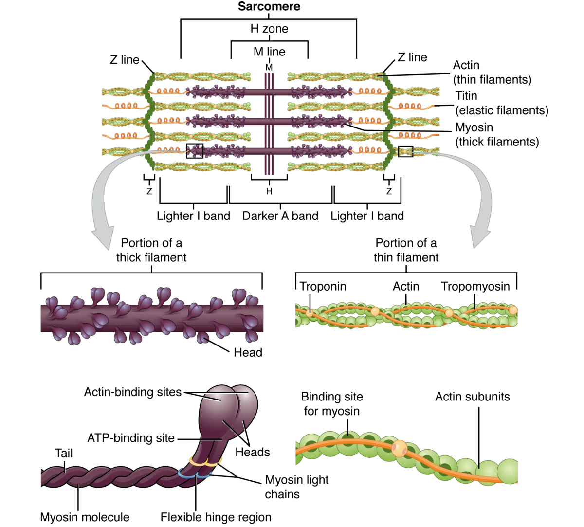
The sarcomere is the repeating unit that makes muscle contraction possible. Each myofibril is a chain of thousands of sarcomeres arranged end to end. The sarcomere is defined as the region between two Z lines (also called Z discs).
Sarcomere Components
The two main filaments:
| Component | Thick Filament | Thin Filament |
|---|---|---|
| Main protein | Myosin (with ATPase heads) | Actin (globular subunits forming F-actin) |
| Regulatory proteins | None on the filament itself | Tropomyosin (covers myosin-binding sites on actin) and Troponin (Ca²⁺ sensor) |
| Location | Center of sarcomere | Anchored to Z lines, extend toward center |
| Diameter | ~15 nm (thick) | ~7 nm (thin) |
The bands and zones:
| Structure | What It Contains | Appearance | Changes During Contraction? |
|---|---|---|---|
| A band | Entire length of thick (myosin) filaments, including any overlap with thin filaments | Dark | NO - stays the same width (A = Always the same) |
| I band | Thin (actin) filaments ONLY (no thick filament overlap) | Light | YES - gets SHORTER |
| H zone | Thick (myosin) filaments ONLY (no thin filament overlap) | Light region within A band | YES - gets SHORTER |
| Z line (Z disc) | Protein disc anchoring thin filaments | Thin dark line | Moves CLOSER to center during contraction |
| M line | Proteins connecting thick filaments at the center | Thin line in center of H zone | Stays in center |
Regulatory Proteins: Troponin and Tropomyosin
These two proteins on the thin filament act as the molecular “safety lock” that prevents contraction when the muscle should be relaxed:
-
Tropomyosin - a long, thread-like protein that wraps around the actin filament, physically covering the myosin-binding sites. When tropomyosin is in the blocking position, myosin heads cannot attach to actin, and contraction cannot occur.
-
Troponin - a regulatory complex that sits on tropomyosin at regular intervals. The key subunit is troponin C, which binds calcium (Ca²⁺). When Ca²⁺ binds to troponin C, the troponin complex changes shape, pulling tropomyosin away from the myosin-binding sites on actin. This exposes the binding sites and allows cross-bridge cycling (contraction) to begin.