
Autonomic NS
Your heart rate, pupil diameter, digestive activity, and bronchiole diameter are all adjusted continuously without a single conscious thought. The autonomic nervous system (ANS) manages these vital functions automatically, keeping your internal environment stable whether you are sprinting from danger or digesting a meal. For the MCAT, you need to know exactly how the ANS is organized, which neurotransmitters it uses at each synapse, and what each division does to every major organ system.
Overview: Two-Neuron Chain
Both divisions of the ANS use a two-neuron chain to connect the CNS to the target organ. The first neuron (preganglionic neuron) has its cell body in the CNS and synapses at an autonomic ganglion. The second neuron (postganglionic neuron) has its cell body in the ganglion and sends its axon to the effector organ.
This two-neuron design differs from the somatic nervous system, which uses a single motor neuron to reach skeletal muscle. The location and length of these neurons differ between the sympathetic and parasympathetic divisions, and this is a commonly tested distinction.
The Sympathetic Division: “Fight or Flight”
The sympathetic nervous system prepares the body for intense physical activity. Think of every change it makes as an answer to the question: “What does the body need to fight or run away?”
Origin: Preganglionic neurons emerge from the thoracolumbar spinal cord (T1-L2). Their cell bodies reside in the lateral horns of the spinal cord grey matter.
Neuron lengths: Preganglionic fibers are short (they synapse in ganglia close to the spinal cord). Postganglionic fibers are long (they must travel from the paravertebral or prevertebral ganglia to the distant target organs).
Neurotransmitters:
- Preganglionic neurons release acetylcholine (ACh) at the ganglion (all preganglionic neurons in both divisions use ACh).
- Postganglionic neurons release norepinephrine (NE) at the target organ. The one major exception: sympathetic postganglionic fibers to sweat glands release ACh, not NE.
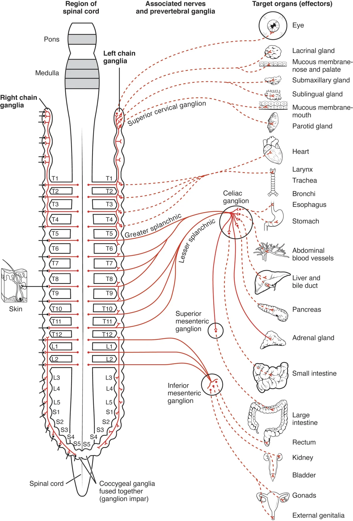
The adrenal medulla deserves special attention. It is a modified sympathetic ganglion. Preganglionic sympathetic fibers synapse directly on chromaffin cells within the adrenal medulla. Instead of sending a postganglionic neuron to a target, these cells release epinephrine (~80%) and norepinephrine (~20%) directly into the bloodstream. This allows the sympathetic response to reach the entire body at once, amplifying the fight-or-flight effect.
The Parasympathetic Division: “Rest and Digest”
The parasympathetic nervous system promotes calming, energy-conserving, and maintenance activities. It dominates during rest, digestion, and recovery.
Origin: Preganglionic neurons emerge from the craniosacral regions - specifically cranial nerves III, VII, IX, and X, plus sacral spinal segments S2-S4.
Neuron lengths: Preganglionic fibers are long (they travel from the brainstem or sacral cord all the way to ganglia near or within the target organ). Postganglionic fibers are short (the ganglion is right next to or embedded in the organ wall).
Neurotransmitters: Both preganglionic and postganglionic neurons release acetylcholine (ACh). This is a key distinction from the sympathetic division, where the postganglionic neurotransmitter is norepinephrine.
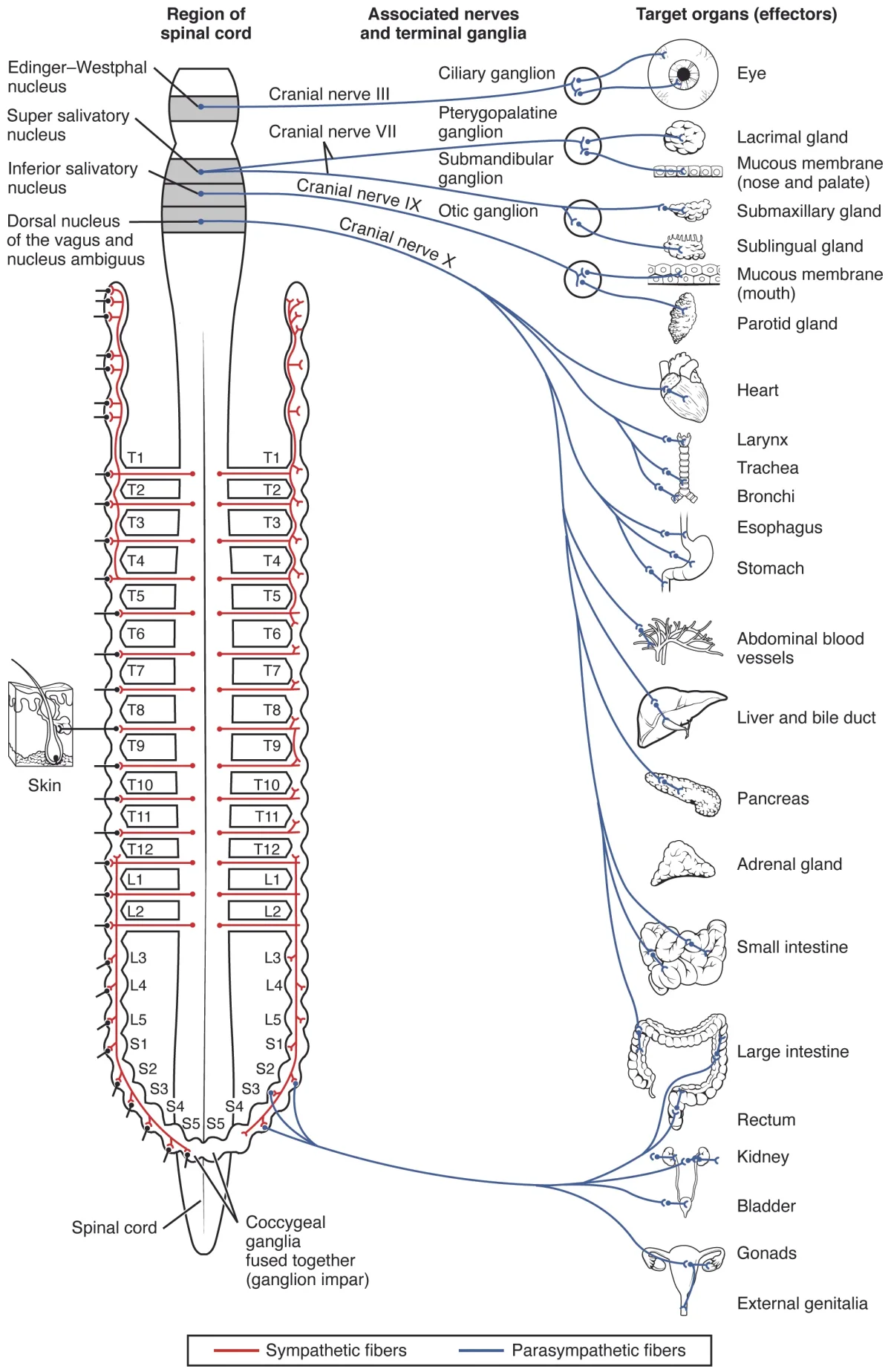
The vagus nerve (cranial nerve X) is by far the most important parasympathetic nerve. It carries approximately 75% of all parasympathetic fibers and innervates the heart, lungs, and virtually the entire gastrointestinal tract. When the MCAT mentions parasympathetic effects on thoracic or abdominal organs, the vagus nerve is almost always the pathway involved.
Dual Innervation and Organ-Specific Effects
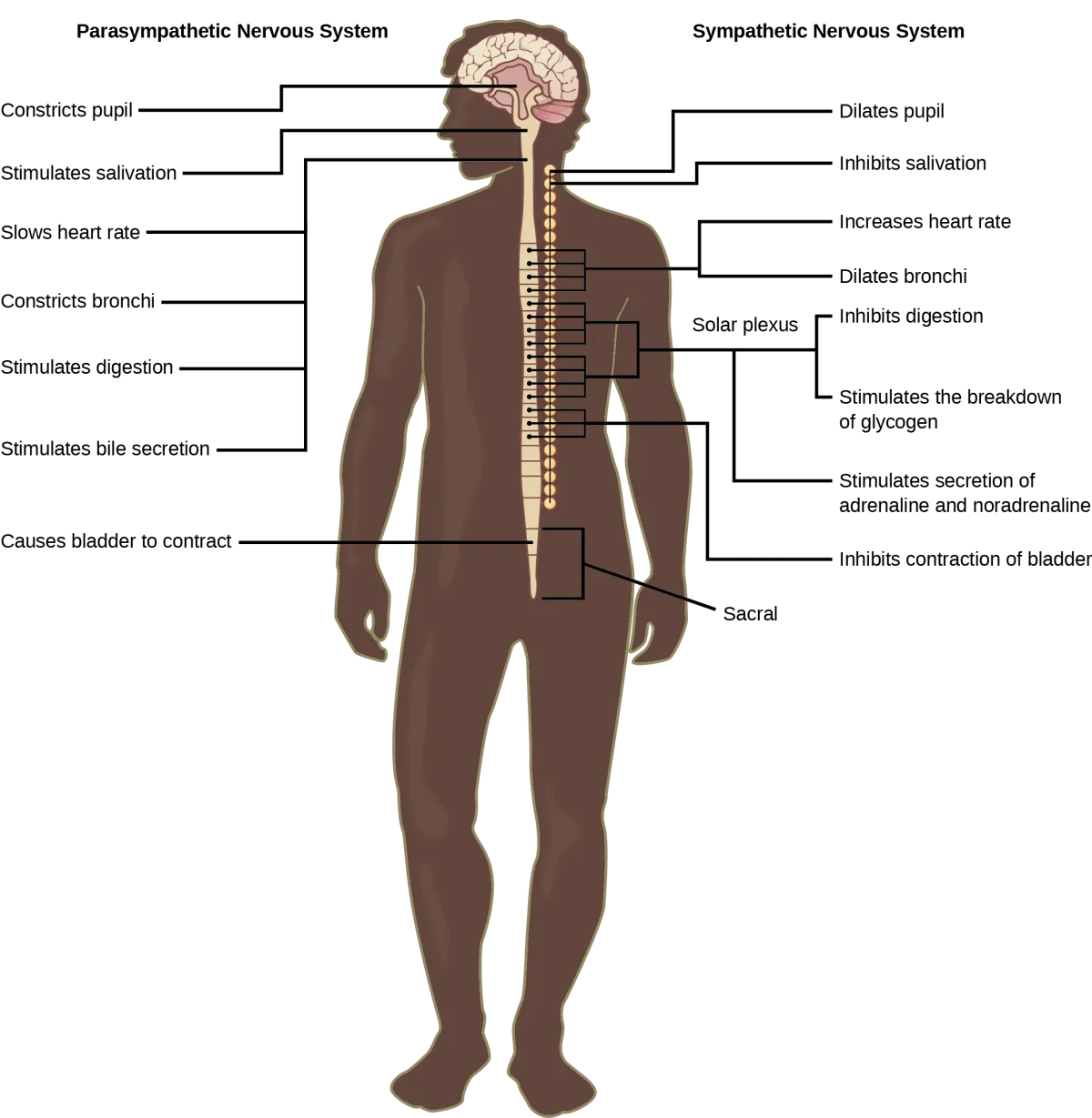
Most organs receive input from both sympathetic and parasympathetic divisions. This dual innervation allows precise control through the balance of opposing signals. The table below summarizes the effects on each organ system:
| Organ/Tissue | Sympathetic Effect | Parasympathetic Effect |
|---|---|---|
| Heart | Increases rate and contractility (cardiac output) | Decreases rate |
| Blood vessels | Constriction (most) | Little direct effect (most vessels lack parasympathetic innervation) |
| Lungs (bronchioles) | Dilation | Constriction |
| Pupils | Dilation (mydriasis) | Constriction (miosis) |
| GI tract (motility) | Decreases motility and secretion | Increases motility and secretion |
| GI sphincters | Contracts (closes) | Relaxes (opens) |
| Bladder (detrusor) | Relaxes (filling) | Contracts (voiding) |
| Bladder sphincter | Contracts (retention) | Relaxes (voiding) |
| Liver | Glycogenolysis (glucose release) | Glycogen synthesis |
| Sweat glands | Stimulates secretion (via ACh) | No innervation |
| Adrenal medulla | Stimulates epinephrine/NE release | No innervation |
Notable exceptions to dual innervation: blood vessels (primarily sympathetic only), sweat glands (sympathetic only via ACh), and the adrenal medulla (sympathetic only).
Receptor Types
The neurotransmitters of the ANS act on specific receptor types. The MCAT expects you to know the major categories.
Cholinergic receptors bind acetylcholine (ACh):
- Nicotinic receptors: found at all autonomic ganglia (both sympathetic and parasympathetic) and at the neuromuscular junction. They are ionotropic (ligand-gated ion channels) and always excitatory.
- Muscarinic receptors: found on target organs innervated by parasympathetic postganglionic fibers (and on sweat glands innervated by sympathetic postganglionic fibers). They are metabotropic (G-protein coupled) and can be excitatory or inhibitory depending on the tissue.
Adrenergic receptors bind norepinephrine and epinephrine:
- Alpha-1 (a1): smooth muscle contraction (vasoconstriction, pupil dilation)
- Alpha-2 (a2): presynaptic inhibition (decreases NE release - negative feedback)
- Beta-1 (b1): heart - increases rate and contractility (“beta-1 = 1 heart”)
- Beta-2 (b2): smooth muscle relaxation - bronchodilation, vasodilation in skeletal muscle (“beta-2 = 2 lungs”)
Neurotransmitter Summary
| Synapse | Sympathetic | Parasympathetic |
|---|---|---|
| Preganglionic neurotransmitter | ACh (nicotinic receptor) | ACh (nicotinic receptor) |
| Postganglionic neurotransmitter | NE (adrenergic receptors) | ACh (muscarinic receptors) |
| Exception | Sweat glands: ACh (muscarinic) | None |
| Special case | Adrenal medulla: Epi + NE into blood | Vagus nerve: ~75% of all parasympathetic output |