
Neural Control of Digestion
Your gut has its own brain. The enteric nervous system (ENS) contains about 100 million neurons - more than the spinal cord - embedded in the walls of the GI tract from esophagus to anus. It can operate completely independently of the brain and spinal cord, earning it the nickname “the second brain.”
The Two Plexuses
The ENS is organized into two interconnected nerve networks (plexuses):
Myenteric plexus (Auerbach’s plexus)
- Located between the inner circular and outer longitudinal muscle layers of the muscularis externa
- Controls motility: peristalsis velocity, contraction intensity, sphincter relaxation
- Stimulation increases gut motility; inhibition relaxes sphincters and slows movement
Submucosal plexus (Meissner’s plexus)
- Located in the submucosa
- Controls secretion and absorption: regulates enzyme release, mucus production, and local blood flow
- Also responds to luminal stretch and chemical stimuli
The ENS Contains All Three Neuron Types
The enteric nervous system is a complete neural circuit:
- Sensory (afferent) neurons - detect mechanical stretch, chemical composition (pH, osmolarity, nutrients), and temperature in the lumen
- Interneurons - process information and coordinate responses
- Motor (efferent) neurons - control smooth muscle contraction, gland secretion, and blood vessel dilation
This means the ENS can carry out complete reflex arcs without any input from the brain or spinal cord. A chemical stimulus in the lumen can trigger enzyme secretion or a peristaltic wave entirely through local ENS circuits.
Autonomic Modulation: Parasympathetic vs. Sympathetic
While the ENS operates autonomously, the central nervous system modulates its activity through the autonomic nervous system:
| Division | Effect on GI | Key Nerve | Neurotransmitter |
|---|---|---|---|
| Parasympathetic (“rest and digest”) | Increases motility, increases secretion, relaxes sphincters | Vagus nerve (CN X) | Acetylcholine (ACh) |
| Sympathetic (“fight or flight”) | Decreases motility, decreases secretion, contracts sphincters | Splanchnic nerves | Norepinephrine (NE) |
The vagus nerve provides parasympathetic innervation to most of the GI tract (esophagus through the transverse colon). The distal colon, rectum, and internal anal sphincter receive parasympathetic input from pelvic splanchnic nerves (S2-S4).
”Rest and Digest” vs. “Fight or Flight”
After a meal, parasympathetic activity dominates:
- Increased salivation
- Increased gastric acid and enzyme secretion
- Increased peristalsis and motility
- Increased blood flow to the GI tract
- This is why you feel drowsy after a big meal - blood is diverted to the gut (“food coma”)
During stress or exercise, sympathetic activity dominates:
- Decreased salivation (dry mouth)
- Decreased secretion and motility
- Blood diverted away from the GI tract to skeletal muscles and brain
- Sphincters contract (no time for digestion when running from danger)
The Defecation Reflex
Defecation involves both the ENS and the CNS:
- Feces distend the rectum, activating stretch receptors
- Sensory signals travel to the sacral spinal cord (S2-S4) via pelvic nerves
- Parasympathetic motor signals return, causing:
- Increased colonic and rectal motility
- Relaxation of the internal anal sphincter (involuntary)
- The external anal sphincter (skeletal muscle, under voluntary somatic control) can be consciously relaxed to allow defecation or kept contracted to delay it
The Three Phases of Gastric Regulation
GI secretion is controlled in three overlapping phases:
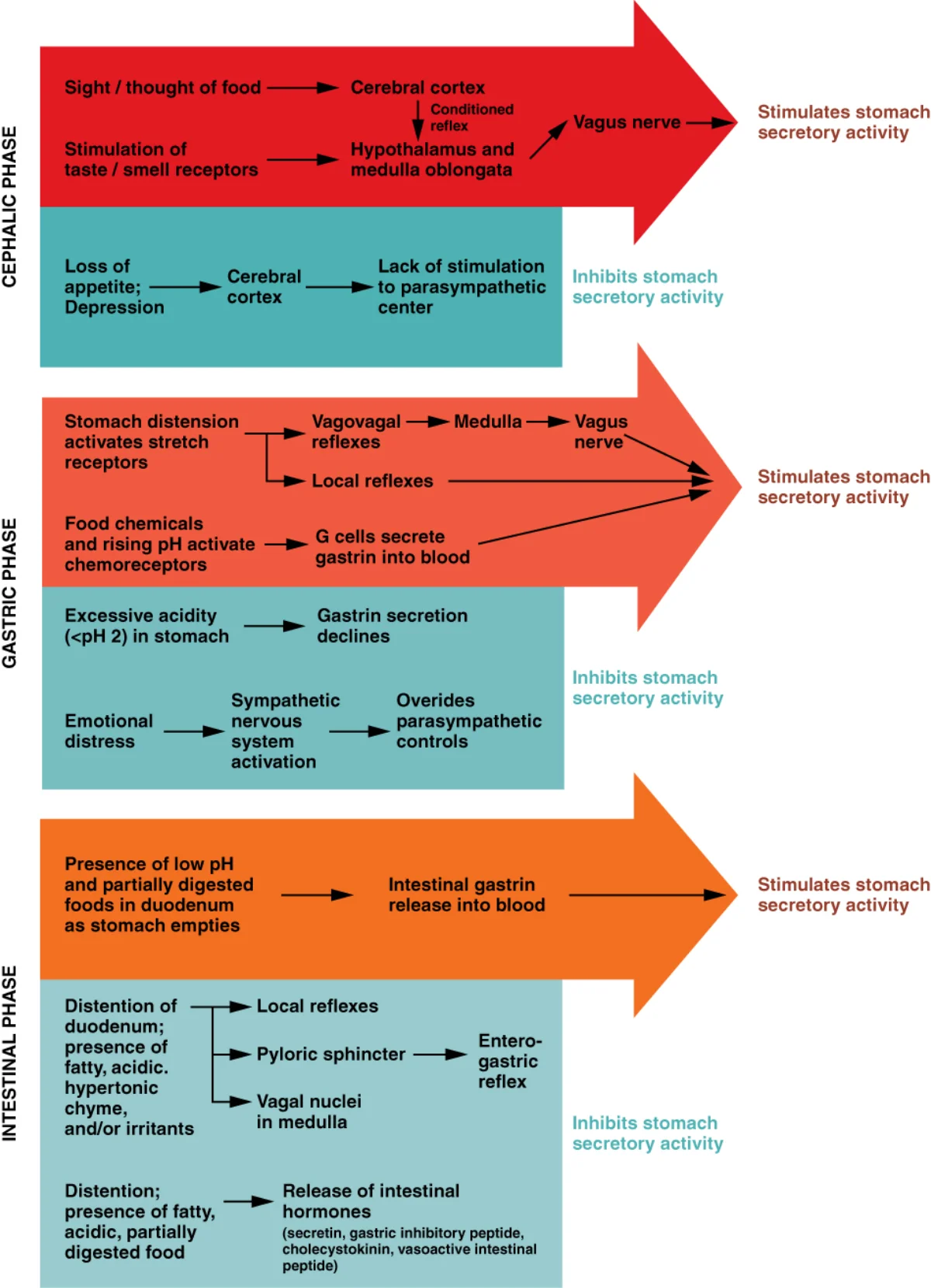
| Phase | Trigger | Mediator | Effect |
|---|---|---|---|
| Cephalic phase | Sight, smell, thought, taste of food | Vagus nerve (parasympathetic) | Stimulates gastric secretion before food arrives (~30% of total) |
| Gastric phase | Food in stomach (distension, peptides, pH changes) | Gastrin, local reflexes | Main stimulation of HCl and pepsinogen (~60% of total) |
| Intestinal phase | Chyme in duodenum (acid, fat, protein) | Secretin, CCK, GIP | Stimulates pancreatic/biliary secretion; inhibits gastric activity |