Three principles. That is the whole theory. If you can remember a three-item list, you know cell theory.
All living things are made of cells. From the tallest redwood tree to the smallest bacterium to the person reading this sentence - every living organism is built from cells.
All cells come from pre-existing cells. No cell pops into existence from nothing. Every cell was made by another cell dividing. Rudolf Virchow summed this up in 1855 with the Latin phrase “Omnis cellula e cellula” - all cells from cells.
The cell is the basic unit of life. It is the smallest thing that can independently carry out the functions of life - metabolism, growth, reproduction, response to stimuli.
A modern addition to cell theory is sometimes called the fourth principle: cells contain hereditary information (DNA) that is passed from parent cell to daughter cell during division. This principle was not part of the original theory because DNA had not yet been discovered, but it is now considered a foundational extension.
The Virus Problem
Here is an important wrinkle the MCAT loves to test: viruses challenge cell theory. Viruses carry genetic material (DNA or RNA) and can evolve, but they cannot reproduce on their own. They must hijack a host cell’s machinery to replicate. Because they lack the ability to independently carry out metabolic processes, viruses are not considered living organisms by most biologists. They exist in a gray zone - not quite alive, not quite dead. We will cover viruses in detail in Section 1.9.
What are the three (or four) tenets of cell theory?
Click to reveal answer
1) All living things are made of cells. 2) All cells arise from pre-existing cells. 3) The cell is the basic unit of life. 4) (Modern addition) Hereditary information (DNA) is passed from parent to daughter cells.
Why do viruses challenge cell theory?
Click to reveal answer
Viruses carry genetic material and can evolve, but they cannot replicate independently or carry out metabolic processes on their own. They require a host cell's machinery, so they do not fit the standard definition of a living organism.
All living organisms (viruses excluded, since they are not alive) fall into one of two cell types: eukaryotic or prokaryotic. The names tell you the key difference if you know a bit of Greek.
Eu- means “true.” Karyon means “kernel” (nucleus). Eukaryotic cells have a true nucleus - their DNA is enclosed in a membrane-bound compartment.
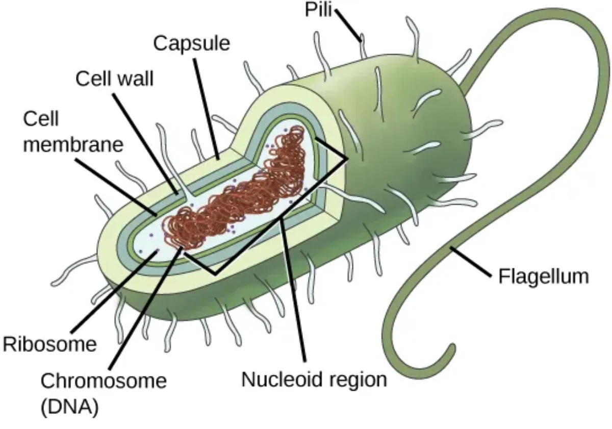
Pro- means “before.” Prokaryotic cells came before eukaryotes in evolutionary history. They are simpler - no true nucleus, no membrane-bound organelles. Their DNA floats freely in a region called the nucleoid.
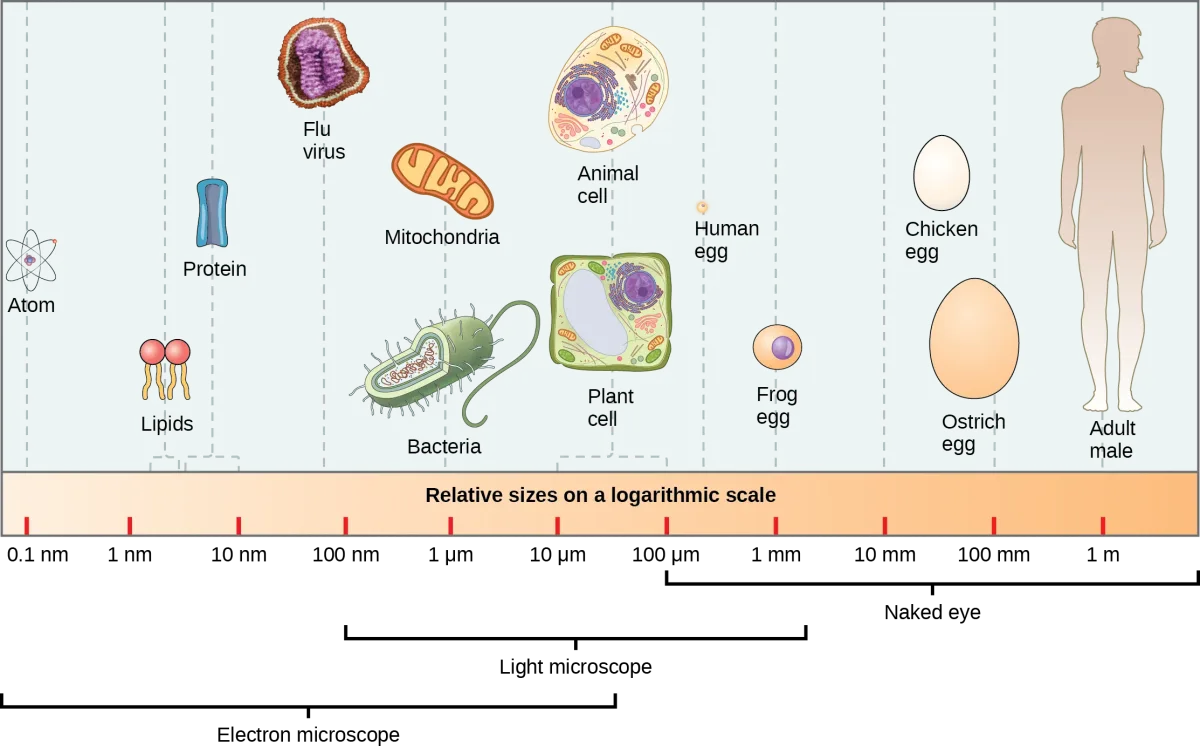
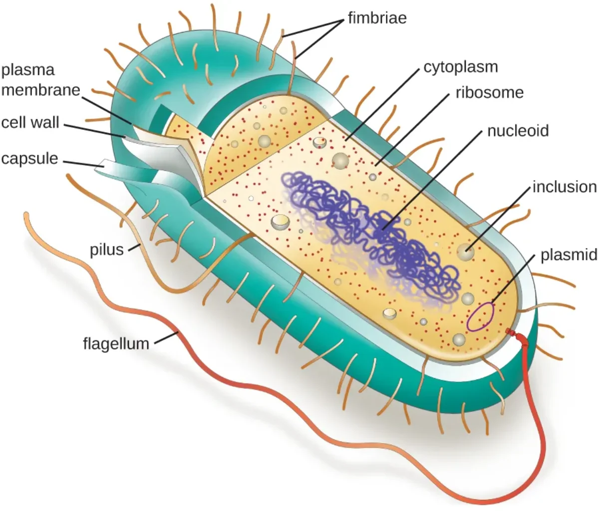
Structure of a typical prokaryotic cell, highlighting the nucleoid region, ribosomes, and external structures. Credit: OpenStax Biology 2e, CC BY 4.0Relative sizes of cells and organisms on a logarithmic scale, from atoms to multicellular organisms. Credit: OpenStax Biology 2e, CC BY 4.0
The Three Domains of Life
When we classify living organisms, they are grouped into three domains: Archaea, Bacteria, and Eukarya. Two of these domains (Archaea and Bacteria) are prokaryotic; one (Eukarya) is eukaryotic.
Why Eukaryotic Cells Have Organelles: Endosymbiotic Theory
Eukaryotic cells are not just “prokaryotes plus a nucleus.” They are prokaryotes that ate other prokaryotes and kept them as internal tenants. The endosymbiotic theory proposes that mitochondria (and, in plants, chloroplasts) are the descendants of free-living prokaryotes that were engulfed by a larger host cell billions of years ago.
Evidence: mitochondria have their own circular DNA, their own 70S ribosomes, a double membrane, and they divide by binary fission. We cover this in depth in Mitochondria.
Archaea in One Minute
Archaea look like bacteria (unicellular, no nucleus, circular DNA) but their molecular machinery resembles eukaryotes. The three features the MCAT wants you to know:
Histones package Archaeal DNA (bacteria lack histones).
Methionine starts translation (bacteria use formylmethionine).
Archaeal RNA polymerases resemble the eukaryotic enzymes.
Many Archaea are extremophiles (hot springs, salt lakes, deep-sea vents) and most are chemosynthetic, pulling energy from inorganic reactions rather than sunlight.
What three features do Archaea share with eukaryotes but NOT with bacteria?
Click to reveal answer
1) Histones for DNA organization. 2) Methionine (not fMet) as the initiator amino acid. 3) Eukaryote-like RNA polymerases. These point to a shared ancestry between Archaea and Eukarya.
What evidence supports the endosymbiotic origin of mitochondria?
Click to reveal answer
Mitochondria have circular DNA, 70S ribosomes, a double membrane, and divide by binary fission - all bacterial features. They are the descendants of an aerobic prokaryote engulfed by an ancestral eukaryotic cell.
Imagine a nightclub with a very picky bouncer at the door. Small, nonpolar molecules (O2, CO2, steroid hormones) are on the VIP list - they walk right in, no questions asked. Large or charged molecules (glucose, amino acids, ions) need a protein escort - a channel or transporter protein that lets them through. And some molecules actually need to be physically dragged inside against their will, which costs energy (ATP). The bouncer does not work for free.
That nightclub is the cell membrane (also called the plasma membrane). It is not just a wall - it is a selectively permeable barrier that controls everything entering and leaving the cell. The specific mechanisms that move molecules across this barrier - from simple diffusion to active pumps - are covered in detail in cell transport.
The Phospholipid Bilayer
The membrane is built from a double layer of phospholipids. Each phospholipid has two parts:
A hydrophilic head (polar, “water-loving”) - faces outward toward water on both sides of the membrane
Two hydrophobic tails (nonpolar, “water-fearing”) - face inward, away from water, creating a greasy interior
This dual nature makes phospholipids amphipathic - they have both a water-loving and a water-fearing region. When you put millions of amphipathic molecules in water, they spontaneously arrange into a bilayer, with heads facing the aqueous environment and tails hiding in the middle. This arrangement creates a hydrophobic core that acts as a barrier to most polar and charged molecules.
The membrane is not rigid. It behaves like a fluid - proteins and lipids can move laterally within the bilayer. This is the basis of the fluid mosaic model: the membrane is a fluid sea of phospholipids with a mosaic of proteins embedded in it.
Cholesterol: the Thermostat of the Membrane
Animal cell membranes also contain cholesterol wedged between phospholipids. Cholesterol is a fluidity buffer:
At high temperatures it restrains phospholipid movement, keeping the membrane from becoming too fluid.
At low temperatures it wedges apart phospholipid tails, preventing the membrane from packing into a rigid gel.
Shorter and more unsaturated (kinked) fatty acid tails also increase fluidity; longer, saturated tails decrease it.
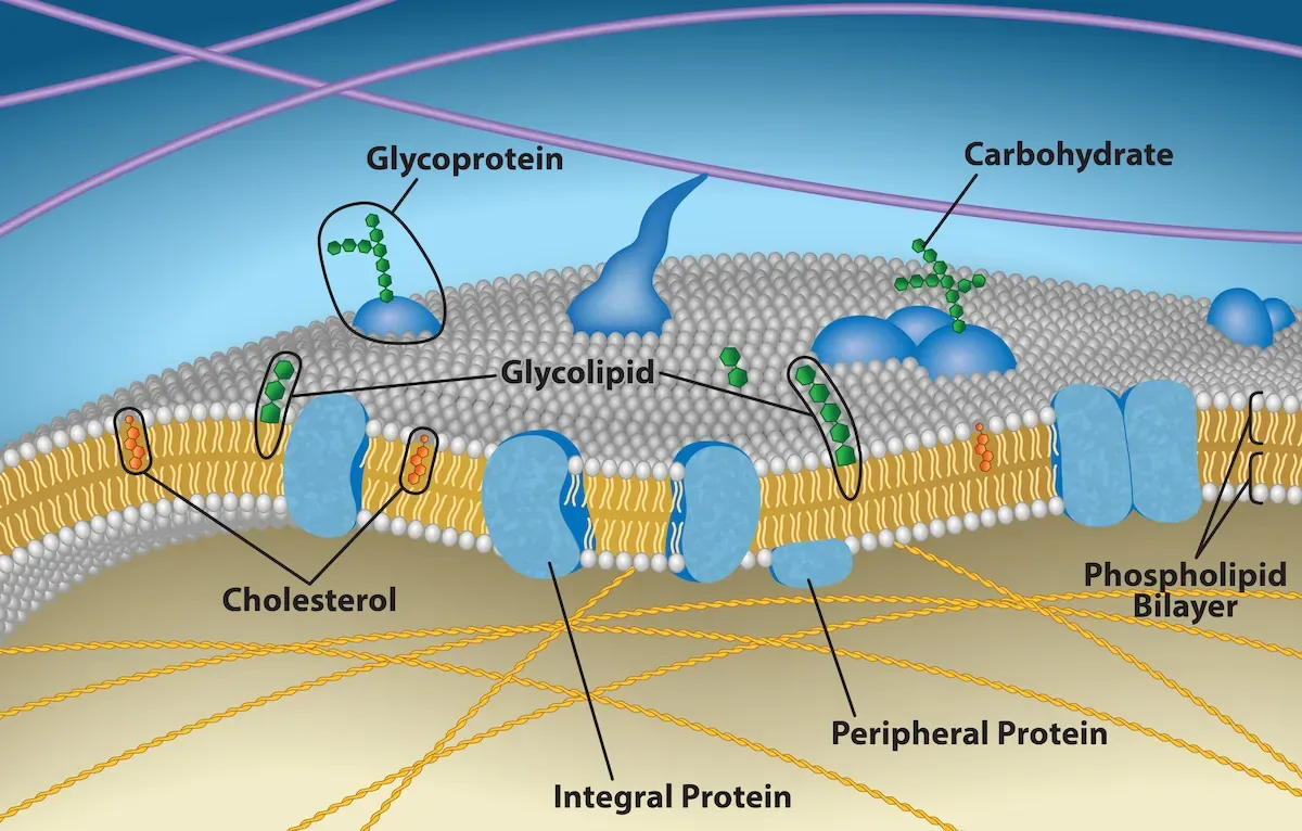
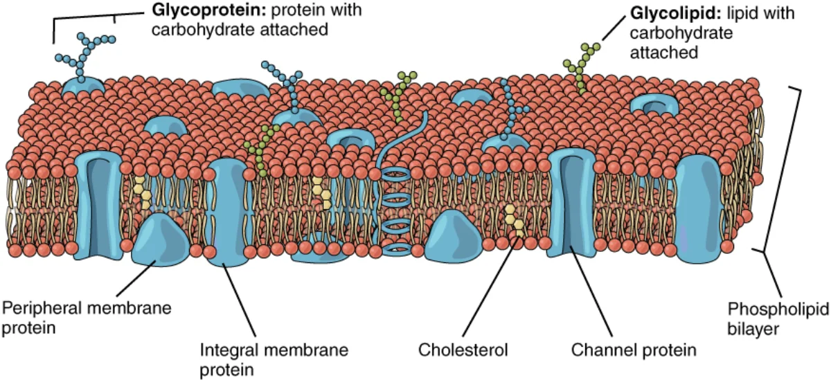
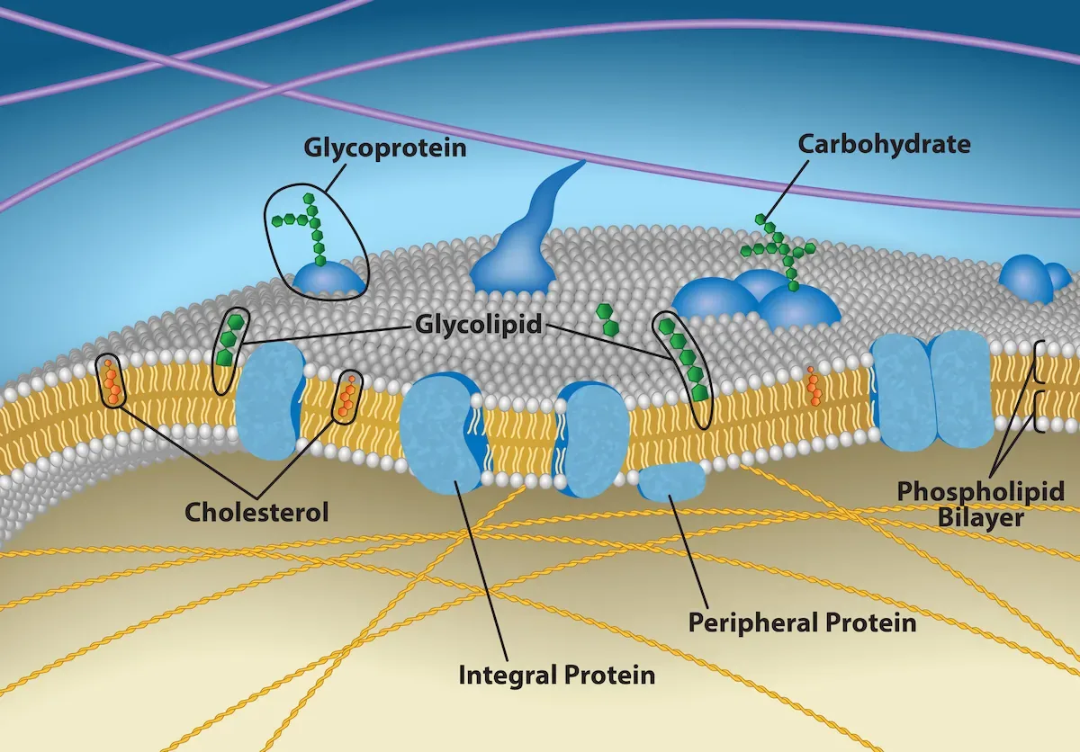
The fluid mosaic model of the cell membrane, with phospholipids, cholesterol, integral proteins, peripheral proteins, and glycoproteins. Credit: OpenStax Biology 2e, CC BY 4.0A closer look at the lipid bilayer showing channel proteins, carrier proteins, and the glycocalyx. Credit: Lumen Learning / OpenStax Anatomy & Physiology, CC BY 4.0
Interactive 3D Cell Membrane. Rotate to explore the phospholipid bilayer, integral proteins, and cholesterol molecules embedded in the membrane.Credit: Lukas Ded via Sketchfab, CC BY
Membrane Proteins and the PERCH Functions
The cell membrane does far more than just act as a barrier. Its embedded proteins give it five major functions, which you can remember with the mnemonic PERCH:
Membrane Protein Types
Integral (transmembrane) proteins span the entire bilayer. They include channels, transporters, and receptors. Because they pass through the hydrophobic core, they have hydrophobic regions in their middle and hydrophilic regions at the ends.
Peripheral proteins sit on the inner or outer surface of the membrane without penetrating the lipid bilayer. They often participate in signaling or provide structural support.
Glycoproteins and glycolipids have carbohydrate chains attached to their extracellular side. These sugar chains form the glycocalyx, which is involved in cell recognition, immune function, and protection.
The Cytosol
The entire internal space enclosed by the cell membrane (excluding organelles) is filled with cytosol - a gel-like aqueous solution. Cytosol is where many metabolic reactions occur, where ribosomes float and translate mRNA, and where the cytoskeleton is anchored. It is about 70% water and contains dissolved ions, small molecules, and proteins. The cytosol plus all organelles together make up the cytoplasm.
Membrane Potential: the Voltage Across the Bilayer
Because the membrane is selectively permeable and because pumps like the Na⁺/K⁺ ATPase unevenly distribute ions, the inside of a typical cell is more negative than the outside (roughly −70 mV in a resting neuron). This resting membrane potential is just stored electrical energy, the way water behind a dam stores gravitational energy.
Every cell has one. When we get to neurons, muscle cells, and secondary active transport, you will keep seeing the same logic: a pump builds an ion gradient, the gradient stores voltage, and opening a channel lets that voltage do work. Details live in Cell Transport and the nervous system chapter.
What does "amphipathic" mean, and why is it important for membrane structure?
Click to reveal answer
Amphipathic means having both hydrophilic (water-loving) and hydrophobic (water-fearing) regions. Phospholipids are amphipathic, which causes them to spontaneously form a bilayer in water - heads face outward toward water, tails face inward away from water. This creates the selectively permeable barrier of the cell membrane.
What does PERCH stand for in the context of cell membrane functions?
How does cholesterol affect membrane fluidity at high vs low temperature?
Click to reveal answer
Cholesterol is a fluidity buffer. At high temperatures it restricts phospholipid motion (decreasing fluidity). At low temperatures it prevents tight packing (increasing fluidity). Net effect: it keeps membrane fluidity in a usable range across temperature changes.
Back to our city analogy. The city (cell) has specialized departments (organelles), each enclosed in its own building (membrane). This compartmentalization is what makes eukaryotic cells so powerful - it allows different chemical reactions to happen simultaneously in isolated environments, without interfering with each other.
Interactive 3D Eukaryotic Cell. Drag to rotate and explore the organelles in three dimensions. Try to identify the nucleus, mitochondria, ER, and Golgi before reading on.Credit: Ebers via Sketchfab, CC BY
The Nucleus - City Hall
The nucleus is the largest organelle and the command center of the cell. It stores the cell’s DNA and controls gene expression - deciding which proteins get made and when.
Structure:
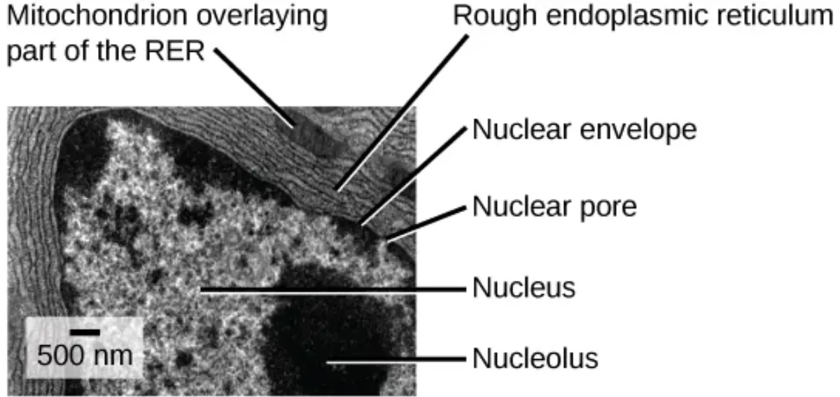
Nuclear envelope: A double membrane (two phospholipid bilayers) that separates the nucleus from the cytoplasm. The outer membrane is continuous with the endoplasmic reticulum.
Nuclear pores: Channels that penetrate the nuclear envelope, acting as security checkpoints. Small molecules diffuse freely, but large molecules (like proteins headed into the nucleus) need a signal sequence that flags them for nuclear import.
Nucleolus: A dense, dark-staining region within the nucleus where ribosomal RNA (rRNA) is synthesized by RNA polymerase I. The nucleolus is essentially a ribosome factory - it assembles the RNA components of ribosomes, which then leave through nuclear pores and get their final protein components added in the cytoplasm.
Chromatin and Chromosomes:
Your DNA does not just float around in a tangled mess. It wraps around proteins called histones, forming a complex called chromatin. Think of it like thread (DNA) wound around spools (histones). This chromatin can be further compacted into visible chromosomes during cell division.
Chromatin exists in two states:
Euchromatin - loosely packed, transcriptionally active (“eu” = true, good - the genes here can be read and expressed)
Heterochromatin - tightly packed, transcriptionally inactive (like a book on a high shelf you cannot reach - the genes are silenced)
Each chromosome is composed of two identical sister chromatids joined at a centromere. The ends of chromosomes are protected by repetitive DNA sequences called telomeres, which act like the plastic tips on shoelaces - they prevent the chromosome from fraying or fusing with neighbors.
Ribosomes - The Construction Workers
Ribosomes are the molecular machines that translate mRNA into proteins. They are NOT membrane-bound organelles - they float freely in the cytoplasm or attach to the rough ER.
Eukaryotic ribosomes: 80S (composed of a 40S small subunit and a 60S large subunit)
Prokaryotic ribosomes: 70S (composed of a 30S small subunit and a 50S large subunit)
The “S” stands for Svedberg units, a measure of sedimentation rate during centrifugation. The numbers do not add up arithmetically because Svedberg units depend on shape and mass, not just mass alone.
Endoplasmic Reticulum - The Factory Floor
The ER is a vast network of membrane-enclosed channels and sacs continuous with the nuclear envelope. It comes in two varieties:
Rough ER (RER):
Studded with ribosomes on its cytoplasmic surface (that is what makes it look “rough” under electron microscopy)
Synthesizes proteins destined for secretion, the cell membrane, or organelles
Folds and modifies these proteins (adding sugar groups, forming disulfide bonds)
Proteins enter the RER lumen through a signal sequence on the growing polypeptide
Smooth ER (SER):
No ribosomes (hence “smooth”)
Synthesizes lipids, phospholipids, and steroid hormones
Detoxifies drugs and poisons (liver cells have abundant SER for this reason)
Stores calcium ions (especially important in muscle cells, where the SER is called the sarcoplasmic reticulum)
Transports proteins from the RER to the Golgi apparatus
Golgi Apparatus - The Post Office
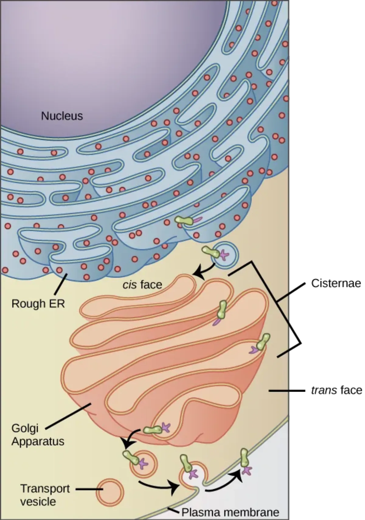
The Golgi is a stack of flattened membrane sacs (cisternae) that receives, modifies, sorts, and ships cellular products. Think of it as the cell’s post office and distribution center.
Cis face (“receiving dock”) - faces the ER. Vesicles from the ER arrive here and fuse with the cis face, dumping their cargo inside.
Trans face (“shipping dock”) - faces the cell membrane. Finished products leave from here in vesicles headed for their final destination.
As proteins and lipids move from cis to trans through the Golgi stack, they undergo modifications: addition of carbohydrate chains (glycosylation), phosphate groups, or sulfate groups. The Golgi also adds signal sequences - molecular address labels that direct each product to the correct destination (lysosome, membrane, or secretion outside the cell).
The endomembrane system: proteins travel from the rough ER through transport vesicles to the Golgi apparatus (cis to trans face), then onward to the plasma membrane or lysosomes. Credit: OpenStax Biology 2e, CC BY 4.0TEM showing the physical continuity between the nuclear envelope and the rough ER - a key feature of the endomembrane system. Nuclear pores, nucleolus, and a nearby mitochondrion are also visible. Credit: OpenStax Biology 2e, CC BY 4.0
Lysosomes - The Recycling and Demolition Crew
Lysosomes are membrane-bound sacs filled with hydrolytic enzymes - molecular scissors that can chop up proteins, carbohydrates, lipids, and nucleic acids. They are the cell’s digestive system and waste disposal unit.
Key facts for the MCAT:
Acidic interior (pH ~5): much lower than cytoplasm (pH 7.4). If a lysosome ruptures, the leaked enzymes drift into neutral pH and go inactive - a built-in safety switch.
Autophagy: lysosomes digest the cell’s own worn-out organelles (damaged mitochondria, old ER).
Apoptosis: lysosomal enzymes help dismantle the cell during programmed cell death.
Phagocytosis cleanup: in macrophages and neutrophils, lysosomes fuse with phagosomes to digest engulfed pathogens.
Peroxisomes - The Hazardous Waste Handlers
Peroxisomes are small, membrane-bound organelles that specialize in oxidation reactions. They are found in both animal and plant cells.
Functions:
Beta-oxidation of very long-chain fatty acids - breaking down fatty acids that are too long for mitochondria to handle
Synthesis of phospholipids - building blocks for cellular membranes
Detoxification - using oxidation reactions that produce hydrogen peroxide (H2O2) as a byproduct
Catalase - the signature enzyme of peroxisomes. It rapidly converts toxic H2O2 into harmless water and oxygen: 2 H2O2 -> 2 H2O + O2
Organelle Summary Table
Organelle
City Analogy
Key Function
Membrane?
Nucleus
City Hall
DNA storage, gene expression
Double membrane
Nucleolus
Blueprint copier in City Hall
rRNA synthesis, ribosome assembly
No membrane (region within nucleus)
Ribosomes
Construction workers
Protein synthesis (translation)
No membrane
Rough ER
Factory floor with workers
Protein synthesis, folding, modification
Single membrane
Smooth ER
Chemical plant
Lipid/steroid synthesis, detox, Ca2+ storage
Single membrane
Golgi apparatus
Post office
Modification, sorting, shipping
Single membrane (stacked)
Lysosomes
Recycling center / demolition crew
Digestion of waste, autophagy, apoptosis
Single membrane
Peroxisomes
Hazardous waste facility
Fatty acid oxidation, H2O2 detox
Single membrane
Mitochondria
Power plant
ATP production
Double membrane
What is the difference between euchromatin and heterochromatin?
Click to reveal answer
Euchromatin is loosely packed and transcriptionally active (genes can be expressed). Heterochromatin is tightly packed and transcriptionally inactive (genes are silenced). Think: Eu = "true" (being used), Hetero = "different" (shut down).
Why do lysosomal enzymes not destroy the cytoplasm if a lysosome ruptures?
Click to reveal answer
Lysosomal enzymes are optimally active at pH ~5 (the acidic interior of the lysosome). The cytoplasm has a neutral pH of ~7.4, so if enzymes leak out, they become inactive and cannot damage the cell. This is a built-in safety mechanism.
What distinguishes the rough ER from the smooth ER in terms of structure and function?
Click to reveal answer
Rough ER has ribosomes on its surface and synthesizes proteins for secretion/membrane insertion. Smooth ER lacks ribosomes and synthesizes lipids/steroids, detoxifies drugs, and stores calcium. "Rough has Ribosomes, Smooth has Steroids."
Mitochondria are the power plants of the cell. Every time you flex a muscle, fire a neuron, or pump a proton, the energy comes from ATP - and the vast majority of your ATP is produced inside mitochondria.
Structure
Mitochondria have a distinctive architecture:
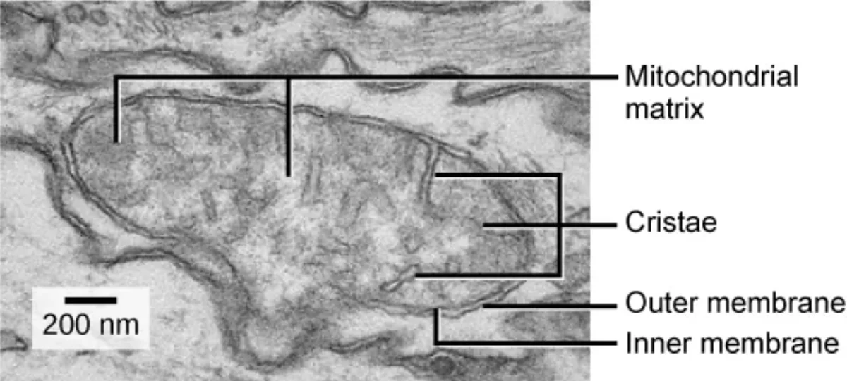
Outer membrane - smooth, contains pores (porins) that allow small molecules to pass freely
Inner membrane - highly folded into structures called cristae. These folds dramatically increase the surface area available for the enzymes of the electron transport chain (ETC) and ATP synthase. The inner membrane is impermeable to most ions and polar molecules - this is critical, because it allows the buildup of a proton gradient.
Intermembrane space - the gap between the outer and inner membranes. Protons (H+) are pumped into this space by the ETC, creating the electrochemical gradient that drives ATP synthesis.
Mitochondrial matrix - the innermost compartment. Contains mitochondrial DNA, mitochondrial ribosomes, and the enzymes of the citric acid cycle (Krebs cycle) and fatty acid beta-oxidation.
TEM of a mitochondrion showing the outer membrane, inner membrane folded into cristae (which increase surface area for the electron transport chain), and the mitochondrial matrix. Credit: OpenStax Biology 2e, CC BY 4.0
Mitochondria as Semi-Autonomous Organelles
Here is something remarkable: mitochondria have their own circular DNA, their own 70S ribosomes (the same size as bacterial ribosomes, not eukaryotic 80S), and they replicate by binary fission - just like bacteria. They even have a double membrane, with the inner membrane resembling a bacterial plasma membrane.
This is not a coincidence. It is evidence for one of the most important theories in biology.
Mitochondria and Apoptosis
Mitochondria are not just about making energy - they are also key players in apoptosis (programmed cell death). When a cell receives the signal to die, the mitochondrial outer membrane becomes permeable, releasing cytochrome c into the cytoplasm. Cytochrome c triggers a cascade that systematically dismantles the cell from the inside. This dual role - sustaining life with ATP and initiating death through apoptosis - makes mitochondria one of the most important organelles in biology.
What four pieces of evidence support the endosymbiotic theory for mitochondria?
Click to reveal answer
1) Circular DNA (like bacteria). 2) 70S ribosomes (like bacteria, not 80S). 3) Double membrane (inner from engulfed bacterium, outer from host vesicle). 4) Replicate by binary fission (like bacteria). These all point to mitochondria being descendants of ancient aerobic prokaryotes.
Where in the mitochondrion does the electron transport chain occur? Why there?
Click to reveal answer
The ETC is located on the inner mitochondrial membrane (cristae). The cristae fold to increase surface area for ETC enzymes and ATP synthase. The inner membrane must be impermeable to protons to maintain the electrochemical gradient that drives ATP synthesis.
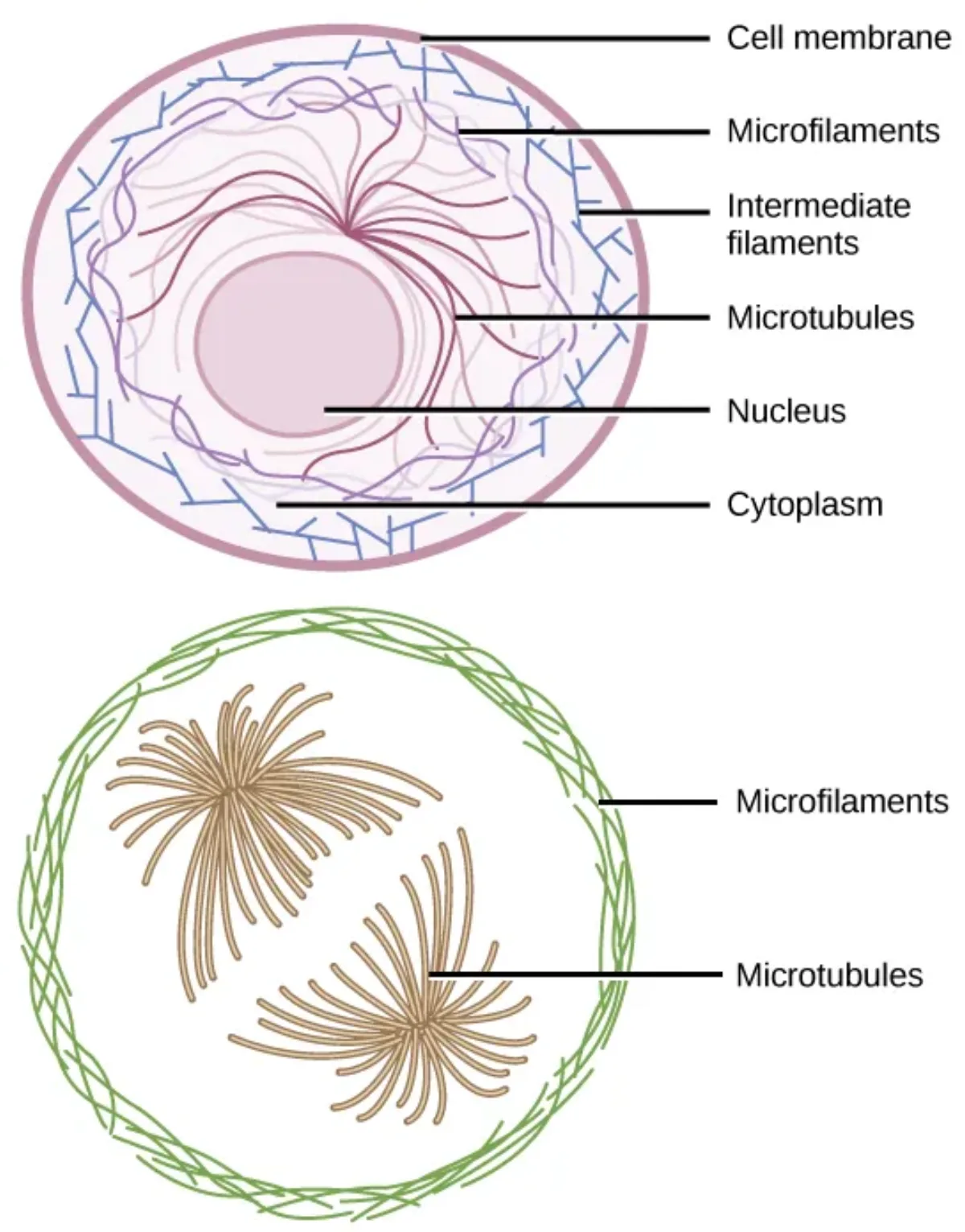
Imagine setting up a large outdoor tent. You need rigid poles to give it height and structure, thin ropes to anchor it and allow some flexibility, and medium-weight straps to hold the fabric in place under tension. The cell has the exact same setup - three types of protein filaments that collectively form the cytoskeleton.
The cytoskeleton does three main jobs:
Structural support - maintains cell shape and resists mechanical stress
Movement - enables cell motility, muscle contraction, and cell division
Intracellular transport - acts as a highway system for motor proteins to carry cargo (vesicles, organelles) to where they are needed
The three components of the cytoskeleton: microfilaments (actin), intermediate filaments, and microtubules, each with distinct sizes and roles. Credit: OpenStax Biology 2e, CC BY 4.0
The Three Filament Types
Microfilaments (Actin Filaments) - 7 nm
Microfilaments are the thinnest filaments, composed of the protein actin polymerized into solid, helical rods. Think of them as the cell’s muscles.
Functions:
Muscle contraction - actin interacts with the motor protein myosin, using ATP to generate force. This actin-myosin interaction is the molecular basis of every muscle contraction in your body.
Cell movement - amoeboid movement, crawling of white blood cells toward infection
Cytokinesis - during cell division, a ring of actin filaments forms the cleavage furrow. The ring contracts like a drawstring on a bag, pinching the cell in two to form two daughter cells.
Microvilli support - microfilaments form the core of microvilli on intestinal epithelial cells, increasing surface area for nutrient absorption
Intermediate Filaments - 10 nm
Intermediate filaments are a diverse family of proteins that provide mechanical strength and tension resistance. They are the toughest of the three filament types - they do not participate in cell movement but are excellent at anchoring structures and resisting pulling forces.
Key types:
Keratin - found in epithelial cells (skin, hair, nails). Gives skin its strength and resilience. This is the protein in all those hair product commercials.
Vimentin - found in connective tissue cells, white blood cells
Desmin - found in muscle cells, helps maintain the structural alignment of sarcomeres
Lamins - found inside the nucleus, forming the nuclear lamina - a meshwork that supports the nuclear envelope from the inside
Microtubules - 25 nm
Microtubules are the largest and most rigid filaments, built from alpha-tubulin and beta-tubulin dimers that polymerize into hollow tubes. They are the railroad tracks of the cell - providing pathways for motor proteins to transport cargo.
Functions:
Intracellular transport - motor proteins kinesin (moves toward the + end, away from the cell center) and dynein (moves toward the - end, toward the cell center) walk along microtubules carrying vesicles, organelles, and other cargo
Cell division - microtubules form the mitotic spindle, which attaches to chromosomes at their kinetochores and pulls sister chromatids apart during mitosis/meiosis
Structural framework for cilia and flagella
Microtubule Organizing Center (MTOC) and Centrioles
Microtubules do not just appear randomly in the cell. They grow outward from a structure called the microtubule organizing center (MTOC), usually located near the nucleus. The MTOC is also called the centrosome in animal cells.
The centrosome contains a pair of centrioles - cylindrical structures made of nine triplets of microtubules arranged in a ring. During cell division, centrioles duplicate and move to opposite poles of the cell, organizing the mitotic spindle.
Microtubules are dynamic - they can rapidly grow (polymerize) by adding tubulin dimers at their + end and shrink (depolymerize) by losing them. This property, called dynamic instability, allows the cell to quickly reorganize its internal structure as needed.
Cilia and Flagella
Both cilia and flagella are extensions of the cell membrane built on a core framework of microtubules. They share the same internal structure: the 9+2 arrangement - nine outer pairs (doublets) of microtubules surrounding two central single microtubules, all enclosed by the plasma membrane.
Cilia are short, hair-like projections that cover the surface of certain cells:
In the respiratory tract, cilia beat in coordinated waves to sweep mucus and trapped debris upward and out of the lungs
In the fallopian tubes, cilia help move eggs from the ovary toward the uterus
Flagella are long, whip-like structures used for propulsion:
In humans, the only flagellated cell is the sperm cell
Flagella use a wave-like motion powered by dynein motor proteins
Important distinction for the MCAT: Eukaryotic flagella (9+2 microtubule structure, powered by dynein/ATP) are completely different from bacterial flagella (made of flagellin protein, rotary motion powered by a proton gradient). Do not confuse them.
What are the three cytoskeletal filaments, their sizes, and their main protein components?
What is the 9+2 arrangement and where is it found?
Click to reveal answer
The 9+2 arrangement is the internal microtubule structure of eukaryotic cilia and flagella: nine outer doublets of microtubules surrounding two central single microtubules. Dynein motor proteins between the doublets power the bending motion.
Think of cells in a tissue like apartments in a building. Some apartments share sealed walls so nothing leaks between them (tight junctions). Some have doorways that let neighbors pass notes back and forth (gap junctions). Some are bolted together to keep the building from falling apart under stress (desmosomes and adherens junctions). And some are screwed into the foundation (hemidesmosomes).
Cells in your body do not exist in isolation. In tissues, neighboring cells are physically connected through specialized structures called cell junctions. Five types matter for the MCAT.
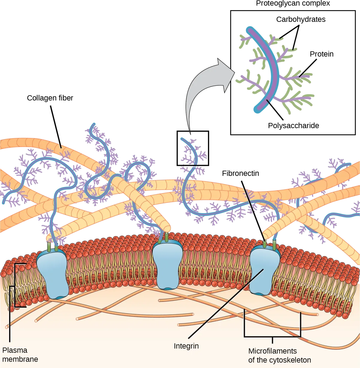
The extracellular matrix and its connection to the cell via integrin proteins, which link collagen and proteoglycan fibers to the internal cytoskeleton. Credit: OpenStax Biology 2e, CC BY 4.0
Tight Junctions - The Waterproof Seal
Tight junctions create a watertight seal between adjacent cells, preventing solutes from leaking through the space between cells (the paracellular route). They force all substances to go through the cells themselves rather than between them.
Where are they critical? In the intestinal epithelium, tight junctions prevent digestive enzymes and bacteria from leaking between cells into the bloodstream. In the blood-brain barrier, tight junctions between endothelial cells prevent most blood-borne substances from entering brain tissue.
Tight junctions must form a continuous band around the entire cell to work. If there are gaps, fluid can leak through, which causes disease. They can be so tight that they create a voltage difference across the epithelium by maintaining different ion concentrations on each side.
Gap Junctions - The Direct Communication Channels
Gap junctions are like tiny tunnels between adjacent cells, allowing direct passage of ions, water, and small signaling molecules. Each gap junction channel is formed by two connexons (one from each cell), and each connexon is made of six connexin protein subunits.
Gap junctions are essential wherever cells need to act in unison:
Cardiac muscle - gap junctions allow the rapid spread of electrical signals (action potentials) between heart cells, ensuring coordinated contractions. This is why the heart beats as a single unit rather than as a collection of individual cells contracting randomly.
Smooth muscle - coordinated contractions in the gut and blood vessels
Embryonic development - allows signaling between neighboring cells during tissue formation
Key limitation: Gap junctions allow small molecules (ions, amino acids, sugars, second messengers like cAMP and IP3) to pass, but they do not transfer large molecules like proteins.
Adherens Junctions - The Belt
Adherens junctions form a continuous belt around a cell just below the tight junctions, gluing neighboring cells together. Like desmosomes, they use cadherin proteins to bridge the intercellular space. The difference is the cytoskeletal anchor: adherens junctions attach to actin microfilaments, while desmosomes attach to intermediate filaments.
Adherens junctions help epithelial sheets maintain shape and distribute contractile force (for example, during embryonic folding).
Desmosomes - The Rivets
Desmosomes are like spot welds or rivets that hold cells together in areas subjected to mechanical stress - stretching, pulling, and shearing forces. They are particularly abundant in skin and cardiac muscle.
Each desmosome consists of cadherin proteins from adjacent cells interlocking with each other in the intercellular space. On the intracellular side, cadherins are anchored to intermediate filaments (typically keratin in epithelial cells or desmin in cardiac cells) through adaptor proteins. This arrangement distributes mechanical force across the entire cytoskeleton rather than concentrating it at one point.
In cardiac muscle, desmosomes are part of intercalated discs - the junctions between heart cells that also contain gap junctions. The desmosomes hold the cells together during the powerful contractions, while the gap junctions coordinate the electrical signals.
Hemidesmosomes - The Anchors
Hemidesmosomes look like half a desmosome (hence “hemi-”), but they serve a different function. Instead of connecting two cells to each other, hemidesmosomes connect the bottom of an epithelial cell to the basement membrane (the extracellular matrix beneath epithelial tissue).
They use integrin proteins (not cadherins like desmosomes) to anchor the cell to structural proteins in the basement membrane. This anchoring prevents epithelial cells from being torn away from the underlying tissue by mechanical forces.
What protein forms gap junction channels, and what is the basic structural unit?
Click to reveal answer
Gap junction channels are formed by connexin proteins. Six connexins assemble into a connexon (hemichannel), and two connexons (one from each cell) dock together to form a complete gap junction channel. They allow passage of ions and small molecules but not proteins.
How do desmosomes and hemidesmosomes differ in what they connect?
Click to reveal answer
Desmosomes connect two adjacent cells to each other using cadherin proteins anchored to intermediate filaments. Hemidesmosomes connect a cell to the underlying basement membrane using integrin proteins. Desmosomes = cell-to-cell; hemidesmosomes = cell-to-matrix.
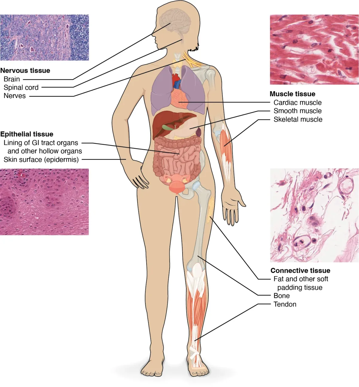
Cells do not work alone. They organize into tissues - groups of similar cells that work together to perform a specific function. There are exactly four tissue types in the human body. Every structure in your body - every organ, every system - is made from some combination of these four.
The four tissue types in the human body: epithelial, connective, muscle, and nervous tissue, with representative examples and microscopy views. Credit: OpenStax Biology 2e, CC BY 4.0
Epithelial Tissue - The Coverings and Linings
Epithelial tissue covers the body’s external surface (skin) and lines internal cavities (digestive tract, blood vessels, airways). It acts as a barrier - protecting against pathogens, preventing dehydration, and regulating absorption and secretion.
Epithelial cells sit on a basement membrane (a thin layer of extracellular matrix) that connects them to underlying connective tissue. They are polarized - the top surface (apical) faces a lumen or the external environment, while the bottom surface (basal) attaches to the basement membrane.
Single layer, but cells of varying heights create the illusion of multiple layers
Respiratory tract (mucus movement)
Classification by cell shape:
Shape
Description
Key Locations
Squamous
Flat, like floor tiles
Blood vessel lining, lung alveoli (thin for diffusion)
Cuboidal
Cube-shaped
Kidney tubules, glands (secretion)
Columnar
Tall, column-shaped
Intestinal lining (absorption, secretion)
You combine layer + shape to classify: simple squamous epithelium lines blood vessels, stratified squamous epithelium forms the outer layer of skin, simple columnar epithelium lines the intestine, pseudostratified columnar epithelium lines the trachea.
The functional cells of an organ (those that perform its core job) are called the parenchyma. In many organs, these are epithelial cells: hepatocytes in the liver, nephrons in the kidney, acid-producing cells in the stomach.
Connective Tissue - The Structural Support
Connective tissue is the architectural framework of the body. While epithelial cells are the “doers” (parenchyma), connective tissue forms the stroma - the support structure that holds everything together.
Key components:
Fibroblasts - the main cell type, responsible for producing the extracellular matrix fibers
Collagen fibers - strong, rope-like fibers that provide tensile strength (resistance to stretching). Collagen is the most abundant protein in the human body.
Elastic fibers - stretchy, rubber band-like fibers that allow tissues to recoil after being stretched (found in lungs, blood vessels, skin)
Reticular fibers - thin, branching fibers that form a delicate net-like framework supporting organs like the spleen and lymph nodes
Extracellular matrix (ECM) - a mix of proteins (fibers) and ground substance (polysaccharides, water, minerals) that surrounds and supports the cells
Types of connective tissue:
Loose connective tissue - flexible packing material found beneath the skin and around organs. Provides cushioning and support.
Dense connective tissue - tightly packed collagen fibers. Found in tendons (connect muscle to bone), ligaments (connect bone to bone), and the dermis of the skin.
Cartilage - firm but flexible tissue. Three types:
Hyaline cartilage - smooth, glassy, found at joint surfaces and in the trachea. Reduces friction during movement.
Elastic cartilage - flexible, found in the ears and epiglottis
Fibrocartilage - tough, shock-absorbing, found in intervertebral discs and the meniscus of the knee
Bone - rigid connective tissue providing structural support and protection for internal organs. Also serves as a mineral reservoir (calcium, phosphate).
Blood - yes, blood is classified as connective tissue. Its “matrix” is plasma (liquid), and its “cells” include red blood cells, white blood cells, and platelets.
Muscle tissue is specialized for contraction. Three types:
Skeletal muscle - voluntary, striated (striped appearance), multinucleated. Attached to bones for movement.
Cardiac muscle - involuntary, striated, uninucleated (or binucleated). Found only in the heart. Connected by intercalated discs (desmosomes + gap junctions).
Smooth muscle - involuntary, non-striated, uninucleated. Found in walls of blood vessels, digestive tract, bladder, and airways. Responsible for peristalsis and vasoconstriction.
Nervous Tissue
Nervous tissue is specialized for transmitting electrical signals. It consists of two main cell types:
Neurons - the electrically excitable cells that generate and transmit nerve impulses (action potentials). Each neuron has a cell body, dendrites (receive signals), and an axon (sends signals).
Glial cells (neuroglia) - supporting cells that nourish, insulate, and protect neurons. Examples include Schwann cells (form myelin in PNS), oligodendrocytes (form myelin in CNS), and astrocytes (maintain the blood-brain barrier).
What is the difference between simple, stratified, and pseudostratified epithelium?
Click to reveal answer
Simple = single layer of cells (good for absorption/exchange). Stratified = multiple layers (good for protection in high-wear areas). Pseudostratified = single layer that looks like multiple layers because cells have different heights (found in respiratory tract).
Why is blood classified as connective tissue?
Click to reveal answer
Blood meets the criteria for connective tissue: it has cells (RBCs, WBCs, platelets) suspended in an extracellular matrix (plasma). Like other connective tissues, it provides support (transporting oxygen, nutrients, and immune cells throughout the body) and originates from mesenchymal stem cells.
Remember the city analogy? A virus is like a carjacker who does not know how to build a car. It has a set of instructions (its genetic material) and maybe a disguise (envelope), but it has absolutely no engine, no factory, no workers.
To reproduce, it must break into your car (cell), hijack the steering wheel (ribosomes and enzymes), and use your gas (ATP and nucleotides) to build copies of itself. When the car is full of copies, it either explodes (lytic cycle) or the virus hides its instructions in your glove compartment and waits (lysogenic cycle).
Viral Structure
Viruses are tiny - ranging from 20 nm to 300 nm, far smaller than even the smallest bacterium (1-10 micrometers). Despite their simplicity, they are incredibly effective at what they do.
Core components:
Genetic material - either DNA or RNA (never both). Can be single-stranded or double-stranded, linear or circular.
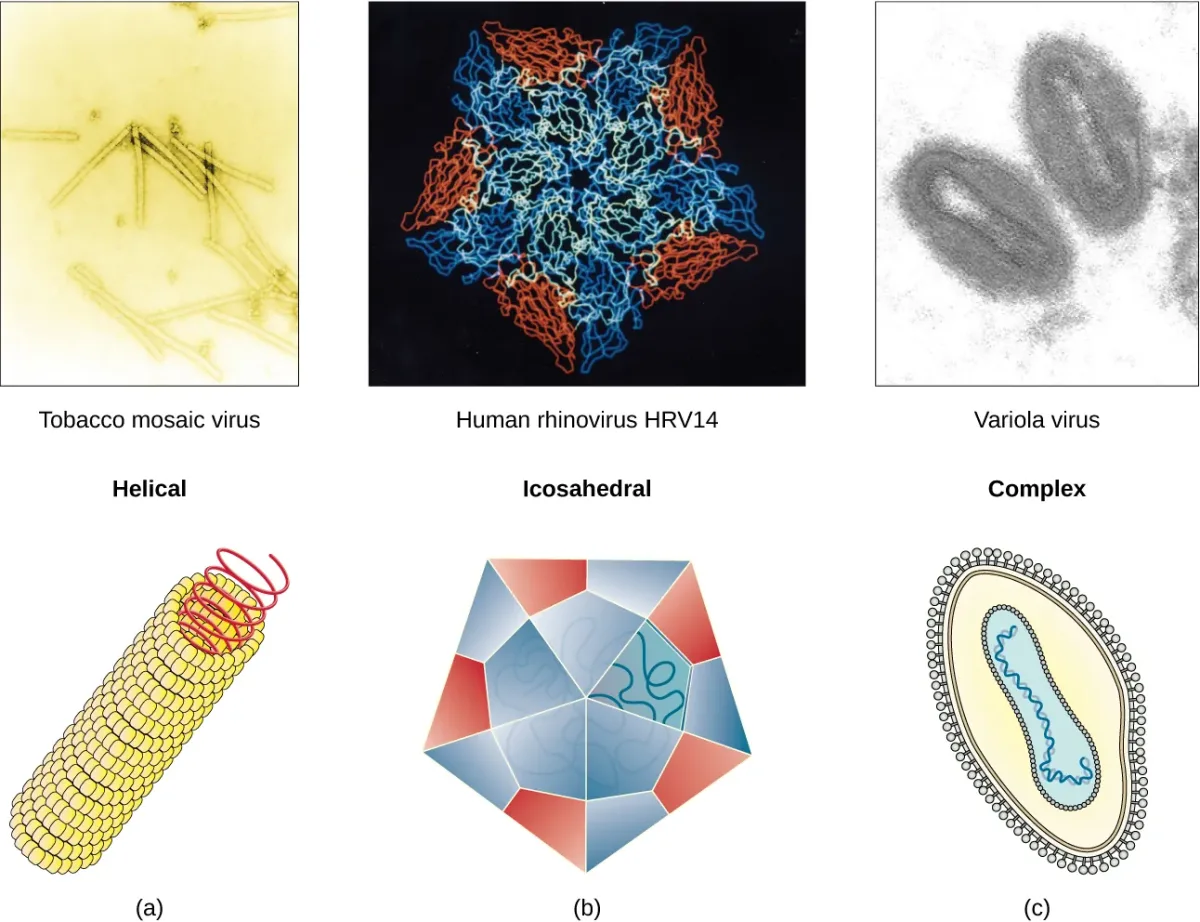
Capsid - a protein shell made of subunits called capsomeres that surrounds and protects the genetic material. Capsids can be helical (spiral), icosahedral (20-sided polyhedron), or complex (like bacteriophages with heads, tails, and tail fibers).
The three major capsid shapes: helical (tobacco mosaic virus), icosahedral (rhinovirus), and complex (variola/smallpox). Credit: OpenStax Biology 2e, CC BY 4.0
Envelope (some viruses) - a lipid bilayer membrane stolen from the host cell during budding. Contains viral glycoproteins used for attachment to host cells.
Naked viruses - lack an envelope. They are generally more resistant to environmental conditions (heat, detergents, desiccation) than enveloped viruses because they do not have a fragile lipid membrane.
Bacteriophage structure (viruses that infect bacteria):
Head (icosahedral capsid containing DNA)
Tail sheath - a contractile tube that injects DNA into the host bacterium, like a molecular syringe
Base plate and tail fibers - structures that recognize and attach to specific receptors on the bacterial surface
Interactive 3D Bacteriophage. Rotate to see the icosahedral head, tail sheath, base plate, and tail fibers that inject DNA into bacteria.Credit: Nima via Sketchfab, CC BY
Viral Classification by Genome
Type
Genome
Example
Key Feature
dsDNA virus
Double-stranded DNA
Herpes simplex virus (HSV), Adenovirus
Most stable; replicates in the nucleus using host machinery
ssDNA virus
Single-stranded DNA
Parvovirus
Must convert to dsDNA before replication
dsRNA virus
Double-stranded RNA
Rotavirus
Carries its own RNA-dependent RNA polymerase
(+) ssRNA virus
Positive-sense single-stranded RNA
Hepatitis C, Coronavirus
RNA can be directly translated by host ribosomes (acts like mRNA)
(-) ssRNA virus
Negative-sense single-stranded RNA
Ebola, Influenza
Must first be converted to (+) sense RNA before translation; carries RNA replicase
Retrovirus
Single-stranded RNA
HIV
Uses reverse transcriptase to convert RNA to DNA, which integrates into host genome
Enveloped vs. Naked Viruses
Enveloped viruses - have a lipid bilayer envelope stolen from the host cell during budding. They are easier to kill outside the body because the envelope is fragile and susceptible to detergents, heat, and drying. They typically enter cells by fusing with the host membrane or by endocytosis.
Naked (non-enveloped) viruses - have only a protein capsid. They are more resistant to environmental stressors and can persist on surfaces longer. They enter cells by endocytosis or by injecting their genome through the cell membrane.
The Viral Life Cycle
All viruses follow a general replication strategy:
Attachment - the virus binds to specific receptors on the host cell surface. This specificity determines which cells and species a virus can infect (tropism).
Entry/Penetration - the viral genome (and sometimes associated enzymes) enters the host cell. Methods vary: membrane fusion (enveloped viruses), endocytosis, or injection (bacteriophages).
Replication and gene expression - the viral genome hijacks the host’s ribosomes, tRNA, amino acids, and enzymes to make viral proteins and copies of the viral genome.
Assembly - new viral capsids are assembled and loaded with copies of the viral genome.
Release - new virions exit the cell. This can happen by lysis (the cell bursts open and dies) or by budding (virions push through the membrane, acquiring an envelope in the process - the cell may survive and continue producing viruses).
Lytic vs. Lysogenic Cycles (Bacteriophages)
Bacteriophages (viruses that infect bacteria) can follow two distinct pathways:
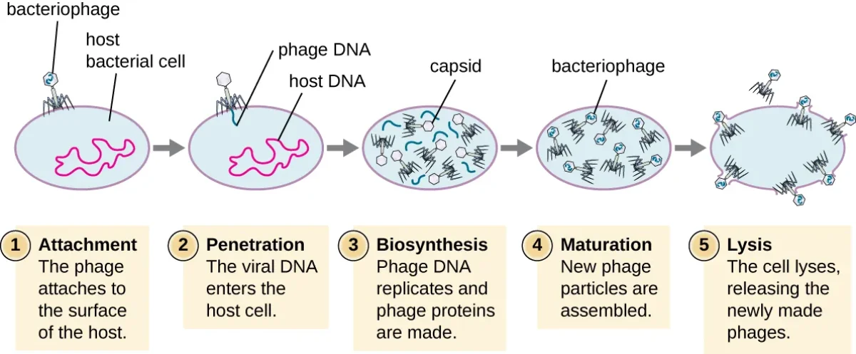
Lytic cycle - the aggressive path. The phage immediately hijacks the bacterial cell, replicates massively, and lyses (bursts) the cell to release hundreds of new phage particles. The host cell is destroyed.
The five stages of the lytic cycle: attachment, penetration, biosynthesis, maturation, and lysis. Credit: OpenStax Microbiology, CC BY 4.0
Lysogenic cycle - the stealth path. Instead of immediately replicating, the phage integrates its DNA into the host bacterium’s chromosome. The integrated viral DNA is called a prophage (or provirus). Every time the bacterium divides, it copies the prophage along with its own DNA, unknowingly passing the viral genes to all its descendants.
The prophage can remain dormant for many generations. But when the bacterium encounters stress (UV radiation, chemical damage, nutrient depletion), the prophage can excise itself from the host chromosome and enter the lytic cycle, producing new phage particles and destroying the host.
Retroviruses
Retroviruses (like HIV) deserve special attention because they violate the “normal” flow of genetic information. Instead of DNA -> RNA -> Protein, retroviruses go RNA -> DNA -> RNA -> Protein.
They carry the enzyme reverse transcriptase, which converts their single-stranded RNA genome into double-stranded DNA. This DNA is then integrated into the host cell’s genome. Once integrated, the viral DNA (called a provirus) is transcribed by the host’s own RNA polymerase, producing new viral RNA and mRNA for viral protein synthesis.
Prions and Viroids - Even Simpler Than Viruses
The MCAT explicitly lists these two subviral agents, so know them:
Prions are infectious misfolded proteins with no nucleic acid at all. The normal prion protein has an alpha-helix-rich fold. The misfolded prion has a beta-pleated sheet-rich fold, and when it contacts a normal copy it templates that normal copy into the misfolded shape. The misfolded proteins clump in neural tissue, producing fatal neurodegenerative disease. Prions challenge the idea that genetic material is required to propagate an infection.
Viroids are short, circular, single-stranded RNA molecules with no protein coat. They infect plants and cause disease by interfering with host gene expression. Hepatitis D virus is a viroid-like human pathogen that can only replicate when Hepatitis B is also present.
What is the difference between the lytic and lysogenic cycles?
Click to reveal answer
Lytic cycle: the phage immediately replicates inside the host, produces many new virions, and lyses (destroys) the host cell. Lysogenic cycle: the phage integrates its DNA into the host chromosome (as a prophage), replicates passively with the host, and can later be triggered to enter the lytic cycle by stress.
How does a retrovirus like HIV replicate?
Click to reveal answer
HIV carries reverse transcriptase, which converts its ssRNA genome into dsDNA. The enzyme integrase then inserts this DNA into the host cell's genome (provirus). The host's RNA polymerase transcribes the provirus into new viral RNA and mRNA for viral protein synthesis. This integration makes HIV extremely difficult to cure.
Why are naked (non-enveloped) viruses harder to kill than enveloped viruses?
Click to reveal answer
Naked viruses lack a lipid envelope, so they are resistant to detergents, heat, and desiccation. Enveloped viruses have a fragile lipid bilayer that is easily disrupted by these agents. This is why norovirus (naked) survives on surfaces much longer than influenza (enveloped).
How do prions and viroids differ from viruses?
Click to reveal answer
Prions are infectious misfolded proteins with no nucleic acid. Viroids are small circular RNAs with no protein coat. Viruses have both protein (capsid) and nucleic acid; prions keep only the protein, viroids keep only the nucleic acid.
Now we return to the nightclub bouncer. The cell membrane is selectively permeable - it lets some things through freely, blocks others entirely, and actively drags certain molecules across. Understanding transport is one of the highest-yield topics on the MCAT because it shows up in biology, biochemistry, and even physics passages.
There are two fundamental categories: passive transport (no energy required, molecules move down their concentration gradient) and active transport (energy required, molecules move against their concentration gradient).
Molecules diffuse down their concentration gradient, from areas of high concentration to areas of low concentration. Credit: OpenStax Biology 2e, CC BY 4.0
Passive Transport
Passive transport is driven entirely by the concentration gradient - molecules naturally move from areas of high concentration to areas of low concentration. No ATP is spent. No energy is needed. It is as natural as a ball rolling downhill.
Simple Diffusion
The simplest form of transport. Small, nonpolar molecules pass directly through the phospholipid bilayer without any help from proteins. No channel needed, no carrier needed - they just dissolve into the membrane and pass through.
Molecules that cross by simple diffusion:
O2 and CO2 (small, nonpolar gases)
Steroid hormones (nonpolar - derived from cholesterol)
Ethanol and other small, uncharged molecules
Molecules that CANNOT cross by simple diffusion:
Ions (Na+, K+, Ca2+, Cl-) - too charged
Glucose, amino acids - too large and polar
Water - technically polar, but it crosses slowly; most water movement uses aquaporins
Osmosis
Osmosis is the diffusion of water across a selectively permeable membrane, from an area of low solute concentration (high water concentration) to an area of high solute concentration (low water concentration). Water moves to dilute the more concentrated solution. The quantitative treatment of osmotic pressure (π = iMRT) is covered in general chemistry.
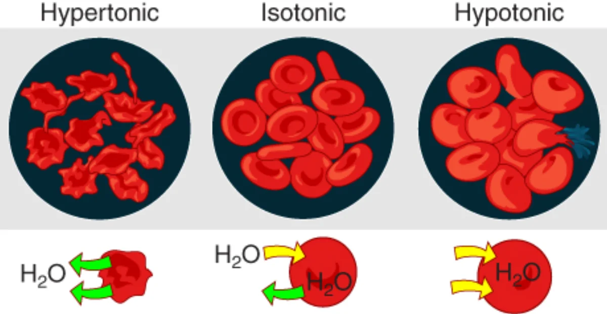
Tonicity describes the effect a solution has on cell volume:
Hypertonic solution - higher solute concentration outside the cell. Water flows OUT of the cell. The cell shrinks (crenation in animal cells, plasmolysis in plant cells).
Hypotonic solution - lower solute concentration outside the cell. Water flows INTO the cell. The cell swells (and may lyse in animal cells; plant cells are protected by their cell wall and become turgid).
Isotonic solution - equal solute concentration inside and outside. No net water movement. Cell volume stays the same.
The effect of tonicity on red blood cells: hypertonic causes crenation, isotonic maintains normal shape, and hypotonic causes lysis. Credit: Lumen Learning / OpenStax Anatomy & Physiology, CC BY 4.0
Facilitated Diffusion
Some molecules are too large, too polar, or too charged to cross the membrane by simple diffusion, but they still move down their concentration gradient (so no energy is needed). They use membrane proteins for help:
Channel proteins - form a hydrophilic pore through the membrane. They can be gated (opened/closed by a signal) or ungated (always open). Ion channels (Na+, K+, Ca2+, Cl-) are the classic examples. Aquaporins are water channels that speed up osmosis.
Carrier proteins (transporters) - bind to a specific molecule, change shape, and release it on the other side of the membrane. Glucose enters most cells via the GLUT transporter - a carrier protein that facilitates glucose diffusion down its concentration gradient.
Facilitated diffusion has two important properties:
Specificity - each channel or carrier is specific for certain molecules
Saturation - there are a limited number of channels/carriers. At high substrate concentrations, all carriers are occupied, and the transport rate reaches a maximum (Vmax). This is analogous to enzyme saturation.
Active Transport
When a molecule needs to move against its concentration gradient (from low concentration to high concentration), the cell must spend energy. This is active transport - it is like pushing a ball uphill.
Primary Active Transport
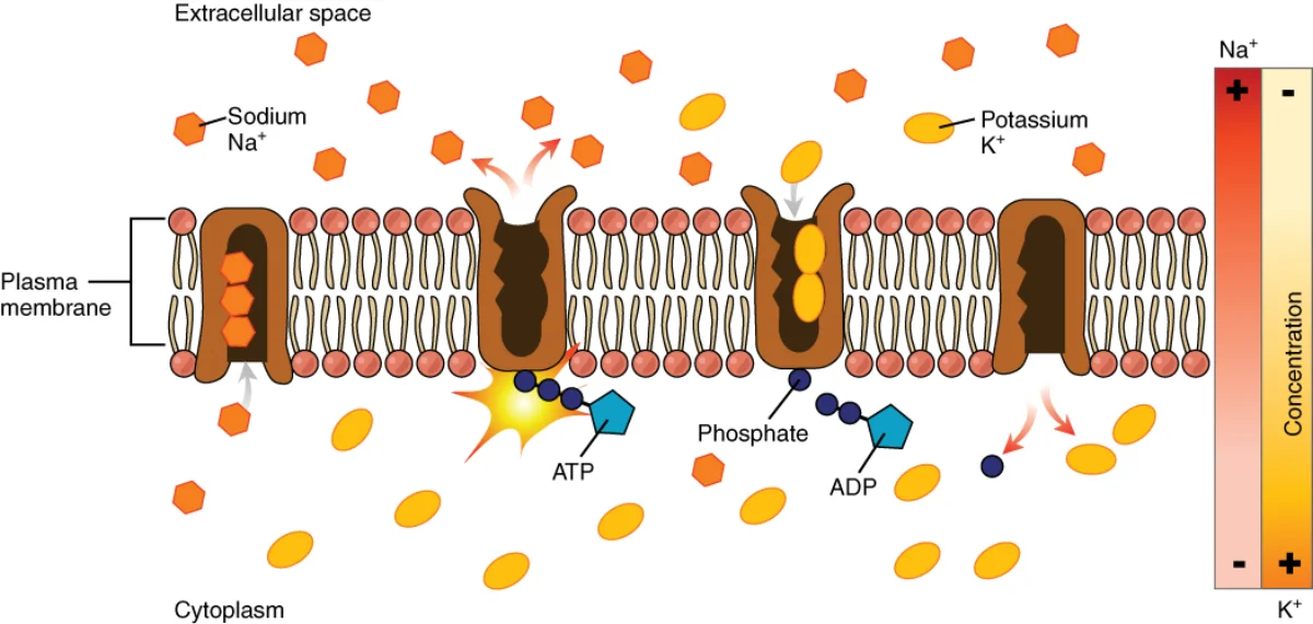
Primary active transport uses ATP directly to move molecules against their gradient. The most important example is the Na+/K+ ATPase (sodium-potassium pump):
Pumps 3 Na+ out of the cell and 2 K+ in per ATP hydrolyzed
Maintains the electrochemical gradient that is essential for nerve impulse transmission, muscle contraction, and secondary active transport
Found in virtually every animal cell
The sodium-potassium pump (Na+/K+ ATPase) pumps 3 Na+ out of the cell and 2 K+ in for each ATP hydrolyzed. Credit: Lumen Learning / OpenStax Anatomy & Physiology, CC BY 4.0
Because 3 positive charges leave and only 2 enter per cycle, the pump is electrogenic - it actively contributes to the negative resting membrane potential, not just the concentration gradient.
Other examples include Ca²⁺ ATPases (pump calcium out of the cytoplasm into the ER or extracellular space) and H⁺/K⁺ ATPase (proton pump in stomach parietal cells that acidifies the stomach).
Secondary Active Transport (Cotransport)
Secondary active transport uses the energy stored in an existing ion gradient (usually the Na+ gradient created by the Na+/K+ ATPase) to drive the transport of another molecule against its gradient. No ATP is directly consumed by the transporter itself, but ATP was used earlier to establish the ion gradient.
Symport (cotransport) - the ion and the transported molecule move in the same direction. Example: SGLT (sodium-glucose linked transporter) in the intestinal and kidney epithelium - Na⁺ flowing down its gradient drags glucose up its gradient into the cell. The Na⁺/K⁺ ATPase on the basolateral side keeps intracellular Na⁺ low, so SGLT has fuel to run.
Antiport (exchange) - the ion and the transported molecule move in opposite directions. Example: Na⁺/H⁺ exchanger - sodium flows in while hydrogen is pumped out.
Bulk Transport - Endocytosis and Exocytosis
For very large molecules, particles, or even entire cells, the membrane uses vesicle-mediated transport - wrapping materials in membrane bubbles.
Endocytosis (bringing things in)
Three types:
Phagocytosis (“cell eating”) - the cell engulfs large particles (bacteria, dead cells) by extending pseudopods around them, forming a phagosome. The phagosome then fuses with a lysosome for digestion. Used by immune cells like macrophages and neutrophils.
Pinocytosis (“cell drinking”) - the cell takes in small droplets of extracellular fluid along with any dissolved molecules. This is nonspecific - it samples the environment.
Receptor-mediated endocytosis - the most selective form. Specific molecules (ligands) bind to receptors on the cell surface, causing the membrane to invaginate and form a coated vesicle (often coated with clathrin protein). This is how cells take up LDL cholesterol, transferrin (iron transport), and many hormones.
Exocytosis (pushing things out)
Vesicles inside the cell fuse with the plasma membrane and release their contents to the exterior. This is how:
Neurotransmitters are released at synapses
Hormones are secreted by endocrine cells
Digestive enzymes are released by pancreatic cells
Antibodies are secreted by plasma cells
Transport Summary Table
Transport Type
Energy?
Direction
Mechanism
Examples
Simple diffusion
No
Down gradient
Directly through bilayer
O2, CO2, steroids
Osmosis
No
Down water gradient
Through membrane/aquaporins
Water
Facilitated diffusion
No
Down gradient
Via channels or carriers
Glucose (GLUT), ions through channels
Primary active transport
Yes (ATP)
Against gradient
ATP-powered pumps
Na+/K+ ATPase, Ca2+ ATPase
Secondary active transport
Yes (indirect)
Against gradient
Uses ion gradient energy
Na+-glucose symporter
Phagocytosis
Yes
Into cell
Pseudopod engulfment
Macrophage eating bacteria
Pinocytosis
Yes
Into cell
Membrane invagination
Nonspecific fluid uptake
Receptor-mediated endocytosis
Yes
Into cell
Clathrin-coated vesicles
LDL cholesterol uptake
Exocytosis
Yes
Out of cell
Vesicle-membrane fusion
Neurotransmitter release
What is the difference between simple diffusion and facilitated diffusion?
Click to reveal answer
Both are passive (no energy needed, move down the gradient). Simple diffusion occurs directly through the lipid bilayer (for small, nonpolar molecules). Facilitated diffusion requires membrane proteins (channels or carriers) to help polar, charged, or large molecules cross. Facilitated diffusion shows saturation kinetics; simple diffusion does not.
A cell is placed in a hypertonic solution. What happens to the cell and why?
Click to reveal answer
The cell shrinks (crenates). A hypertonic solution has higher solute concentration outside the cell, so water moves OUT of the cell by osmosis to try to equalize concentrations. The cell loses water and decreases in volume.
How does the Na+/K+ ATPase work, and why is it important?
Click to reveal answer
It pumps 3 Na+ out and 2 K+ in per ATP hydrolyzed, creating an electrochemical gradient. This gradient is essential for nerve impulses, muscle contraction, cell volume regulation, and powering secondary active transport (like Na+-glucose cotransport). It is found in virtually every animal cell.
Before we close this chapter, we need to cover bacteria in more detail. We introduced prokaryotic cells in Section 1.2, but bacteria have unique structural features and reproductive strategies that are high-yield MCAT topics.
Detailed diagram of a prokaryotic bacterial cell with its key structural components. Credit: OpenStax Microbiology, CC BY 4.0
Bacterial Cell Structure
Bacteria are single-celled organisms with a relatively simple architecture, but every structure serves a critical function.
Cell wall - a rigid layer outside the plasma membrane, composed of peptidoglycan (a polymer of sugars and amino acids). The cell wall provides structural support, maintains cell shape, and protects against osmotic lysis. Some antibiotics target peptidoglycan synthesis, which is why cell wall structure matters for understanding antibiotic selectivity.
Gram-positive vs. Gram-negative bacteria:
The Gram stain is one of the most important classification tools in microbiology. The procedure uses crystal violet dye followed by a safranin counterstain. The result depends on cell wall structure:
Feature
Gram-Positive
Gram-Negative
Peptidoglycan layer
Thick (90-95% of wall)
Thin (in periplasmic space)
Outer membrane
Absent
Present (contains LPS)
Gram stain color
Purple/violet (retains crystal violet)
Pink/red (loses crystal violet, picks up safranin)
Lipoteichoic acid (LTA)
Present
Absent
Lipopolysaccharide (LPS)
Absent
Present (endotoxin - lipid A is toxic)
Antibiotic susceptibility
Generally more susceptible to cell-wall-targeting antibiotics
More resistant (outer membrane barrier)
Other key bacterial structures:
Plasma membrane - phospholipid bilayer, same basic structure as eukaryotic membranes
Nucleoid - region of the cytoplasm where the single circular chromosome resides (not enclosed by a membrane)
Plasmids - small, circular, extrachromosomal DNA molecules that replicate independently. They often carry genes for antibiotic resistance, toxin production, or novel metabolic capabilities. Plasmids can be transferred between bacteria.
Ribosomes - 70S (30S + 50S subunits). Smaller than eukaryotic 80S ribosomes. This difference allows antibiotics to selectively target bacterial ribosomes without harming human cells.
Flagella - long, whip-like appendages for motility. Bacterial flagella are structurally different from eukaryotic flagella - they are made of flagellin protein, rotate like a propeller driven by a proton gradient (not ATP), and consist of a filament, hook, and basal body. Bacteria use their flagella to perform chemotaxis: by alternating smooth “runs” (counter-clockwise rotation) with random “tumbles” (clockwise rotation), they bias their movement toward attractants (nutrients) and away from repellents (toxins). Surface chemoreceptors detect the gradient and adjust the ratio of running to tumbling.
Pili (fimbriae) - short, hair-like projections used for adhesion to surfaces and other cells. Sex pili (conjugation pili) are specialized pili that form a bridge between two bacteria during conjugation for DNA transfer.
Capsule - a polysaccharide layer outside the cell wall found in some bacteria. It protects against phagocytosis by the immune system and helps bacteria adhere to surfaces (like forming biofilms on medical devices).
Endospores - some bacteria (like Clostridium and Bacillus) can form highly resistant dormant structures called endospores when conditions become harsh. Endospores can survive extreme heat, radiation, desiccation, and chemicals. When conditions improve, the endospore germinates back into a vegetative cell.
Bacterial Classification
Beyond Gram staining, bacteria can be classified by shape, oxygen requirement, nutritional strategy, and temperature tolerance.
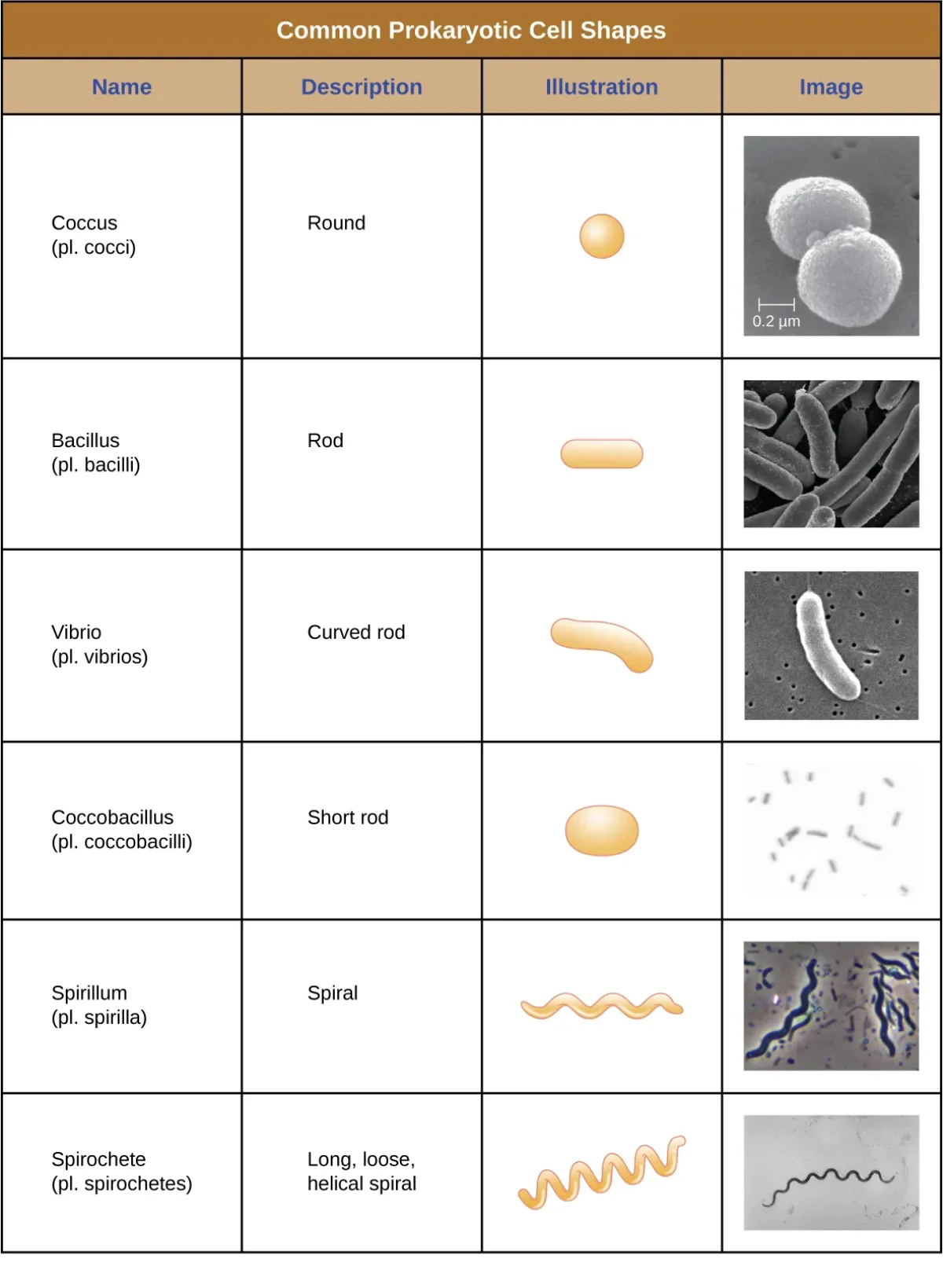
Common prokaryotic cell shapes: cocci, bacilli, vibrios, spirilla, and spirochetes. Credit: OpenStax Microbiology, CC BY 4.0
Bacteria can also be arranged in patterns: diplo- (pairs), strepto- (chains), staphylo- (clusters), tetrads (groups of four).
By oxygen requirement:
Obligate aerobes - must have O2 for metabolism
Obligate anaerobes - O2 is toxic and kills them (produces reactive oxygen species they cannot detoxify)
Facultative anaerobes - prefer O2 but can switch to fermentation without it (e.g., E. coli)
Aerotolerant anaerobes - do not use O2 for metabolism but are not harmed by its presence
By nutritional strategy:
Chemoautotrophs - get energy from chemical reactions and carbon from CO2 (e.g., Nitrosomonas - oxidizes ammonia)
Chemoheterotrophs - get both energy and carbon from organic molecules (e.g., E. coli - most pathogens)
Photoautotrophs - get energy from sunlight and carbon from CO2 (e.g., cyanobacteria)
Photoheterotrophs - get energy from sunlight but need organic carbon sources
Bacterial Reproduction and Genetic Exchange
Bacteria reproduce asexually by binary fission - the cell copies its circular chromosome, elongates, and splits into two identical daughter cells. This is fast (some bacteria divide every 20 minutes) but produces genetic clones.
To generate genetic diversity (without sexual reproduction), bacteria use three mechanisms of horizontal gene transfer:
Transformation - a bacterium picks up free-floating DNA fragments from its environment (usually from dead, lysed bacteria nearby). The foreign DNA is incorporated into the bacterium’s genome. This is most common in Gram-negative rod-shaped bacteria.
Conjugation - bacterial “mating.” An F+ bacterium (donor, carrying the F plasmid / fertility factor) extends a sex pilus to connect with an F- bacterium (recipient). The F plasmid is replicated and a copy is transferred through the conjugation bridge to the recipient, converting it to F+. This is how antibiotic resistance genes spread rapidly through bacterial populations.
Sometimes the F plasmid integrates into the bacterial chromosome itself. A bacterium with an integrated F factor is called Hfr (high frequency of recombination). When an Hfr cell conjugates, it attempts to transfer its entire chromosome (not just the plasmid) to the recipient. The bridge usually breaks before the full transfer is complete, but the recipient acquires some chromosomal genes from the donor.
Transduction - accidental gene transfer mediated by a bacteriophage (virus). During phage replication inside a bacterium, the phage may accidentally package some of the host’s DNA into a new phage particle. When this phage infects another bacterium, it delivers the previous host’s DNA to the new host, potentially adding new genes to its genome.
Transposons - mobile genetic elements (sometimes called “jumping genes”) that can move from one location to another within a genome, or between genomes. Transposons are found in both prokaryotes and eukaryotes. They contribute to genetic diversity and can disrupt genes when they insert into coding regions. In bacteria, transposons are a major mechanism for spreading antibiotic resistance genes between the chromosome and plasmids.
Bacterial Growth Curve
When bacteria are placed in a new environment with available nutrients, the population grows in a predictable pattern with four phases:
Lag phase - bacteria adapt to the new environment, synthesize enzymes and proteins needed for growth. Cell division has not yet begun.
Log (exponential) phase - bacteria divide at a maximum, constant rate. The population doubles at regular intervals. This is the phase where bacteria are most susceptible to antibiotics.
Stationary phase - resources become limited and waste products accumulate. The rate of cell division equals the rate of cell death, so the population size plateaus.
Death (decline) phase - nutrients are exhausted and waste builds up. The death rate exceeds the division rate, and the population declines.
What is the structural difference between Gram-positive and Gram-negative bacterial cell walls?
Click to reveal answer
Gram-positive: thick peptidoglycan layer (90-95%), lipoteichoic acid, no outer membrane. Gram-negative: thin peptidoglycan layer in the periplasmic space, an outer membrane containing lipopolysaccharide (LPS/endotoxin). Gram-positive retains crystal violet (purple); Gram-negative picks up safranin (pink).
What are the three mechanisms of horizontal gene transfer in bacteria?
Click to reveal answer
1) Transformation - uptake of free DNA from the environment. 2) Conjugation - direct transfer via sex pilus (F+ to F- cell). 3) Transduction - phage accidentally packages host DNA and delivers it to a new bacterium. Remember: TranCT.
How does a bacterium move toward a nutrient (chemotaxis)?
Click to reveal answer
It alternates "runs" (straight swimming via counter-clockwise flagellar rotation) with "tumbles" (random reorientations via clockwise rotation). When surface chemoreceptors sense the attractant concentration rising, the bacterium lengthens its runs. This biased random walk moves the population up the gradient.