
Organelles
Back to our city analogy. The city (cell) has specialized departments (organelles), each enclosed in its own building (membrane). This compartmentalization is what makes eukaryotic cells so powerful - it allows different chemical reactions to happen simultaneously in isolated environments, without interfering with each other.
The Nucleus - City Hall
The nucleus is the largest organelle and the command center of the cell. It stores the cell’s DNA and controls gene expression - deciding which proteins get made and when.
Structure:
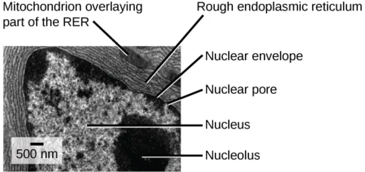
- Nuclear envelope: A double membrane (two phospholipid bilayers) that separates the nucleus from the cytoplasm. The outer membrane is continuous with the endoplasmic reticulum.
- Nuclear pores: Channels that penetrate the nuclear envelope, acting as security checkpoints. Small molecules diffuse freely, but large molecules (like proteins headed into the nucleus) need a signal sequence that flags them for nuclear import.
- Nucleolus: A dense, dark-staining region within the nucleus where ribosomal RNA (rRNA) is synthesized by RNA polymerase I. The nucleolus is essentially a ribosome factory - it assembles the RNA components of ribosomes, which then leave through nuclear pores and get their final protein components added in the cytoplasm.
Chromatin and Chromosomes:
Your DNA does not just float around in a tangled mess. It wraps around proteins called histones, forming a complex called chromatin. Think of it like thread (DNA) wound around spools (histones). This chromatin can be further compacted into visible chromosomes during cell division.
Chromatin exists in two states:
- Euchromatin - loosely packed, transcriptionally active (“eu” = true, good - the genes here can be read and expressed)
- Heterochromatin - tightly packed, transcriptionally inactive (like a book on a high shelf you cannot reach - the genes are silenced)
Each chromosome is composed of two identical sister chromatids joined at a centromere. The ends of chromosomes are protected by repetitive DNA sequences called telomeres, which act like the plastic tips on shoelaces - they prevent the chromosome from fraying or fusing with neighbors.
Ribosomes - The Construction Workers
Ribosomes are the molecular machines that translate mRNA into proteins. They are NOT membrane-bound organelles - they float freely in the cytoplasm or attach to the rough ER.
- Eukaryotic ribosomes: 80S (composed of a 40S small subunit and a 60S large subunit)
- Prokaryotic ribosomes: 70S (composed of a 30S small subunit and a 50S large subunit)
The “S” stands for Svedberg units, a measure of sedimentation rate during centrifugation. The numbers do not add up arithmetically because Svedberg units depend on shape and mass, not just mass alone.
Endoplasmic Reticulum - The Factory Floor
The ER is a vast network of membrane-enclosed channels and sacs continuous with the nuclear envelope. It comes in two varieties:
Rough ER (RER):
- Studded with ribosomes on its cytoplasmic surface (that is what makes it look “rough” under electron microscopy)
- Synthesizes proteins destined for secretion, the cell membrane, or organelles
- Folds and modifies these proteins (adding sugar groups, forming disulfide bonds)
- Proteins enter the RER lumen through a signal sequence on the growing polypeptide
Smooth ER (SER):
- No ribosomes (hence “smooth”)
- Synthesizes lipids, phospholipids, and steroid hormones
- Detoxifies drugs and poisons (liver cells have abundant SER for this reason)
- Stores calcium ions (especially important in muscle cells, where the SER is called the sarcoplasmic reticulum)
- Transports proteins from the RER to the Golgi apparatus
Golgi Apparatus - The Post Office
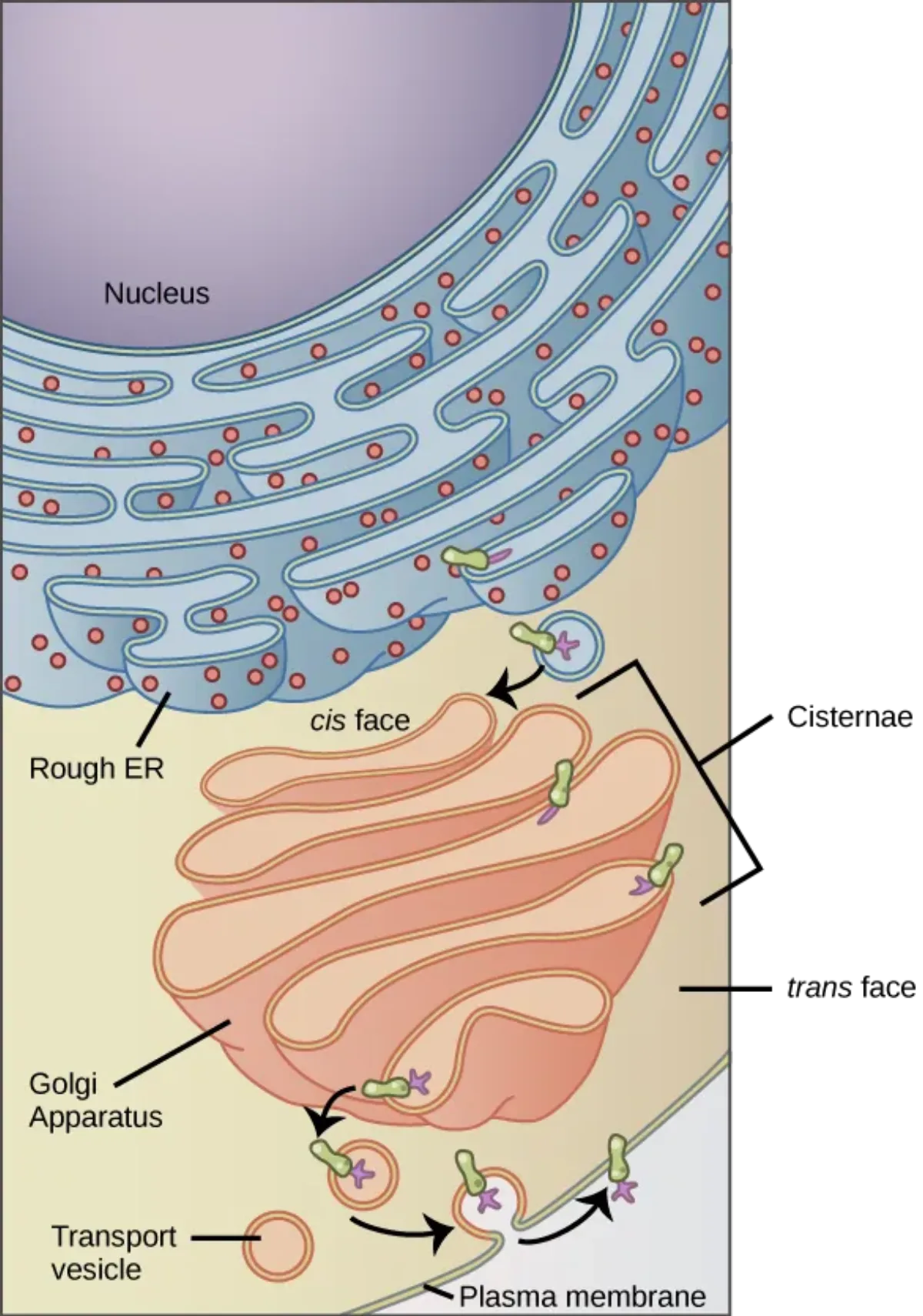
The Golgi is a stack of flattened membrane sacs (cisternae) that receives, modifies, sorts, and ships cellular products. Think of it as the cell’s post office and distribution center.
- Cis face (“receiving dock”) - faces the ER. Vesicles from the ER arrive here and fuse with the cis face, dumping their cargo inside.
- Trans face (“shipping dock”) - faces the cell membrane. Finished products leave from here in vesicles headed for their final destination.
As proteins and lipids move from cis to trans through the Golgi stack, they undergo modifications: addition of carbohydrate chains (glycosylation), phosphate groups, or sulfate groups. The Golgi also adds signal sequences - molecular address labels that direct each product to the correct destination (lysosome, membrane, or secretion outside the cell).
Lysosomes - The Recycling and Demolition Crew
Lysosomes are membrane-bound sacs filled with hydrolytic enzymes - molecular scissors that can chop up proteins, carbohydrates, lipids, and nucleic acids. They are the cell’s digestive system and waste disposal unit.
Key facts for the MCAT:
- Acidic interior (pH ~5): much lower than cytoplasm (pH 7.4). If a lysosome ruptures, the leaked enzymes drift into neutral pH and go inactive - a built-in safety switch.
- Autophagy: lysosomes digest the cell’s own worn-out organelles (damaged mitochondria, old ER).
- Apoptosis: lysosomal enzymes help dismantle the cell during programmed cell death.
- Phagocytosis cleanup: in macrophages and neutrophils, lysosomes fuse with phagosomes to digest engulfed pathogens.
Peroxisomes - The Hazardous Waste Handlers
Peroxisomes are small, membrane-bound organelles that specialize in oxidation reactions. They are found in both animal and plant cells.
Functions:
- Beta-oxidation of very long-chain fatty acids - breaking down fatty acids that are too long for mitochondria to handle
- Synthesis of phospholipids - building blocks for cellular membranes
- Detoxification - using oxidation reactions that produce hydrogen peroxide (H2O2) as a byproduct
- Catalase - the signature enzyme of peroxisomes. It rapidly converts toxic H2O2 into harmless water and oxygen: 2 H2O2 -> 2 H2O + O2
Organelle Summary Table
| Organelle | City Analogy | Key Function | Membrane? |
|---|---|---|---|
| Nucleus | City Hall | DNA storage, gene expression | Double membrane |
| Nucleolus | Blueprint copier in City Hall | rRNA synthesis, ribosome assembly | No membrane (region within nucleus) |
| Ribosomes | Construction workers | Protein synthesis (translation) | No membrane |
| Rough ER | Factory floor with workers | Protein synthesis, folding, modification | Single membrane |
| Smooth ER | Chemical plant | Lipid/steroid synthesis, detox, Ca2+ storage | Single membrane |
| Golgi apparatus | Post office | Modification, sorting, shipping | Single membrane (stacked) |
| Lysosomes | Recycling center / demolition crew | Digestion of waste, autophagy, apoptosis | Single membrane |
| Peroxisomes | Hazardous waste facility | Fatty acid oxidation, H2O2 detox | Single membrane |
| Mitochondria | Power plant | ATP production | Double membrane |