
Cell Junctions
Think of cells in a tissue like apartments in a building. Some apartments share sealed walls so nothing leaks between them (tight junctions). Some have doorways that let neighbors pass notes back and forth (gap junctions). Some are bolted together to keep the building from falling apart under stress (desmosomes and adherens junctions). And some are screwed into the foundation (hemidesmosomes).
Cells in your body do not exist in isolation. In tissues, neighboring cells are physically connected through specialized structures called cell junctions. Five types matter for the MCAT.
Tight Junctions - The Waterproof Seal
Tight junctions create a watertight seal between adjacent cells, preventing solutes from leaking through the space between cells (the paracellular route). They force all substances to go through the cells themselves rather than between them.
Where are they critical? In the intestinal epithelium, tight junctions prevent digestive enzymes and bacteria from leaking between cells into the bloodstream. In the blood-brain barrier, tight junctions between endothelial cells prevent most blood-borne substances from entering brain tissue.
Tight junctions must form a continuous band around the entire cell to work. If there are gaps, fluid can leak through, which causes disease. They can be so tight that they create a voltage difference across the epithelium by maintaining different ion concentrations on each side.
Gap Junctions - The Direct Communication Channels
Gap junctions are like tiny tunnels between adjacent cells, allowing direct passage of ions, water, and small signaling molecules. Each gap junction channel is formed by two connexons (one from each cell), and each connexon is made of six connexin protein subunits.
Gap junctions are essential wherever cells need to act in unison:
- Cardiac muscle - gap junctions allow the rapid spread of electrical signals (action potentials) between heart cells, ensuring coordinated contractions. This is why the heart beats as a single unit rather than as a collection of individual cells contracting randomly.
- Smooth muscle - coordinated contractions in the gut and blood vessels
- Embryonic development - allows signaling between neighboring cells during tissue formation
Key limitation: Gap junctions allow small molecules (ions, amino acids, sugars, second messengers like cAMP and IP3) to pass, but they do not transfer large molecules like proteins.
Adherens Junctions - The Belt
Adherens junctions form a continuous belt around a cell just below the tight junctions, gluing neighboring cells together. Like desmosomes, they use cadherin proteins to bridge the intercellular space. The difference is the cytoskeletal anchor: adherens junctions attach to actin microfilaments, while desmosomes attach to intermediate filaments.
Adherens junctions help epithelial sheets maintain shape and distribute contractile force (for example, during embryonic folding).
Desmosomes - The Rivets
Desmosomes are like spot welds or rivets that hold cells together in areas subjected to mechanical stress - stretching, pulling, and shearing forces. They are particularly abundant in skin and cardiac muscle.
Each desmosome consists of cadherin proteins from adjacent cells interlocking with each other in the intercellular space. On the intracellular side, cadherins are anchored to intermediate filaments (typically keratin in epithelial cells or desmin in cardiac cells) through adaptor proteins. This arrangement distributes mechanical force across the entire cytoskeleton rather than concentrating it at one point.
In cardiac muscle, desmosomes are part of intercalated discs - the junctions between heart cells that also contain gap junctions. The desmosomes hold the cells together during the powerful contractions, while the gap junctions coordinate the electrical signals.
Hemidesmosomes - The Anchors
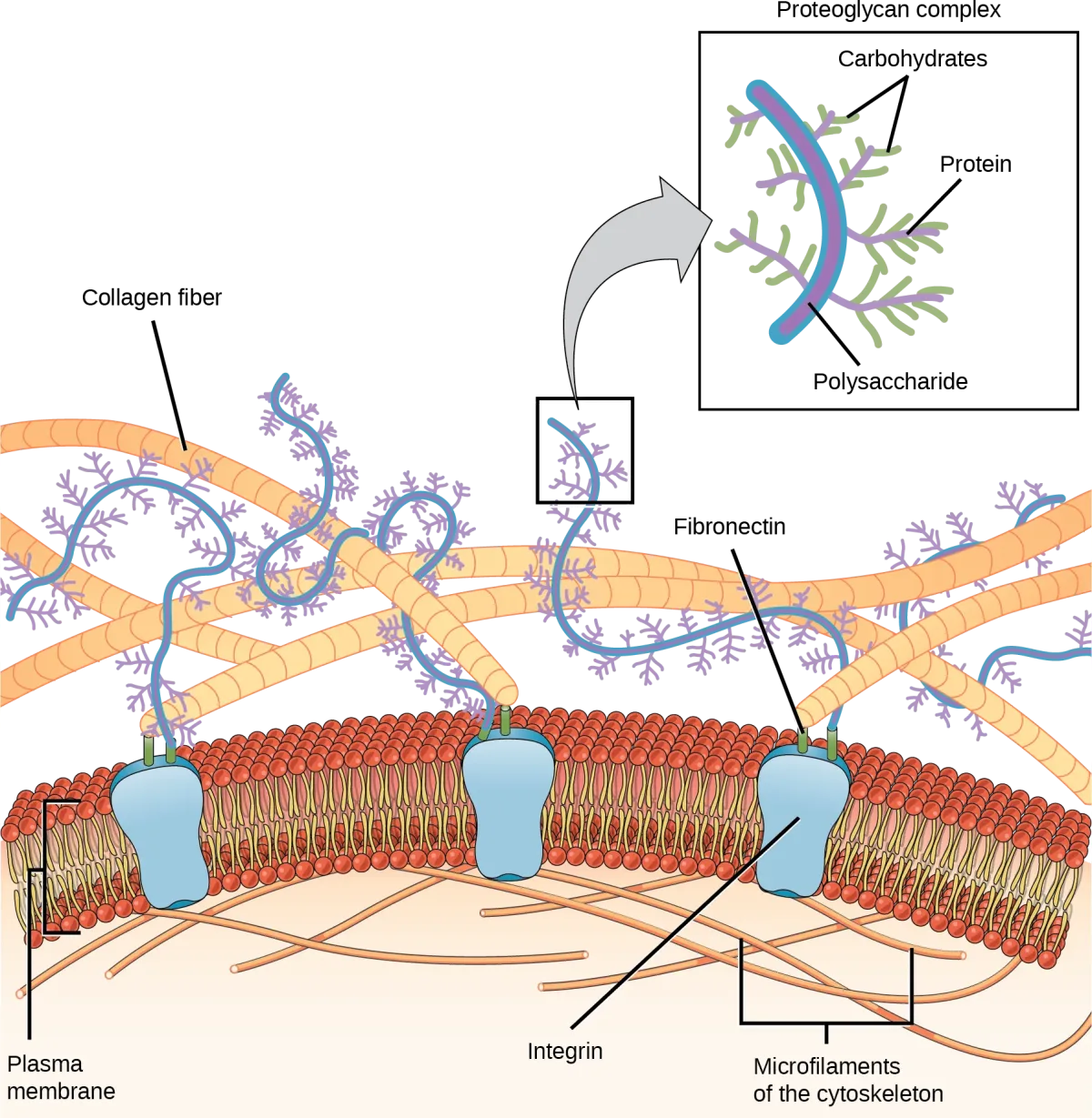
Hemidesmosomes look like half a desmosome (hence “hemi-”), but they serve a different function. Instead of connecting two cells to each other, hemidesmosomes connect the bottom of an epithelial cell to the basement membrane (the extracellular matrix beneath epithelial tissue).
They use integrin proteins (not cadherins like desmosomes) to anchor the cell to structural proteins in the basement membrane. This anchoring prevents epithelial cells from being torn away from the underlying tissue by mechanical forces.