
Cell Membrane
Imagine a nightclub with a very picky bouncer at the door. Small, nonpolar molecules (O2, CO2, steroid hormones) are on the VIP list - they walk right in, no questions asked. Large or charged molecules (glucose, amino acids, ions) need a protein escort - a channel or transporter protein that lets them through. And some molecules actually need to be physically dragged inside against their will, which costs energy (ATP). The bouncer does not work for free.
That nightclub is the cell membrane (also called the plasma membrane). It is not just a wall - it is a selectively permeable barrier that controls everything entering and leaving the cell. The specific mechanisms that move molecules across this barrier - from simple diffusion to active pumps - are covered in detail in cell transport.
The Phospholipid Bilayer
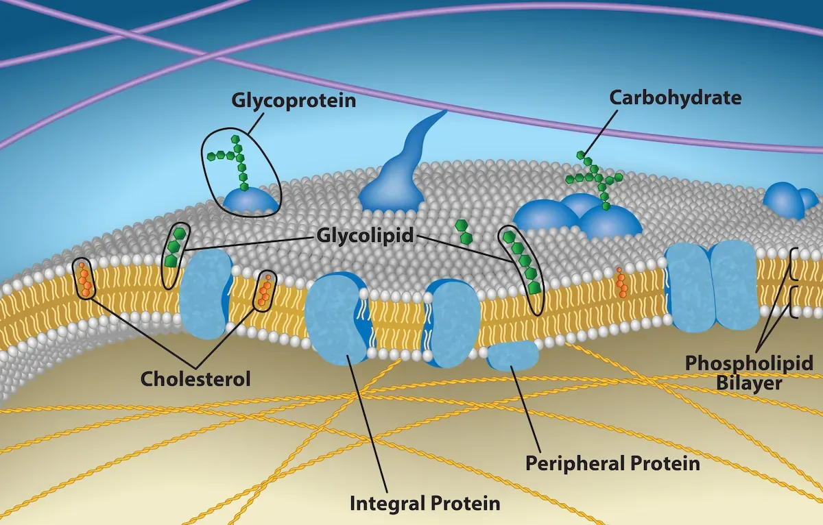
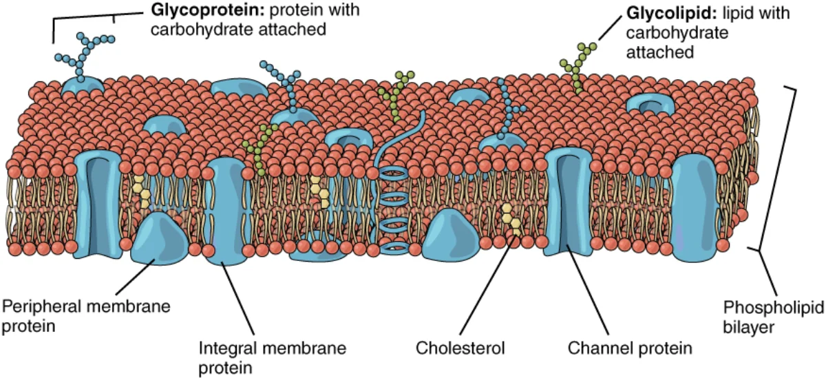
The membrane is built from a double layer of phospholipids. Each phospholipid has two parts:
- A hydrophilic head (polar, “water-loving”) - faces outward toward water on both sides of the membrane
- Two hydrophobic tails (nonpolar, “water-fearing”) - face inward, away from water, creating a greasy interior
This dual nature makes phospholipids amphipathic - they have both a water-loving and a water-fearing region. When you put millions of amphipathic molecules in water, they spontaneously arrange into a bilayer, with heads facing the aqueous environment and tails hiding in the middle. This arrangement creates a hydrophobic core that acts as a barrier to most polar and charged molecules.
The membrane is not rigid. It behaves like a fluid - proteins and lipids can move laterally within the bilayer. This is the basis of the fluid mosaic model: the membrane is a fluid sea of phospholipids with a mosaic of proteins embedded in it.
Cholesterol: the Thermostat of the Membrane
Animal cell membranes also contain cholesterol wedged between phospholipids. Cholesterol is a fluidity buffer:
- At high temperatures it restrains phospholipid movement, keeping the membrane from becoming too fluid.
- At low temperatures it wedges apart phospholipid tails, preventing the membrane from packing into a rigid gel.
Shorter and more unsaturated (kinked) fatty acid tails also increase fluidity; longer, saturated tails decrease it.
Membrane Proteins and the PERCH Functions
The cell membrane does far more than just act as a barrier. Its embedded proteins give it five major functions, which you can remember with the mnemonic PERCH:
Membrane Protein Types
- Integral (transmembrane) proteins span the entire bilayer. They include channels, transporters, and receptors. Because they pass through the hydrophobic core, they have hydrophobic regions in their middle and hydrophilic regions at the ends.
- Peripheral proteins sit on the inner or outer surface of the membrane without penetrating the lipid bilayer. They often participate in signaling or provide structural support.
- Glycoproteins and glycolipids have carbohydrate chains attached to their extracellular side. These sugar chains form the glycocalyx, which is involved in cell recognition, immune function, and protection.
The Cytosol
The entire internal space enclosed by the cell membrane (excluding organelles) is filled with cytosol - a gel-like aqueous solution. Cytosol is where many metabolic reactions occur, where ribosomes float and translate mRNA, and where the cytoskeleton is anchored. It is about 70% water and contains dissolved ions, small molecules, and proteins. The cytosol plus all organelles together make up the cytoplasm.
Membrane Potential: the Voltage Across the Bilayer
Because the membrane is selectively permeable and because pumps like the Na⁺/K⁺ ATPase unevenly distribute ions, the inside of a typical cell is more negative than the outside (roughly −70 mV in a resting neuron). This resting membrane potential is just stored electrical energy, the way water behind a dam stores gravitational energy.
Every cell has one. When we get to neurons, muscle cells, and secondary active transport, you will keep seeing the same logic: a pump builds an ion gradient, the gradient stores voltage, and opening a channel lets that voltage do work. Details live in Cell Transport and the nervous system chapter.