
Cytoskeleton
Imagine setting up a large outdoor tent. You need rigid poles to give it height and structure, thin ropes to anchor it and allow some flexibility, and medium-weight straps to hold the fabric in place under tension. The cell has the exact same setup - three types of protein filaments that collectively form the cytoskeleton.
The cytoskeleton does three main jobs:
- Structural support - maintains cell shape and resists mechanical stress
- Movement - enables cell motility, muscle contraction, and cell division
- Intracellular transport - acts as a highway system for motor proteins to carry cargo (vesicles, organelles) to where they are needed
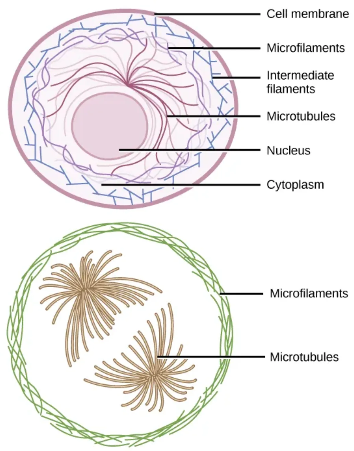
The Three Filament Types
Microfilaments (Actin Filaments) - 7 nm
Microfilaments are the thinnest filaments, composed of the protein actin polymerized into solid, helical rods. Think of them as the cell’s muscles.
Functions:
- Muscle contraction - actin interacts with the motor protein myosin, using ATP to generate force. This actin-myosin interaction is the molecular basis of every muscle contraction in your body.
- Cell movement - amoeboid movement, crawling of white blood cells toward infection
- Cytokinesis - during cell division, a ring of actin filaments forms the cleavage furrow. The ring contracts like a drawstring on a bag, pinching the cell in two to form two daughter cells.
- Microvilli support - microfilaments form the core of microvilli on intestinal epithelial cells, increasing surface area for nutrient absorption
Intermediate Filaments - 10 nm
Intermediate filaments are a diverse family of proteins that provide mechanical strength and tension resistance. They are the toughest of the three filament types - they do not participate in cell movement but are excellent at anchoring structures and resisting pulling forces.
Key types:
- Keratin - found in epithelial cells (skin, hair, nails). Gives skin its strength and resilience. This is the protein in all those hair product commercials.
- Vimentin - found in connective tissue cells, white blood cells
- Desmin - found in muscle cells, helps maintain the structural alignment of sarcomeres
- Lamins - found inside the nucleus, forming the nuclear lamina - a meshwork that supports the nuclear envelope from the inside
Microtubules - 25 nm
Microtubules are the largest and most rigid filaments, built from alpha-tubulin and beta-tubulin dimers that polymerize into hollow tubes. They are the railroad tracks of the cell - providing pathways for motor proteins to transport cargo.
Functions:
- Intracellular transport - motor proteins kinesin (moves toward the + end, away from the cell center) and dynein (moves toward the - end, toward the cell center) walk along microtubules carrying vesicles, organelles, and other cargo
- Cell division - microtubules form the mitotic spindle, which attaches to chromosomes at their kinetochores and pulls sister chromatids apart during mitosis/meiosis
- Structural framework for cilia and flagella
Microtubule Organizing Center (MTOC) and Centrioles
Microtubules do not just appear randomly in the cell. They grow outward from a structure called the microtubule organizing center (MTOC), usually located near the nucleus. The MTOC is also called the centrosome in animal cells.
The centrosome contains a pair of centrioles - cylindrical structures made of nine triplets of microtubules arranged in a ring. During cell division, centrioles duplicate and move to opposite poles of the cell, organizing the mitotic spindle.
Microtubules are dynamic - they can rapidly grow (polymerize) by adding tubulin dimers at their + end and shrink (depolymerize) by losing them. This property, called dynamic instability, allows the cell to quickly reorganize its internal structure as needed.
Cilia and Flagella
Both cilia and flagella are extensions of the cell membrane built on a core framework of microtubules. They share the same internal structure: the 9+2 arrangement - nine outer pairs (doublets) of microtubules surrounding two central single microtubules, all enclosed by the plasma membrane.
Cilia are short, hair-like projections that cover the surface of certain cells:
- In the respiratory tract, cilia beat in coordinated waves to sweep mucus and trapped debris upward and out of the lungs
- In the fallopian tubes, cilia help move eggs from the ovary toward the uterus
Flagella are long, whip-like structures used for propulsion:
- In humans, the only flagellated cell is the sperm cell
- Flagella use a wave-like motion powered by dynein motor proteins
Important distinction for the MCAT: Eukaryotic flagella (9+2 microtubule structure, powered by dynein/ATP) are completely different from bacterial flagella (made of flagellin protein, rotary motion powered by a proton gradient). Do not confuse them.