
Bacteria
Before we close this chapter, we need to cover bacteria in more detail. We introduced prokaryotic cells in Section 1.2, but bacteria have unique structural features and reproductive strategies that are high-yield MCAT topics.
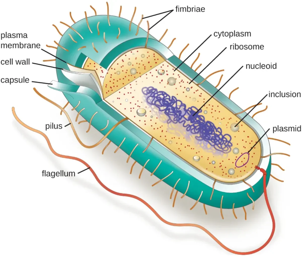
Bacterial Cell Structure
Bacteria are single-celled organisms with a relatively simple architecture, but every structure serves a critical function.
Cell wall - a rigid layer outside the plasma membrane, composed of peptidoglycan (a polymer of sugars and amino acids). The cell wall provides structural support, maintains cell shape, and protects against osmotic lysis. Some antibiotics target peptidoglycan synthesis, which is why cell wall structure matters for understanding antibiotic selectivity.
Gram-positive vs. Gram-negative bacteria:
The Gram stain is one of the most important classification tools in microbiology. The procedure uses crystal violet dye followed by a safranin counterstain. The result depends on cell wall structure:
| Feature | Gram-Positive | Gram-Negative |
|---|---|---|
| Peptidoglycan layer | Thick (90-95% of wall) | Thin (in periplasmic space) |
| Outer membrane | Absent | Present (contains LPS) |
| Gram stain color | Purple/violet (retains crystal violet) | Pink/red (loses crystal violet, picks up safranin) |
| Lipoteichoic acid (LTA) | Present | Absent |
| Lipopolysaccharide (LPS) | Absent | Present (endotoxin - lipid A is toxic) |
| Antibiotic susceptibility | Generally more susceptible to cell-wall-targeting antibiotics | More resistant (outer membrane barrier) |
Other key bacterial structures:
- Plasma membrane - phospholipid bilayer, same basic structure as eukaryotic membranes
- Nucleoid - region of the cytoplasm where the single circular chromosome resides (not enclosed by a membrane)
- Plasmids - small, circular, extrachromosomal DNA molecules that replicate independently. They often carry genes for antibiotic resistance, toxin production, or novel metabolic capabilities. Plasmids can be transferred between bacteria.
- Ribosomes - 70S (30S + 50S subunits). Smaller than eukaryotic 80S ribosomes. This difference allows antibiotics to selectively target bacterial ribosomes without harming human cells.
- Flagella - long, whip-like appendages for motility. Bacterial flagella are structurally different from eukaryotic flagella - they are made of flagellin protein, rotate like a propeller driven by a proton gradient (not ATP), and consist of a filament, hook, and basal body. Bacteria use their flagella to perform chemotaxis: by alternating smooth “runs” (counter-clockwise rotation) with random “tumbles” (clockwise rotation), they bias their movement toward attractants (nutrients) and away from repellents (toxins). Surface chemoreceptors detect the gradient and adjust the ratio of running to tumbling.
- Pili (fimbriae) - short, hair-like projections used for adhesion to surfaces and other cells. Sex pili (conjugation pili) are specialized pili that form a bridge between two bacteria during conjugation for DNA transfer.
- Capsule - a polysaccharide layer outside the cell wall found in some bacteria. It protects against phagocytosis by the immune system and helps bacteria adhere to surfaces (like forming biofilms on medical devices).
- Endospores - some bacteria (like Clostridium and Bacillus) can form highly resistant dormant structures called endospores when conditions become harsh. Endospores can survive extreme heat, radiation, desiccation, and chemicals. When conditions improve, the endospore germinates back into a vegetative cell.
Bacterial Classification
Beyond Gram staining, bacteria can be classified by shape, oxygen requirement, nutritional strategy, and temperature tolerance.
By shape:
- Cocci - spherical (e.g., Staphylococcus, Streptococcus)
- Bacilli - rod-shaped (e.g., E. coli, Bacillus)
- Spirilla - spiral-shaped (e.g., Treponema, Borrelia)
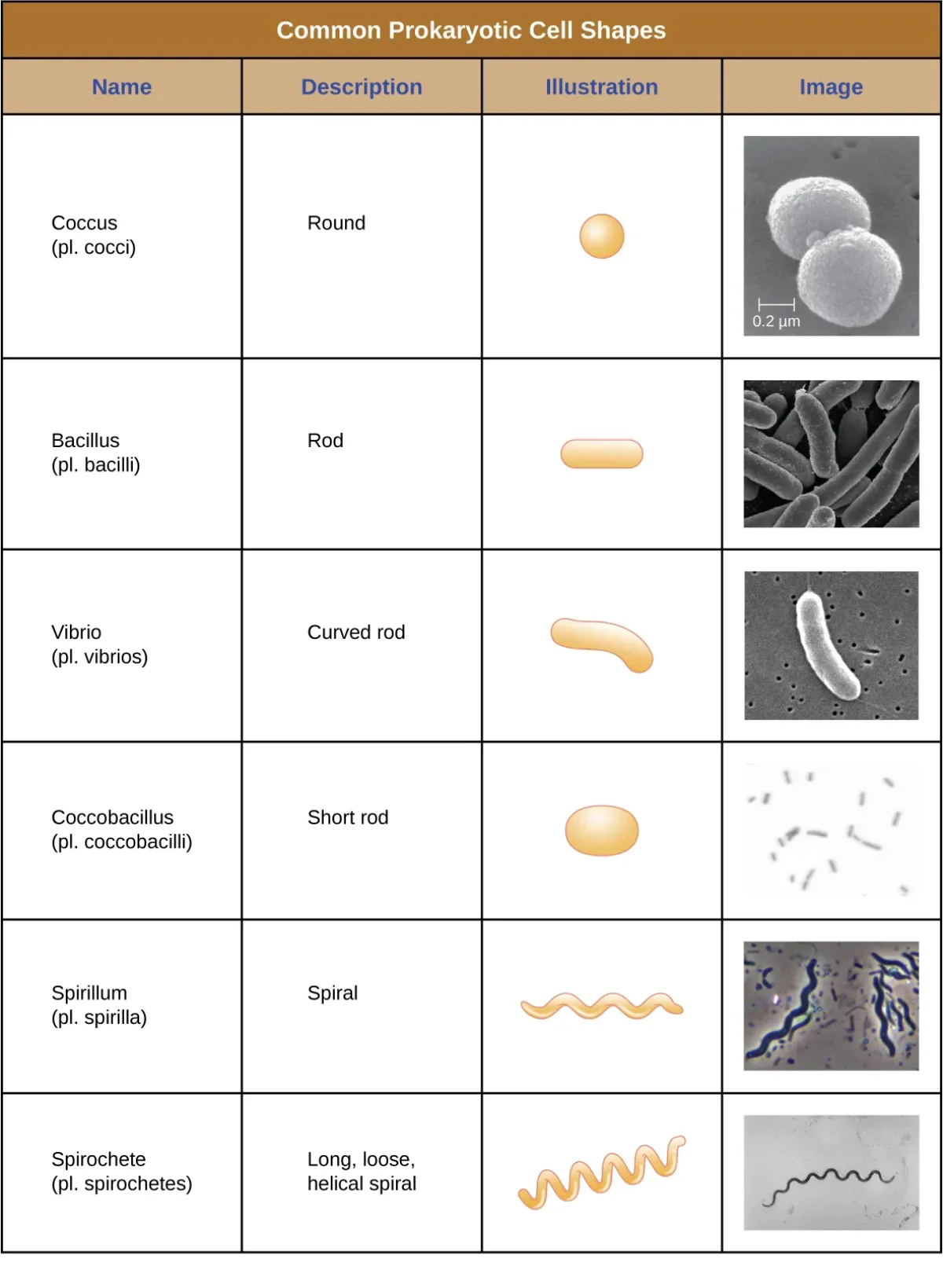
Bacteria can also be arranged in patterns: diplo- (pairs), strepto- (chains), staphylo- (clusters), tetrads (groups of four).
By oxygen requirement:
- Obligate aerobes - must have O2 for metabolism
- Obligate anaerobes - O2 is toxic and kills them (produces reactive oxygen species they cannot detoxify)
- Facultative anaerobes - prefer O2 but can switch to fermentation without it (e.g., E. coli)
- Aerotolerant anaerobes - do not use O2 for metabolism but are not harmed by its presence
By nutritional strategy:
- Chemoautotrophs - get energy from chemical reactions and carbon from CO2 (e.g., Nitrosomonas - oxidizes ammonia)
- Chemoheterotrophs - get both energy and carbon from organic molecules (e.g., E. coli - most pathogens)
- Photoautotrophs - get energy from sunlight and carbon from CO2 (e.g., cyanobacteria)
- Photoheterotrophs - get energy from sunlight but need organic carbon sources
Bacterial Reproduction and Genetic Exchange
Bacteria reproduce asexually by binary fission - the cell copies its circular chromosome, elongates, and splits into two identical daughter cells. This is fast (some bacteria divide every 20 minutes) but produces genetic clones.
To generate genetic diversity (without sexual reproduction), bacteria use three mechanisms of horizontal gene transfer:
Transformation - a bacterium picks up free-floating DNA fragments from its environment (usually from dead, lysed bacteria nearby). The foreign DNA is incorporated into the bacterium’s genome. This is most common in Gram-negative rod-shaped bacteria.
Conjugation - bacterial “mating.” An F+ bacterium (donor, carrying the F plasmid / fertility factor) extends a sex pilus to connect with an F- bacterium (recipient). The F plasmid is replicated and a copy is transferred through the conjugation bridge to the recipient, converting it to F+. This is how antibiotic resistance genes spread rapidly through bacterial populations.
Sometimes the F plasmid integrates into the bacterial chromosome itself. A bacterium with an integrated F factor is called Hfr (high frequency of recombination). When an Hfr cell conjugates, it attempts to transfer its entire chromosome (not just the plasmid) to the recipient. The bridge usually breaks before the full transfer is complete, but the recipient acquires some chromosomal genes from the donor.
Transduction - accidental gene transfer mediated by a bacteriophage (virus). During phage replication inside a bacterium, the phage may accidentally package some of the host’s DNA into a new phage particle. When this phage infects another bacterium, it delivers the previous host’s DNA to the new host, potentially adding new genes to its genome.
Transposons - mobile genetic elements (sometimes called “jumping genes”) that can move from one location to another within a genome, or between genomes. Transposons are found in both prokaryotes and eukaryotes. They contribute to genetic diversity and can disrupt genes when they insert into coding regions. In bacteria, transposons are a major mechanism for spreading antibiotic resistance genes between the chromosome and plasmids.
Bacterial Growth Curve
When bacteria are placed in a new environment with available nutrients, the population grows in a predictable pattern with four phases:
- Lag phase - bacteria adapt to the new environment, synthesize enzymes and proteins needed for growth. Cell division has not yet begun.
- Log (exponential) phase - bacteria divide at a maximum, constant rate. The population doubles at regular intervals. This is the phase where bacteria are most susceptible to antibiotics.
- Stationary phase - resources become limited and waste products accumulate. The rate of cell division equals the rate of cell death, so the population size plateaus.
- Death (decline) phase - nutrients are exhausted and waste builds up. The death rate exceeds the division rate, and the population declines.