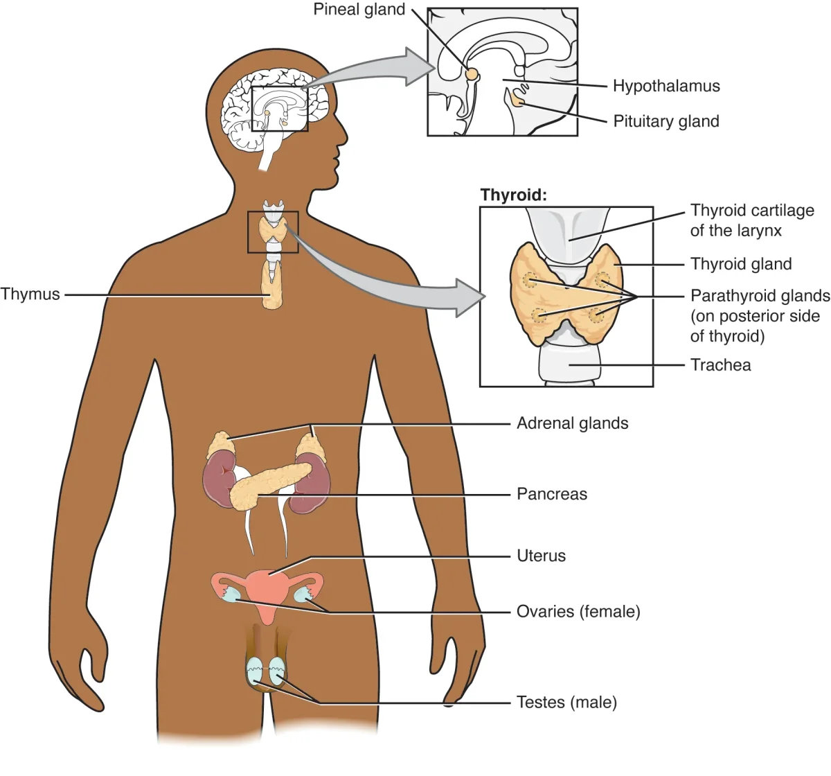
↗The major endocrine glands are distributed throughout the body, communicating via hormones released into the bloodstream. Credit: OpenStax Anatomy and Physiology 2e, CC BY 4.0
Not all hormones work the same way. A peptide hormone like insulin cannot cross a cell membrane - it has to ring the doorbell and wait for someone inside to relay the message. A steroid hormone like cortisol walks right through the front door and goes straight to the nucleus to change which genes get turned on. Understanding this difference is one of the most frequently tested concepts on the MCAT.
The key principle: a hormone’s chemical structure determines everything about how it works - how it travels in the blood, whether it can cross cell membranes, where its receptor lives, how fast it acts, and how long its effects last.
The Three Major Hormone Classes
Hormones fall into three structural categories. Each category has a predictable set of properties that you can reason through from first principles.
1. Peptide hormones are chains of amino acids - essentially small proteins. They are water-soluble (hydrophilic) because of their polar amino acid side chains. Examples include insulin, growth hormone, ADH, oxytocin, FSH, LH, ACTH, TSH, and prolactin.
2. Steroid hormones are derived from cholesterol. They are lipid-soluble (hydrophobic) because of their four-ring hydrocarbon structure. Examples include cortisol, aldosterone, testosterone, estrogen, and progesterone.
3. Amino acid-derived hormones are synthesized from single amino acids, primarily tyrosine and tryptophan. This class is tricky because it includes hormones that behave like peptide hormones AND hormones that behave like steroid hormones. Catecholamines (epinephrine, norepinephrine, dopamine) are water-soluble and act like peptide hormones. Thyroid hormones (T3, T4) are lipid-soluble and act like steroid hormones.
Peptide Hormone Signaling
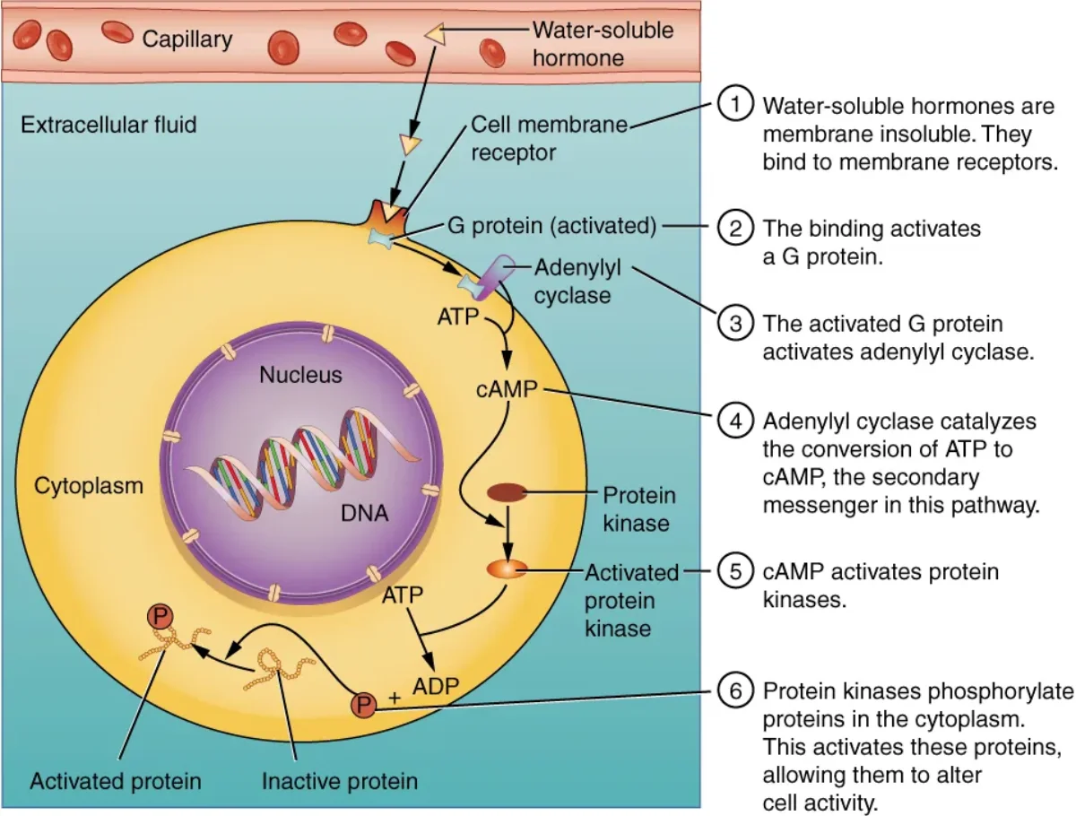
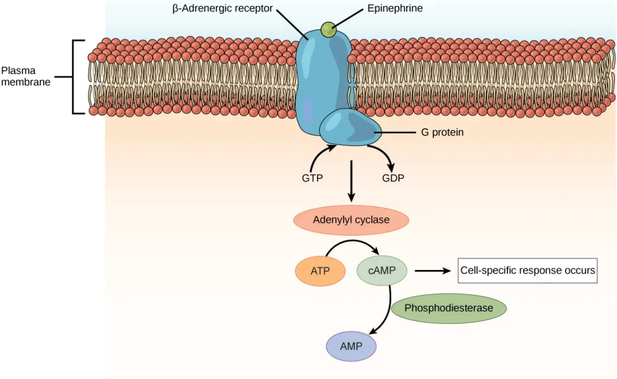
Peptide hormones bind to surface receptors and trigger intracellular second messenger cascades. Credit: OpenStax Anatomy and Physiology 2e, CC BY 4.0
Because peptide hormones are water-soluble, they dissolve freely in blood plasma and do not need carrier proteins. However, they cannot cross the lipid bilayer of a target cell. Instead, they bind to receptors on the cell surface.
Most peptide hormone receptors are G-protein coupled receptors (GPCRs). When the hormone binds, the receptor activates a G-protein, which activates an enzyme (like adenylyl cyclase), which produces a second messenger (like cAMP), which activates downstream kinases (like protein kinase A). This cascade amplifies the signal enormously.
Other peptide hormones (like insulin) use receptor tyrosine kinases (RTKs) - the receptor itself has enzymatic activity and directly phosphorylates intracellular proteins when the hormone binds.
Key features of peptide hormone signaling:
Fast onset (seconds to minutes) - no gene transcription required
Short duration - effects stop when the signal cascade is deactivated
Signal amplification - one hormone molecule can produce thousands of second messenger molecules
Stored in vesicles - pre-made and released by exocytosis when needed
Steroid Hormone Signaling
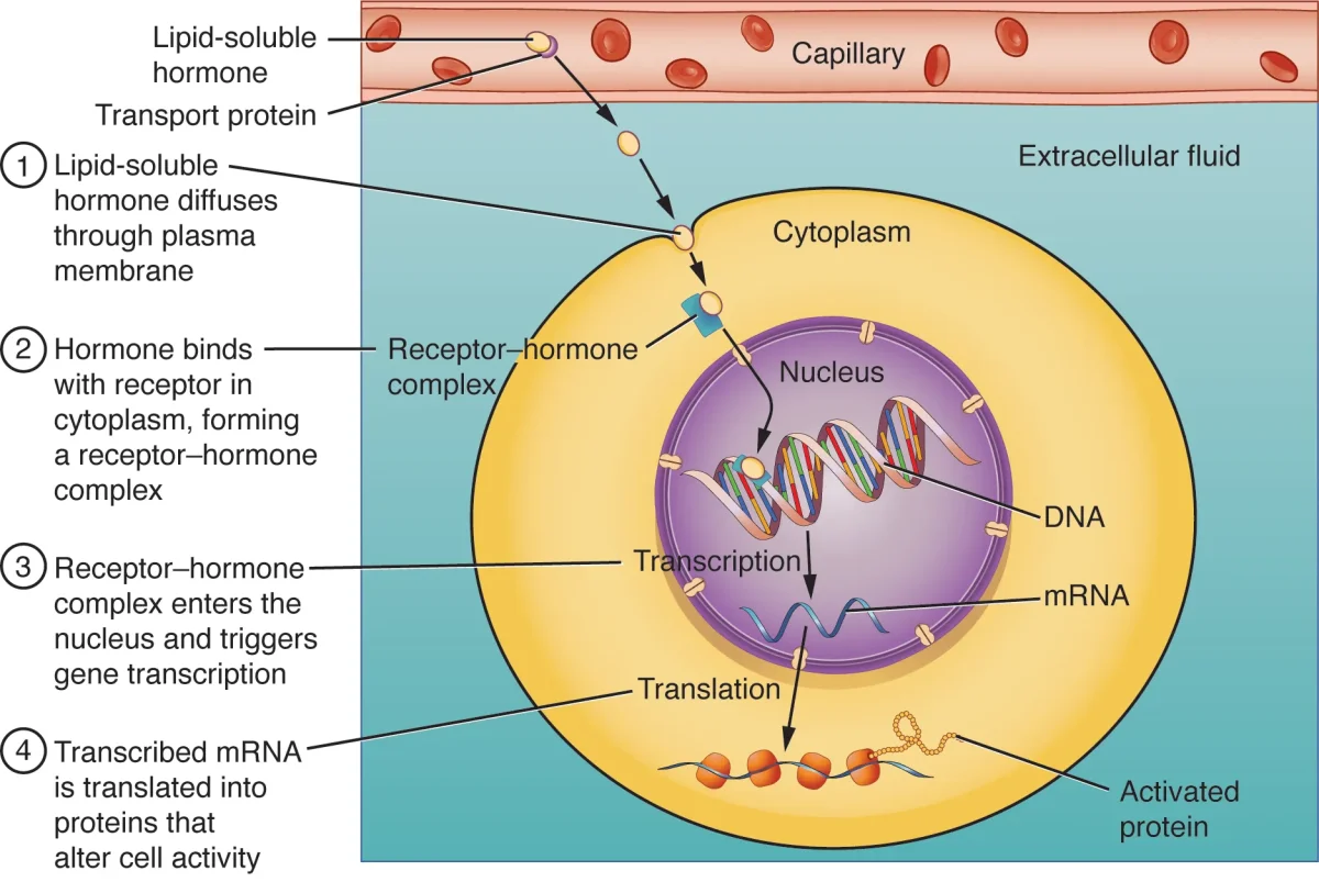
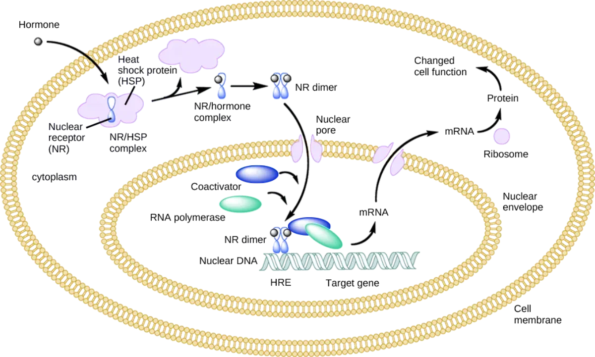
Steroid hormones cross the membrane, bind intracellular receptors, and directly alter gene transcription. Credit: OpenStax Anatomy and Physiology 2e, CC BY 4.0
Steroid hormones are lipid-soluble, so they can diffuse directly through the cell membrane. But this lipid solubility creates a problem: they cannot dissolve in blood. They must travel bound to carrier proteins like albumin or specific binding globulins (e.g., sex hormone-binding globulin, cortisol-binding globulin).
Once a steroid hormone dissociates from its carrier and enters a target cell, it binds to an intracellular receptor - either in the cytoplasm or directly in the nucleus. The hormone-receptor complex then binds to specific DNA sequences called hormone response elements (HREs) and directly modulates gene transcription.
Key features of steroid hormone signaling:
Slow onset (hours to days) - requires gene transcription and protein synthesis
Long duration - effects persist because new proteins have been made
No signal amplification at the receptor level - but gene transcription itself amplifies the response
Not stored - synthesized on demand from cholesterol and released immediately (they diffuse right through the membrane)
Amino Acid-Derived Hormones: The Exception Class
This class is the MCAT’s favorite trick because the two subgroups behave completely differently:
Catecholamines (epinephrine, norepinephrine, dopamine) - derived from tyrosine in the adrenal medulla. Despite being modified amino acids, they are water-soluble. They bind to surface receptors (adrenergic receptors, which are GPCRs) and use second messenger cascades. They act fast - exactly like peptide hormones.
Thyroid hormones (T3, T4) - also derived from tyrosine, but in the thyroid gland with iodine incorporation. They are lipid-soluble (unusual for an amino acid derivative). They enter cells via transporter proteins, bind to nuclear receptors, and alter gene expression. They act slowly - like steroid hormones.
Feature
Peptide Hormones
Steroid Hormones
Catecholamines
Thyroid Hormones
Structure
Amino acid chains
Cholesterol-derived
Tyrosine-derived
Tyrosine + iodine
Solubility
Water-soluble
Lipid-soluble
Water-soluble
Lipid-soluble
Blood transport
Free in plasma
Carrier proteins
Free in plasma
Carrier proteins
Receptor location
Cell surface
Intracellular
Cell surface
Nuclear
Mechanism
Second messengers
Gene transcription
Second messengers
Gene transcription
Speed of action
Fast (sec-min)
Slow (hrs-days)
Fast (sec-min)
Slow (hrs-days)
Duration
Short
Long
Short
Long
Storage
Vesicles
Not stored
Vesicles
Stored as thyroglobulin
Types of Signaling
Hormones are classified not just by structure but also by how far their signal travels:
Endocrine signaling - hormones released into the bloodstream travel to distant target organs. This is the classic “endocrine” pathway. Example: insulin from the pancreas targeting liver and muscle cells.
Paracrine signaling - chemical messengers act on nearby cells without entering the bloodstream. Example: neurotransmitters at a synapse, or histamine released by mast cells affecting nearby blood vessels.
Autocrine signaling - a cell releases a signal that acts on itself. Example: immune cells releasing cytokines that bind to receptors on their own surface, amplifying their own activation.
Juxtacrine signaling - requires direct cell-to-cell contact. The signaling molecule stays anchored in the membrane of one cell and binds to a receptor on an adjacent cell. Example: Notch-Delta signaling during embryonic development, or antigen presentation between immune cells.
Direct vs. tropic hormones - direct hormones act on non-endocrine target tissues (insulin targets liver/muscle). Tropic hormones target other endocrine glands to stimulate hormone release (TSH targets the thyroid, ACTH targets the adrenal cortex). Tropic hormones create the hierarchical chains that the MCAT loves to test.
A hormone derived from cholesterol binds to an intracellular receptor and alters gene transcription. What class does it belong to, and why can't it be stored in vesicles?
Click to reveal answer
It is a steroid hormone. Because steroid hormones are lipid-soluble, they would diffuse right through the membrane of any storage vesicle. Instead, they are synthesized on demand from cholesterol and released immediately by diffusion through the cell membrane.
Epinephrine and T3 are both derived from tyrosine. Why does epinephrine act in seconds while T3 takes hours?
Click to reveal answer
Epinephrine is water-soluble and binds to surface GPCRs, activating a rapid second messenger cascade (cAMP pathway). T3 is lipid-soluble, enters the nucleus, and alters gene transcription - a process that requires mRNA synthesis and protein translation, taking hours to days.
A researcher discovers a new hormone that requires a carrier protein in the blood, has effects lasting several days, and directly alters gene expression. Is this most likely a peptide, steroid, or catecholamine?
Click to reveal answer
Steroid hormone (or thyroid hormone). Needing a carrier protein means it is lipid-soluble. Long-lasting effects and direct gene expression changes point to intracellular receptor signaling. Peptide hormones and catecholamines are water-soluble, travel freely in plasma, and act quickly via second messengers.
If the endocrine system is a corporation, the hypothalamus is the CEO and the pituitary gland is middle management. The hypothalamus receives information from the nervous system - body temperature, blood osmolarity, stress signals, circadian rhythms - and translates that neural information into hormonal commands. The pituitary gland receives those commands and delegates them to the appropriate endocrine organ.
This hypothalamus-pituitary axis is the master control system for nearly every hormonal pathway in the body. Understanding it is not optional for the MCAT - it is the framework that connects thyroid, adrenal, gonadal, and growth hormone regulation into a single logical system.
The Hypothalamus: Where Nervous Meets Endocrine
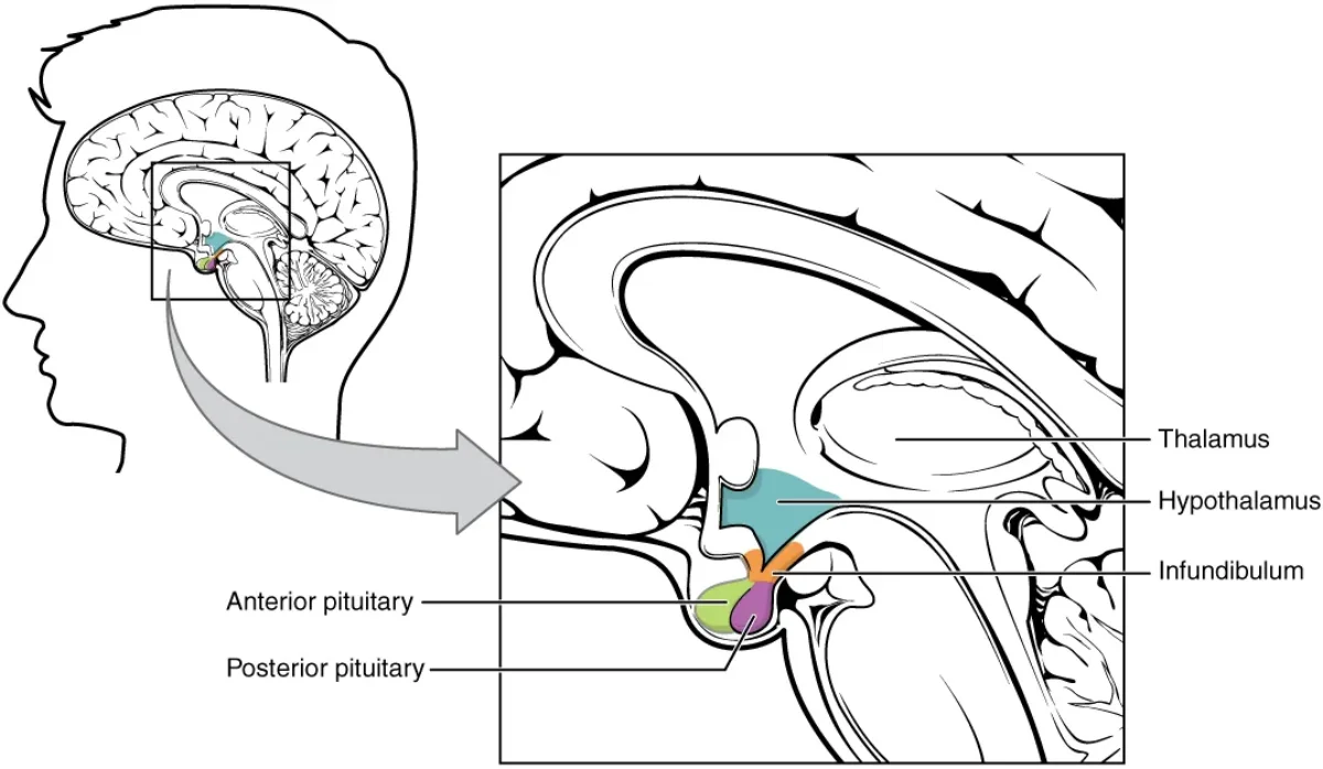
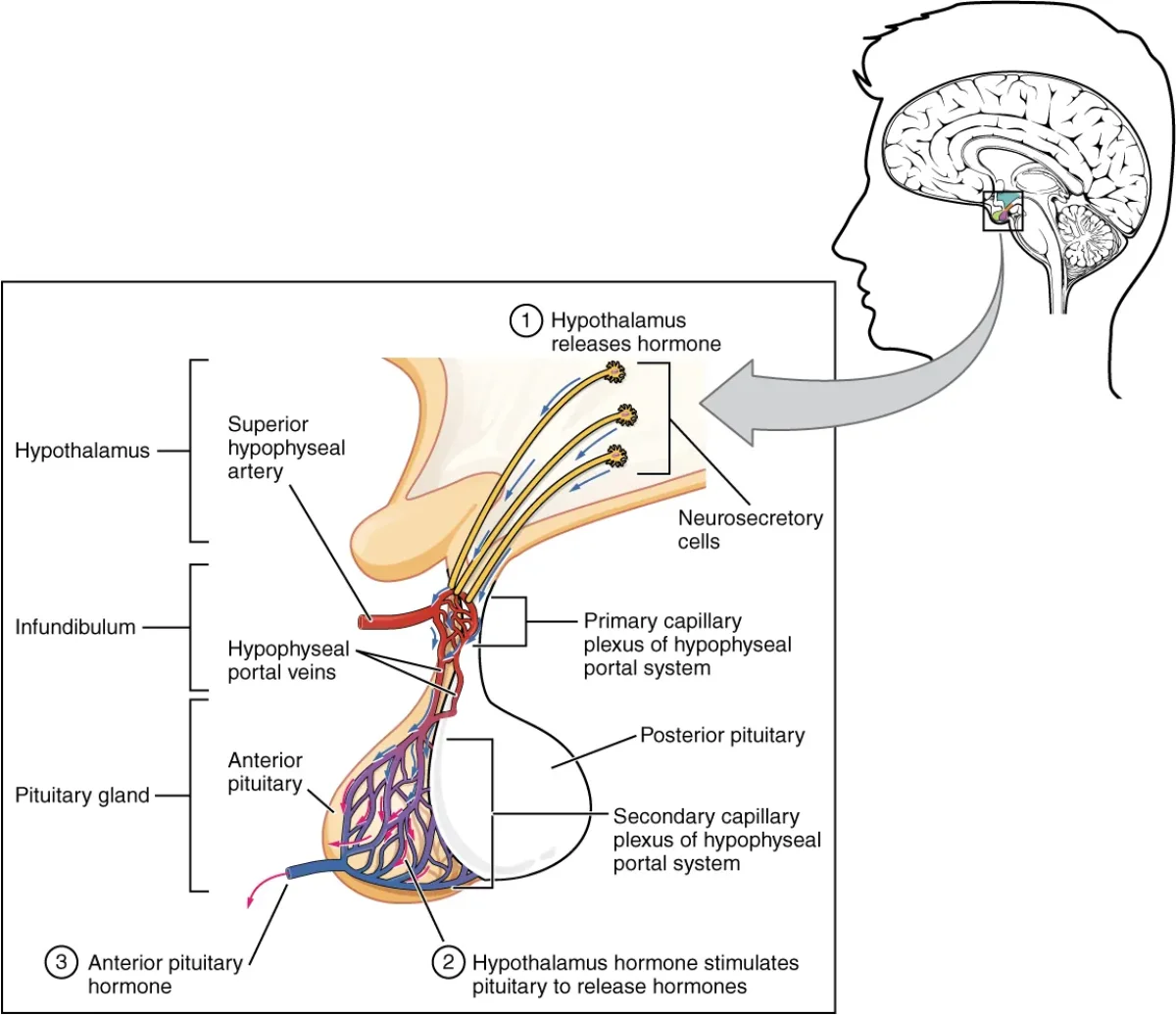
The hypothalamus-pituitary complex sits at the base of the brain, connecting neural and hormonal control. Credit: OpenStax Anatomy and Physiology 2e, CC BY 4.0
The hypothalamus is a small region at the base of the brain, sitting just above the pituitary gland. It is the critical link between the nervous system and the endocrine system - a neuroendocrine organ.
The hypothalamus produces releasing hormones and inhibiting hormones that travel to the anterior pituitary through a specialized blood vessel network called the hypophyseal portal system. This portal system is a direct vascular connection - hypothalamic hormones travel a very short distance through these portal vessels and arrive at the anterior pituitary in high concentrations, ensuring precise control.
Key hypothalamic hormones:
GnRH (gonadotropin-releasing hormone) - stimulates FSH and LH release
TRH (thyrotropin-releasing hormone) - stimulates TSH release
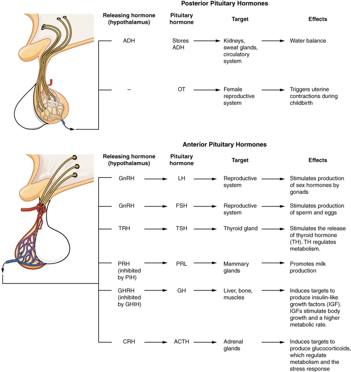
The hypophyseal portal system delivers hypothalamic hormones directly to the anterior pituitary. Credit: OpenStax Anatomy and Physiology 2e, CC BY 4.0The anterior pituitary produces seven hormones (FLAT PEG); the posterior pituitary stores and releases two (oxytocin and ADH). Credit: OpenStax Anatomy and Physiology 2e, CC BY 4.0
The anterior pituitary (adenohypophysis) contains five types of secretory cells, each producing specific hormones. The anterior pituitary synthesizes its own hormones in response to hypothalamic signals.
The Tropic Hormones (FLAT)
FSH (Follicle-Stimulating Hormone) - targets the gonads. In females, FSH stimulates growth of ovarian follicles and estrogen production by granulosa cells. In males, FSH acts on Sertoli cells in the seminiferous tubules to support spermatogenesis. Regulated by GnRH from the hypothalamus, with negative feedback from estrogen and inhibin.
LH (Luteinizing Hormone) - also targets the gonads. In females, an LH surge triggers ovulation at mid-cycle and supports the corpus luteum (which produces progesterone). In males, LH stimulates Leydig cells in the interstitial tissue of the testes to produce testosterone. Regulated by GnRH, with negative feedback from sex hormones.
ACTH (Adrenocorticotropic Hormone) - targets the adrenal cortex, specifically the zona fasciculata, to stimulate cortisol production. ACTH binds to GPCRs on adrenal cortical cells and activates the cAMP pathway. Regulated by CRH from the hypothalamus, with negative feedback from cortisol. This is the HPA axis (Hypothalamic-Pituitary-Adrenal axis).
TSH (Thyroid-Stimulating Hormone) - targets the thyroid gland to stimulate production and release of T3 and T4. Regulated by TRH from the hypothalamus, with negative feedback from circulating thyroid hormones. This is the HPT axis (Hypothalamic-Pituitary-Thyroid axis).
The Direct Hormones (PEG)
Prolactin - acts directly on mammary gland tissue to stimulate milk production. Under tonic inhibitory control by dopamine from the hypothalamus. During breastfeeding, suckling reduces dopamine release, allowing prolactin to rise - a positive feedback loop that continues as long as the infant nurses.
Endorphins - peptide hormones that bind to opioid receptors in the brain to reduce pain perception and produce feelings of well-being. Released during exercise, stress, and pain. The “runner’s high” is largely mediated by endorphins.
Growth Hormone (GH) - also called somatotropin. Acts on the liver to stimulate production of IGF-1 (insulin-like growth factor 1), which mediates most of GH’s growth-promoting effects: bone growth at epiphyseal plates, muscle mass increase, protein synthesis. GH also has direct metabolic effects - it raises blood glucose (anti-insulin effect), promotes lipolysis, and stimulates gluconeogenesis.
GH release is pulsatile - it peaks during deep sleep, exercise, puberty, and hypoglycemia. It is stimulated by GHRH and inhibited by somatostatin and IGF-1 (negative feedback).
The Posterior Pituitary: Oxytocin and ADH
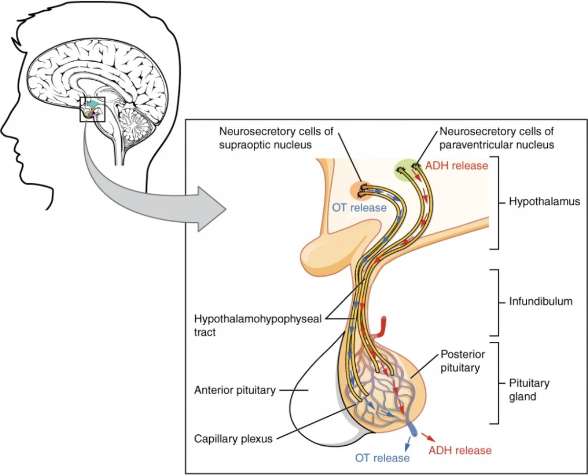
The posterior pituitary does not synthesize hormones. Hypothalamic neurons make oxytocin and ADH in their cell bodies, then transport them down axons for storage and release. Credit: Lumen Learning / OpenStax Anatomy and Physiology, CC BY 4.0
The posterior pituitary (neurohypophysis) is fundamentally different from the anterior pituitary. It does not synthesize any hormones. Instead, it stores and releases hormones that were made in the hypothalamus.
Neurons in the hypothalamus (specifically in the supraoptic and paraventricular nuclei) synthesize oxytocin and ADH in their cell bodies. These hormones travel down the axons of these neurons and are stored in the axon terminals within the posterior pituitary. When the neurons fire, the hormones are released directly into the bloodstream.
Oxytocin - stimulates uterine contractions during labor and milk ejection (letdown reflex) during breastfeeding. Also involved in social bonding and trust. During labor, oxytocin operates via a positive feedback loop: contractions push the baby’s head against the cervix, which signals more oxytocin release, which causes stronger contractions, which pushes harder on the cervix. The loop breaks when the baby is delivered and the stimulus is removed.
ADH (Antidiuretic Hormone) - also called vasopressin. Released in response to high blood osmolarity (detected by osmoreceptors in the hypothalamus) or low blood volume. ADH acts on the kidneys’ collecting ducts by binding to V2 receptors, which triggers insertion of aquaporin-2 channels into the cell membranes. This increases water reabsorption, concentrates the urine, and dilutes the blood. ADH also causes vasoconstriction (via V1 receptors on blood vessels), raising blood pressure. Alcohol inhibits ADH release - which is why drinking alcohol increases urine output and can lead to dehydration.
Gland
Hormones
Targets
Key Regulators
Anterior pituitary
FSH, LH
Gonads
GnRH (+), sex hormones (-)
Anterior pituitary
ACTH
Adrenal cortex
CRH (+), cortisol (-)
Anterior pituitary
TSH
Thyroid
TRH (+), T3/T4 (-)
Anterior pituitary
GH
Liver, bone, muscle
GHRH (+), somatostatin (-), IGF-1 (-)
Anterior pituitary
Prolactin
Mammary glands
Dopamine (-), suckling (+)
Anterior pituitary
Endorphins
Brain (opioid receptors)
Stress, exercise
Posterior pituitary
Oxytocin
Uterus, mammary glands
Cervical stretch, suckling
Posterior pituitary
ADH
Kidneys, blood vessels
High osmolarity (+), low volume (+)
A tumor compresses the pituitary stalk, severing the connection between the hypothalamus and pituitary. Which anterior pituitary hormone will paradoxically INCREASE?
Click to reveal answer
Prolactin. All other anterior pituitary hormones are primarily under stimulatory control and will decrease when the hypothalamic connection is lost. Prolactin is unique - it is under tonic inhibitory control by dopamine. Without dopamine from the hypothalamus, the inhibition is removed, and prolactin levels rise.
What is the key structural difference between how the anterior and posterior pituitary receive their hormonal instructions from the hypothalamus?
Click to reveal answer
The anterior pituitary receives releasing/inhibiting hormones via the hypophyseal portal system (a blood vessel network). The posterior pituitary receives hormones via direct neuronal axon terminals - hypothalamic neurons synthesize oxytocin and ADH in their cell bodies and transport them down axons into the posterior pituitary for storage and release.
Your thyroid gland sets the speed at which your entire body runs. Every cell in your body has receptors for thyroid hormones. When thyroid levels are high, your metabolism runs hot - your heart beats faster, you burn more calories, you feel wired and restless. When thyroid levels are low, everything slows - your heart rate drops, you gain weight, you feel sluggish and cold. The thyroid is the body’s thermostat dial, and the MCAT expects you to understand exactly how it is turned up and turned down.
Sitting right behind the thyroid, the four tiny parathyroid glands handle a completely different job: keeping blood calcium in an extremely narrow range. Calcium is not just for bones - it is essential for muscle contraction, nerve transmission, blood clotting, and enzyme function. Even small deviations in blood calcium can be life-threatening.
Thyroid Hormones: T3 and T4
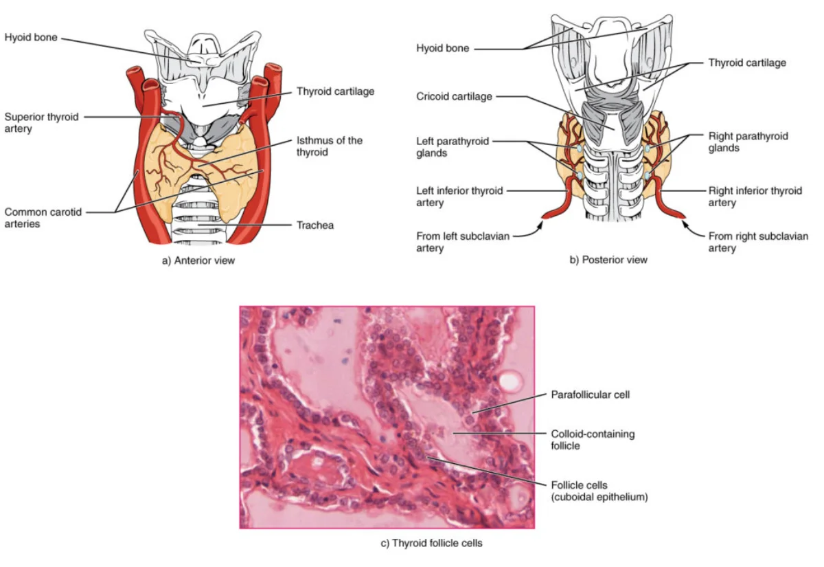
The thyroid gland wraps around the trachea. The histological inset shows follicles filled with colloid (thyroglobulin), where T3/T4 are stored before release. Credit: Lumen Learning / OpenStax Anatomy and Physiology, CC BY 4.0
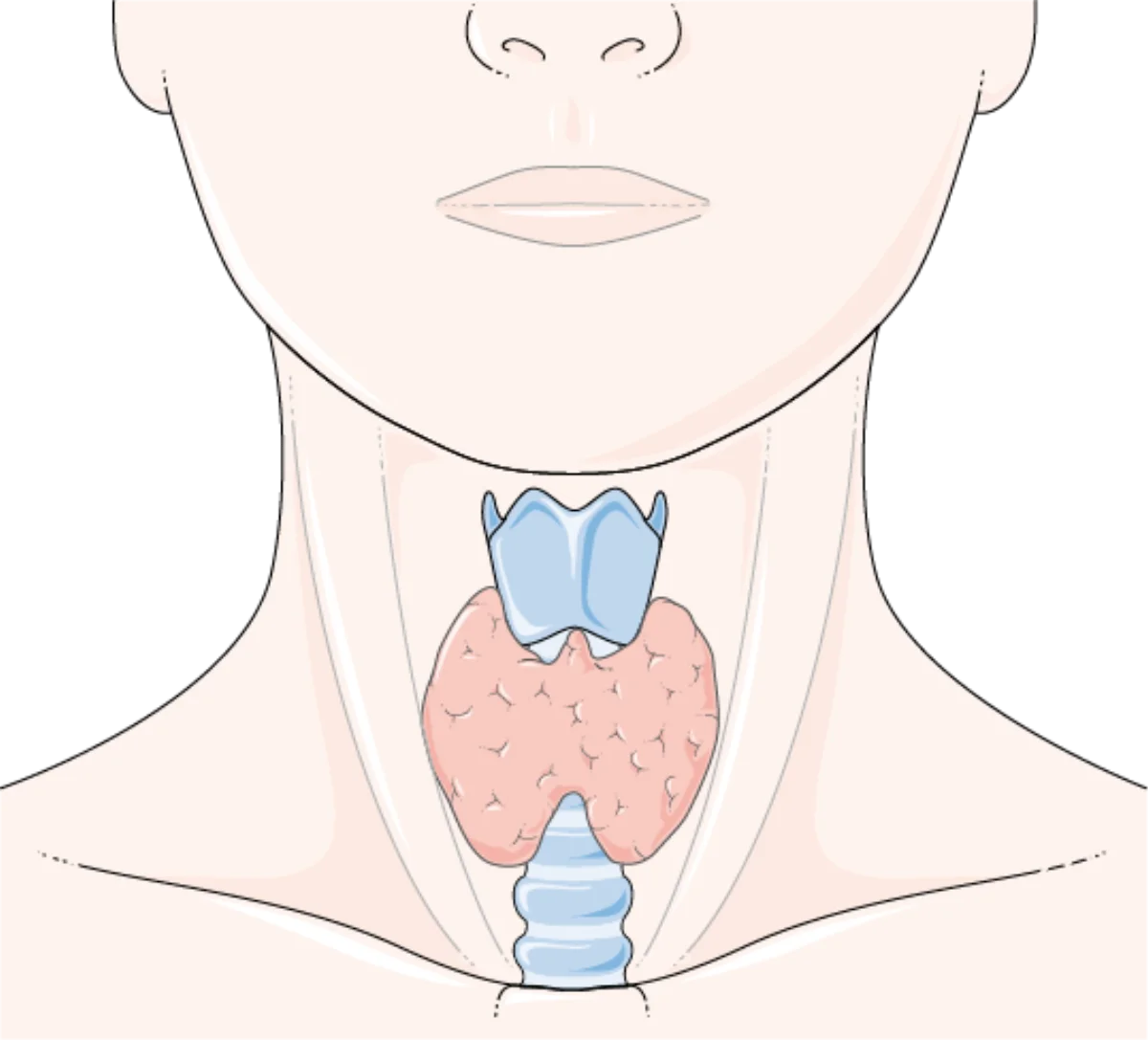
The thyroid gland is a butterfly-shaped gland located in the front of the neck, wrapped around the trachea. It produces two closely related hormones:
T4 (thyroxine) - contains four iodine atoms. T4 is the primary hormone released by the thyroid, but it is relatively inactive. Think of T4 as the “storage form.”
T3 (triiodothyronine) - contains three iodine atoms. T3 is the biologically active form - about 3-5 times more potent than T4. Most T3 is produced by conversion of T4 to T3 in peripheral tissues (primarily the liver and kidneys) by removing one iodine atom.
Both T3 and T4 are derived from the amino acid tyrosine, but they incorporate iodine during synthesis, making them lipid-soluble - unusual for amino acid derivatives. They travel in the blood bound to carrier proteins (thyroxine-binding globulin) and, once inside a target cell, bind to nuclear receptors to alter gene transcription.
Thyroid Hormone Regulation: The HPT Axis
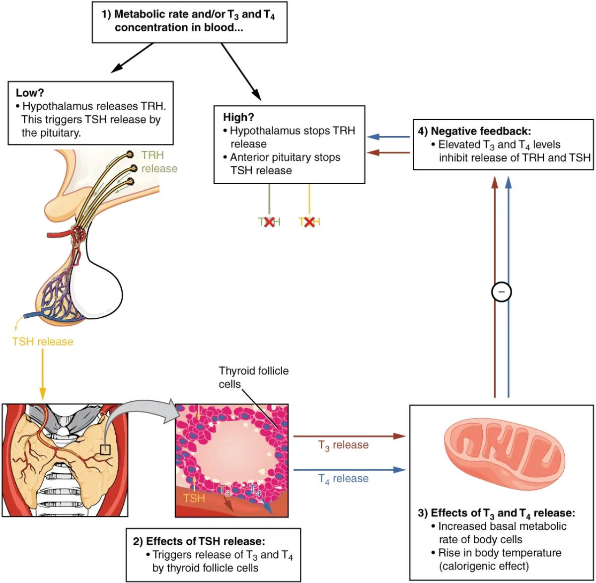
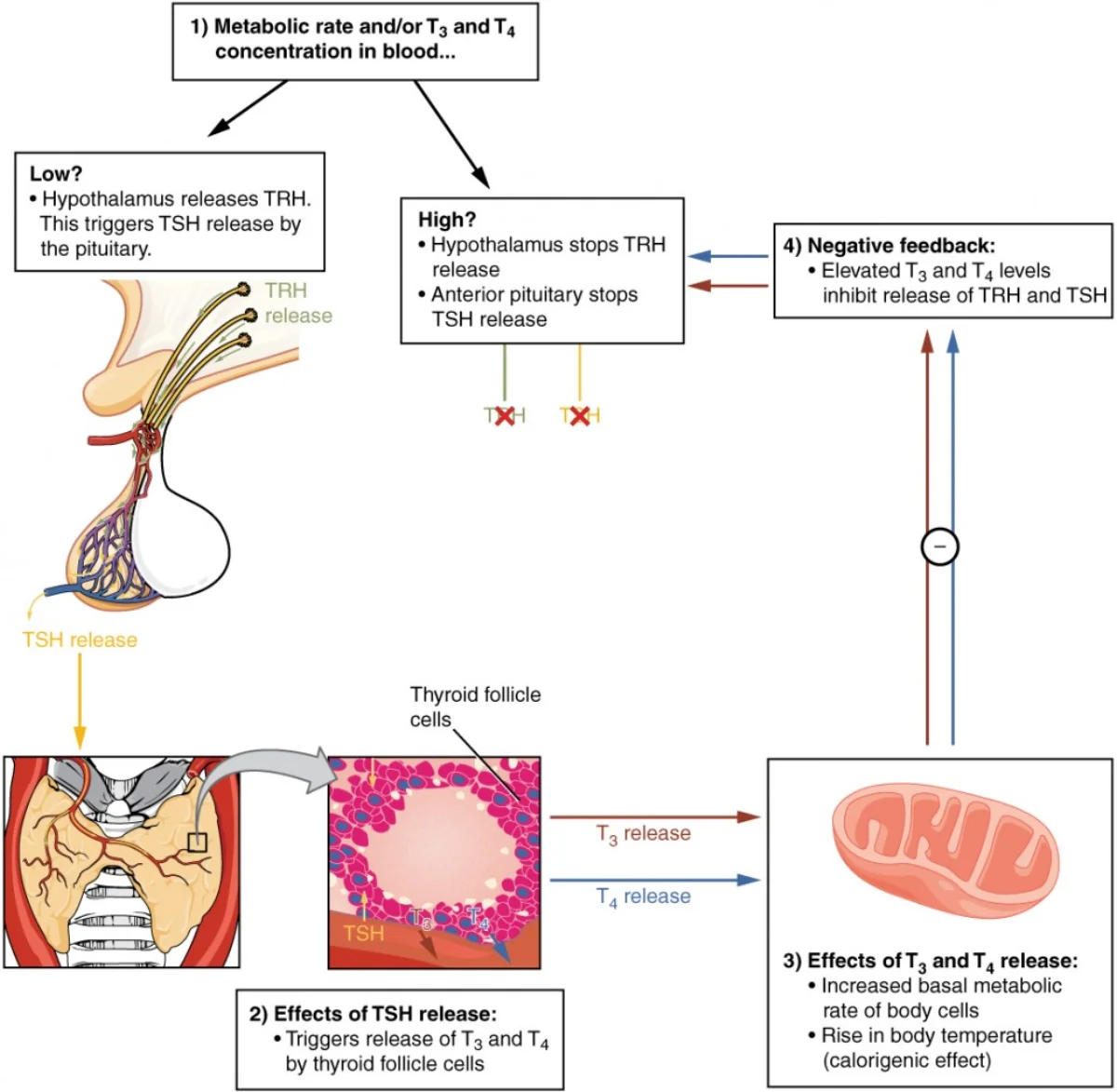
The HPT axis: TRH stimulates TSH, TSH stimulates T3/T4, and rising T3/T4 inhibit both TRH and TSH via negative feedback. Credit: OpenStax Anatomy and Physiology 2e, CC BY 4.0The butterfly-shaped thyroid gland wraps around the trachea in the front of the neck. Credit: Servier Medical Art, CC BY 4.0
The hypothalamic-pituitary-thyroid axis is the classic example of a negative feedback loop:
The hypothalamus detects low thyroid hormone levels and releases TRH (thyrotropin-releasing hormone)
TRH stimulates the anterior pituitary to release TSH (thyroid-stimulating hormone)
TSH stimulates the thyroid gland to produce and release T3 and T4
Rising T3/T4 levels inhibit both the hypothalamus (reducing TRH) and the anterior pituitary (reducing TSH)
With less TSH stimulation, the thyroid reduces hormone production
When T3/T4 levels drop, the inhibition is lifted and the cycle restarts
This feedback loop keeps thyroid hormones in a tight range. On the MCAT, you must be able to predict what happens at every level when one component is disrupted.
Effects of Thyroid Hormones
Thyroid hormones affect virtually every organ system:
Metabolism - increase basal metabolic rate, oxygen consumption, and heat production
Cardiovascular - increase heart rate and cardiac output
Nervous system - essential for brain development in fetuses and infants (deficiency during development causes cretinism - severe intellectual disability and growth failure); in adults, affect mood and alertness
Growth - required for normal growth and development (work synergistically with growth hormone)
GI tract - increase gut motility and nutrient absorption
Bone - promote bone remodeling
Calcitonin
The thyroid gland also produces calcitonin from parafollicular cells (C cells). Calcitonin has one job: lower blood calcium when it gets too high.
Calcitonin works by:
Stimulating osteoblasts (bone-building cells) to deposit calcium into bone
Inhibiting osteoclasts (bone-destroying cells) to prevent calcium release from bone
Increasing calcium excretion by the kidneys
Decreasing calcium absorption in the gut
Parathyroid Hormone (PTH)
Four tiny parathyroid glands sit on the posterior surface of the thyroid. They produce parathyroid hormone (PTH), which does the exact opposite of calcitonin: it raises blood calcium.
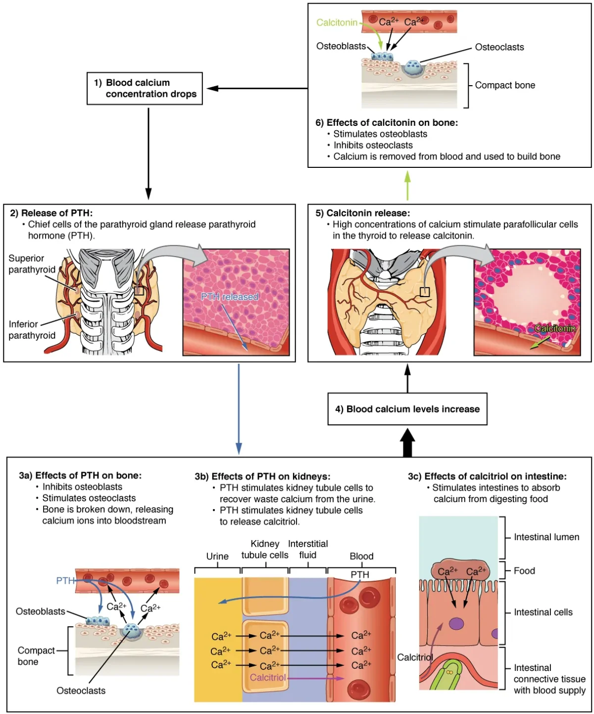
PTH is released when blood calcium drops too low (hypocalcemia). It raises calcium through three mechanisms:
Bone - stimulates osteoclasts to break down bone and release calcium into the blood (opposite of calcitonin)
Kidneys - increases calcium reabsorption (less calcium lost in urine) and stimulates activation of vitamin D (converting 25-hydroxyvitamin D to active calcitriol in the proximal tubule)
Gut - indirectly increases calcium absorption from food via calcitriol (active vitamin D)
When blood calcium is restored, elevated Ca2+ directly inhibits further PTH secretion from the parathyroid glands.
Calcitonin vs. PTH: The Calcium Seesaw
PTH and calcitonin work in opposition to maintain blood calcium in a narrow range. Credit: OpenStax Anatomy and Physiology 2e, CC BY 4.0
These two hormones work in opposition to keep blood calcium in its narrow optimal range (~8.5-10.5 mg/dL):
Feature
Calcitonin
PTH
Source
Thyroid C cells
Parathyroid glands
Released when
Ca2+ too HIGH
Ca2+ too LOW
Effect on blood Ca2+
Decreases
Increases
Effect on osteoblasts
Stimulates (build bone)
Inhibits
Effect on osteoclasts
Inhibits (stop breakdown)
Stimulates (break bone)
Kidney effect
Increases Ca2+ excretion
Decreases Ca2+ excretion, activates vitamin D
Gut effect
Decreases Ca2+ absorption
Increases Ca2+ absorption (via vitamin D)
Thyroid Disorders
Hyperthyroidism (overactive thyroid) - too much T3/T4. Symptoms: weight loss, rapid heart rate, anxiety, tremor, heat intolerance, increased sweating. The most common cause is Graves’ disease, an autoimmune condition where antibodies mimic TSH and stimulate the thyroid. In Graves’ disease, TSH levels are actually LOW (because high T3/T4 suppress TSH via negative feedback, but the antibodies bypass this control).
Hypothyroidism (underactive thyroid) - too little T3/T4. Symptoms: weight gain, fatigue, cold intolerance, constipation, depression, dry skin. Can be caused by iodine deficiency, autoimmune destruction (Hashimoto’s thyroiditis), or surgical removal. In primary hypothyroidism, TSH levels are HIGH (because low T3/T4 cannot suppress TSH).
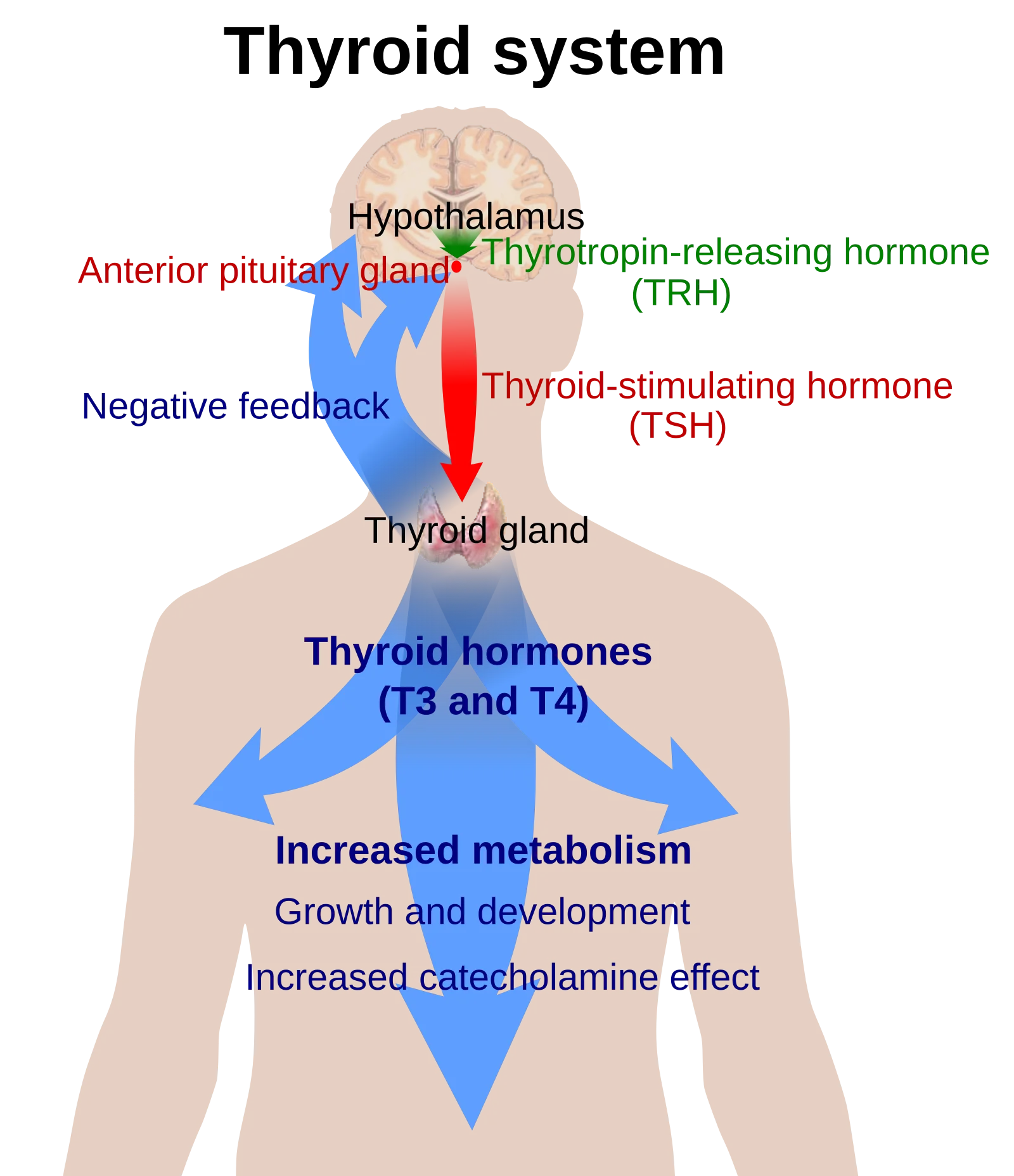
The HPT axis feedback loop. The hypothalamus releases TRH, which stimulates TSH from the anterior pituitary, which stimulates T3/T4 from the thyroid. T3/T4 feeds back to inhibit both TRH and TSH. In hypothyroidism, low T3/T4 means high TSH; in hyperthyroidism, high T3/T4 means low TSH. Credit: Wikimedia Commons, CC0 Public Domain
Goiter - enlargement of the thyroid gland. Can occur in both hyper- and hypothyroidism. In iodine deficiency, the thyroid enlarges as it tries to compensate for inadequate hormone production under constant TSH stimulation.
A patient has very high T4 levels but very low TSH. What is the most likely explanation?
Click to reveal answer
The thyroid is overproducing T4 independently of TSH stimulation (hyperthyroidism). The elevated T4 suppresses TSH via negative feedback on the pituitary. The most common cause is Graves' disease, where autoantibodies mimic TSH and directly stimulate the thyroid, bypassing normal pituitary control.
A patient's blood calcium is dangerously low. Which hormone is primarily responsible for correcting this, and what are its three mechanisms?
Click to reveal answer
PTH (parathyroid hormone) is the primary corrector. Its three mechanisms: (1) stimulates osteoclasts to release calcium from bone, (2) increases calcium reabsorption in the kidneys and activates vitamin D, (3) increases calcium absorption from the gut (indirectly via calcitriol/active vitamin D).
Sitting like small hats on top of each kidney, the adrenal glands are two organs in one. The outer layer (cortex) is an endocrine gland that produces steroid hormones on a timescale of hours to days. The inner core (medulla) is essentially a modified extension of the sympathetic nervous system that produces catecholamines for immediate fight-or-flight responses measured in seconds.
This dual identity makes the adrenal glands a bridge between chronic stress management and acute survival responses. The MCAT tests both layers extensively - and expects you to know exactly which hormones come from which zone.
Adrenal Cortex: Three Zones, Three Hormone Classes
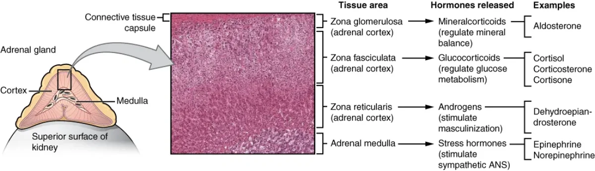
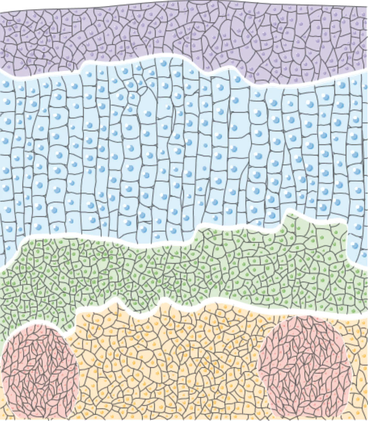
The adrenal gland has three cortical zones (each producing a different steroid class) surrounding the catecholamine-producing medulla. Credit: OpenStax Anatomy and Physiology 2e, CC BY 4.0The adrenal cortex wraps around the medulla in concentric layers. Credit: Servier Medical Art, CC BY 4.0
The adrenal cortex is organized into three concentric layers, each producing a different class of steroid hormone. All three classes are synthesized from cholesterol.
Interactive 3D Adrenal Gland. Rotate to see the three cortex zones (glomerulosa, fasciculata, reticularis) wrapping around the inner medulla.Credit: Nima via Sketchfab, CC BY
Zona Glomerulosa - Mineralocorticoids (Salt)
The outermost layer produces aldosterone, the primary mineralocorticoid. Aldosterone acts on the distal convoluted tubule and collecting duct of the kidney nephron to:
Increase sodium reabsorption (Na+ moves from urine back into blood)
Increase potassium secretion (K+ moves from blood into urine)
Increase water retention (water follows sodium osmotically)
The net effect: blood volume increases, blood pressure rises.
Aldosterone is primarily regulated by the renin-angiotensin-aldosterone system (RAAS), not by ACTH. When blood pressure drops or sodium levels fall, the kidneys release renin, which converts angiotensinogen to angiotensin I. ACE (angiotensin-converting enzyme, primarily in the lungs) converts angiotensin I to angiotensin II. Angiotensin II is a potent vasoconstrictor AND stimulates aldosterone release from the zona glomerulosa.
High potassium levels also directly stimulate aldosterone release - this is a safety mechanism to prevent dangerous hyperkalemia.
Zona Fasciculata - Glucocorticoids (Sugar)
The middle (and largest) layer produces cortisol, the primary glucocorticoid. Cortisol is the body’s long-term stress hormone, regulated by the HPA axis (CRH from hypothalamus - ACTH from anterior pituitary - cortisol from adrenal cortex).
Cortisol’s effects:
Raises blood glucose - stimulates gluconeogenesis in the liver, reduces glucose uptake in peripheral tissues
Protein catabolism - breaks down muscle protein to provide amino acid substrates for gluconeogenesis
Lipolysis - mobilizes fatty acids from adipose tissue
Immunosuppressive - chronic elevation weakens the immune system
Cardiovascular - increases vascular sensitivity to catecholamines, maintaining blood pressure
Cortisol follows a diurnal rhythm - highest in the early morning (preparing the body for the day) and lowest at night. Stress overrides this rhythm.
Zona Reticularis - Androgens (Sex)
The innermost cortical layer produces androgens - primarily DHEA (dehydroepiandrosterone) and androstenedione. These are weak sex hormones that serve as precursors. In peripheral tissues (especially the gonads and skin), they are converted into more potent forms: testosterone and estrogen.
In males, adrenal androgens are a minor source compared to testicular testosterone. In females, adrenal androgens are a significant source of circulating androgens and contribute to libido, pubic hair growth, and axillary hair growth.
Adrenal Medulla - Catecholamines (Fight or Flight)
The adrenal medulla is the inner core of the gland. It is not really a typical endocrine organ - it is a collection of modified postganglionic sympathetic neurons called chromaffin cells. These cells are directly innervated by preganglionic sympathetic neurons via the splanchnic nerves.
When the sympathetic nervous system is activated (stress, danger, exercise), acetylcholine from preganglionic neurons stimulates the chromaffin cells to release catecholamines directly into the bloodstream:
Epinephrine (adrenaline) - ~80% of medullary output
Norepinephrine (noradrenaline) - ~20% of medullary output
Both are derived from the amino acid tyrosine through the pathway: Tyrosine - L-DOPA - Dopamine - Norepinephrine - Epinephrine.
Redirects blood from skin and viscera to muscles and brain
Zone/Region
Hormone Class
Key Hormones
Primary Regulator
Main Effects
Zona Glomerulosa
Mineralocorticoids
Aldosterone
RAAS, K+ levels
Na+/H2O retention, K+ excretion
Zona Fasciculata
Glucocorticoids
Cortisol
ACTH (HPA axis)
Raise glucose, anti-inflammatory
Zona Reticularis
Androgens
DHEA, androstenedione
ACTH
Sex hormone precursors
Adrenal Medulla
Catecholamines
Epinephrine, norepinephrine
Sympathetic nervous system
Fight-or-flight response
A patient has elevated aldosterone but normal cortisol. Which zone of the adrenal cortex is most likely affected, and what electrolyte abnormalities would you expect?
Click to reveal answer
The zona glomerulosa (outermost layer) produces aldosterone. Excess aldosterone causes increased sodium reabsorption and potassium excretion - expect hypernatremia (high Na+), hypokalemia (low K+), and hypertension from water retention.
Why is the adrenal medulla considered a modified part of the sympathetic nervous system rather than a typical endocrine gland?
Click to reveal answer
The chromaffin cells of the adrenal medulla are embryologically derived from neural crest cells (same origin as sympathetic ganglia neurons). They are directly innervated by preganglionic sympathetic neurons and release catecholamines into the blood in response to acetylcholine - functioning as modified postganglionic sympathetic neurons that release their "neurotransmitters" into the bloodstream rather than onto a target organ.
You just finished a massive pasta dinner. Your blood glucose is spiking. Without intervention, that sugar would damage your blood vessels, nerves, and organs. But within minutes, your pancreas has detected the rise, released insulin, and directed cells throughout your body to absorb the excess glucose. By the time you feel sleepy on the couch, your blood sugar is already heading back to normal.
Now imagine it is 6 a.m. and you have not eaten in 10 hours. Your blood glucose is dropping. Your brain - which runs almost entirely on glucose - is at risk. Your pancreas detects the drop, releases glucagon, and your liver begins converting stored glycogen back into glucose, pushing your blood sugar back up.
This insulin-glucagon seesaw is one of the most elegant regulatory systems in the body - and one of the most heavily tested topics on the MCAT.
Dual Function: Exocrine and Endocrine
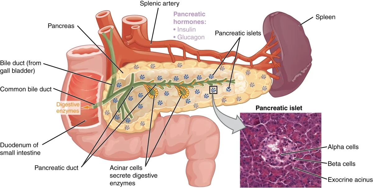
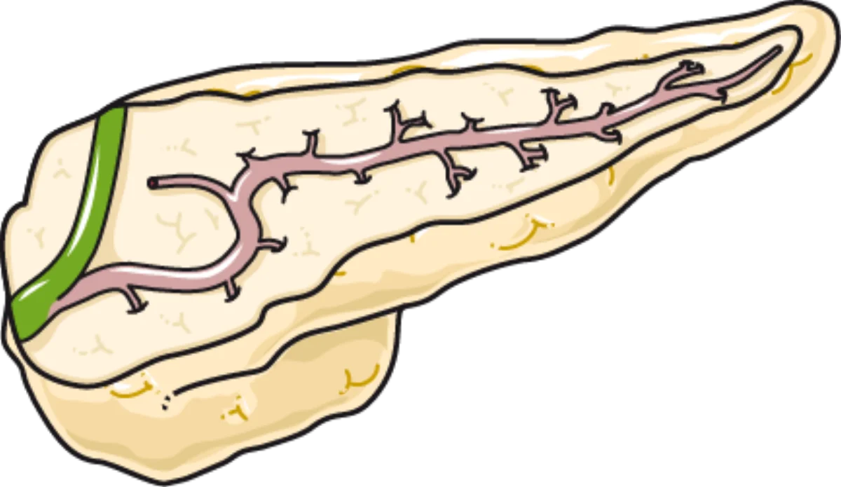
The pancreas is unique because it functions as both an exocrine and an endocrine gland. The exocrine portion (about 99% of the pancreas) consists of acinar cells that produce digestive enzymes (amylase, lipase, trypsin, chymotrypsin) and secrete them through the pancreatic duct into the duodenum. The digestive role of these enzymes is covered in detail in the pancreas section of the digestive system chapter.
The pancreatic islets of Langerhans contain alpha, beta, and delta cells, each producing a different hormone. Credit: OpenStax Anatomy and Physiology 2e, CC BY 4.0The pancreas is a dual-function organ with exocrine (digestive enzyme) and endocrine (hormone-producing) components. Credit: Servier Medical Art, CC BY 4.0
The endocrine portion consists of small clusters of hormone-producing cells called the islets of Langerhans (about 1% of pancreatic mass, but critical for metabolic regulation). Each islet contains several cell types:
Cell Type
Hormone
% of Islet
Function
Beta (B) cells
Insulin
~60-70%
Lowers blood glucose
Alpha (A) cells
Glucagon
~20-30%
Raises blood glucose
Delta (D) cells
Somatostatin
~5-10%
Inhibits insulin AND glucagon
PP (F) cells
Pancreatic polypeptide
~1-2%
Regulates pancreatic secretions
Epsilon cells
Ghrelin
<1%
Stimulates appetite
Insulin: The Storage Hormone
Insulin is a peptide hormone released by beta cells in response to elevated blood glucose (the primary stimulus). It is also stimulated by elevated amino acids, certain GI hormones (incretins like GLP-1), and parasympathetic stimulation (via the vagus nerve during eating).
Insulin’s key metabolic effects:
In the liver:
Stimulates glycogenesis (glucose to glycogen storage)
Inhibits gluconeogenesis (prevents new glucose production)
Promotes lipogenesis (fatty acid synthesis)
In muscle:
Increases glucose uptake by promoting GLUT4 transporter insertion into the cell membrane
Stimulates glycogenesis and protein synthesis
In adipose tissue:
Increases glucose uptake via GLUT4
Stimulates lipogenesis (glucose to triglycerides)
Inhibits lipolysis (prevents fat breakdown)
The net effect: blood glucose drops as glucose is taken up by cells and stored as glycogen or fat.
Glucagon: The Mobilization Hormone
Glucagon is a peptide hormone released by alpha cells in response to low blood glucose (the primary stimulus). It is also stimulated by elevated amino acids (which makes physiological sense - a high-protein, low-carb meal needs glucagon to prevent hypoglycemia) and by epinephrine during stress.
Glucagon’s effects are essentially the opposite of insulin, and its primary target is the liver:
Stimulates glycogenolysis (glycogen breakdown to glucose)
Stimulates gluconeogenesis (production of new glucose from amino acids, lactate, and glycerol)
Stimulates lipolysis (fat breakdown, releasing fatty acids and glycerol)
Stimulates ketogenesis (conversion of fatty acids to ketone bodies during prolonged fasting)
The net effect: blood glucose rises as the liver dumps glucose into the bloodstream.
Somatostatin: The Brake Pedal
Somatostatin from delta cells inhibits both insulin and glucagon secretion. It also inhibits the release of growth hormone from the anterior pituitary (where it is called Growth Hormone-Inhibiting Hormone, or GHIH). In the GI tract, somatostatin slows digestion by reducing gut motility, enzyme secretion, and nutrient absorption.
Somatostatin prevents wild swings in blood glucose by dampening both sides of the seesaw simultaneously. It is released after meals when GI hormone levels and nutrient concentrations are high.
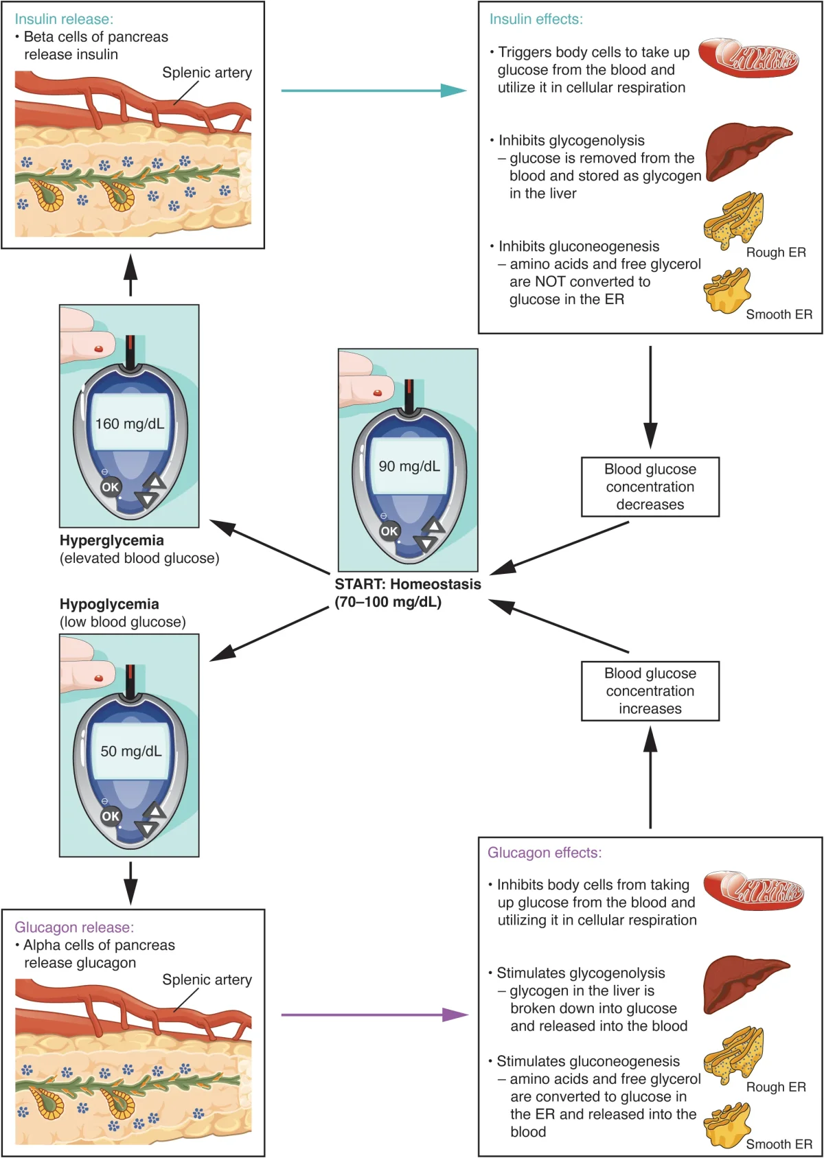
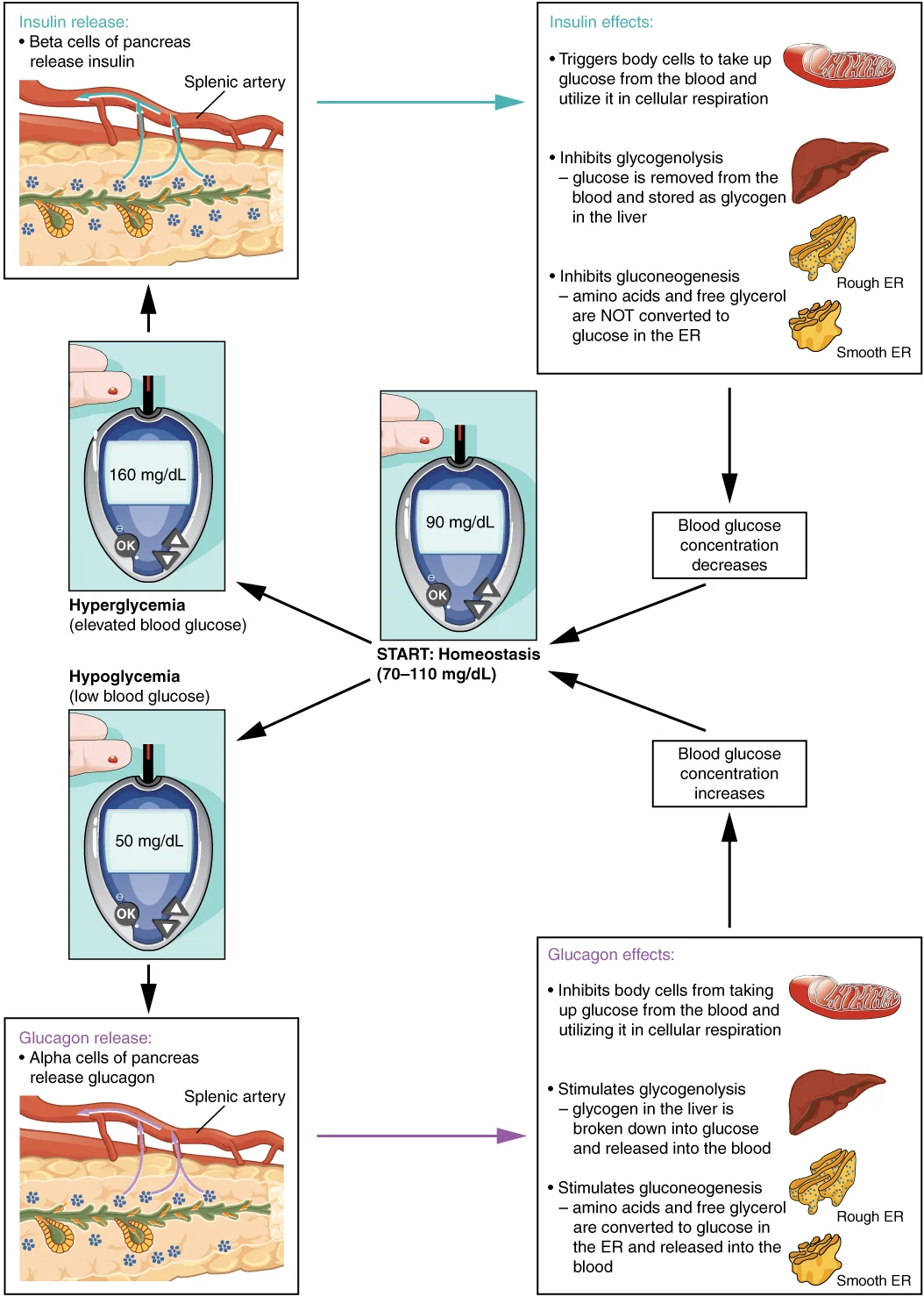
The insulin-glucagon seesaw maintains blood glucose within a narrow range through opposing metabolic effects. Credit: OpenStax Anatomy and Physiology 2e, CC BY 4.0
Blood glucose homeostasis. After a meal, high glucose triggers insulin from beta cells (promotes uptake and glycogenesis). During fasting, low glucose triggers glucagon from alpha cells (promotes glycogenolysis and gluconeogenesis). Credit: OpenStax College, CC BY 3.0
Type 1 Diabetes - autoimmune destruction of beta cells. The body produces little to no insulin. Blood glucose stays high because cells cannot take it up. Patients require exogenous insulin injections. Onset typically in childhood/adolescence.
Type 2 Diabetes - target cells become resistant to insulin. The pancreas initially compensates by producing more insulin, but over time the beta cells become exhausted and insulin production declines. Associated with obesity, physical inactivity, and genetic factors. Most common form (~90-95% of diabetes cases). Managed with lifestyle changes, oral medications that improve insulin sensitivity, and sometimes insulin.
During a 24-hour fast, which hormone predominates and what two hepatic pathways does it activate to maintain blood glucose?
Click to reveal answer
Glucagon predominates during fasting. It activates (1) glycogenolysis (breaking down liver glycogen into glucose) and (2) gluconeogenesis (synthesizing new glucose from amino acids, lactate, and glycerol). As the fast extends, glycogen stores deplete and gluconeogenesis becomes the primary source of blood glucose.
Why does insulin promote GLUT4 transporter insertion specifically in muscle and adipose tissue, but not in the brain or liver?
Click to reveal answer
The brain uses GLUT1 and GLUT3 transporters, which are constitutively expressed (always present) and do not require insulin. This ensures the brain always has access to glucose, even during fasting. The liver uses GLUT2, which is also insulin-independent but has a high Km - it takes up glucose passively in proportion to blood glucose concentration. GLUT4 (insulin-dependent) is specific to muscle and adipose tissue, making these tissues "insulin-sensitive."
The gonads - testes in males and ovaries in females - are the body’s reproductive endocrine organs. They produce gametes (sperm and eggs) and secrete sex hormones that drive puberty, maintain reproductive function, and influence everything from bone density to mood. The MCAT frequently tests the hypothalamic-pituitary-gonadal (HPG) axis and the feedback loops that regulate these hormones.
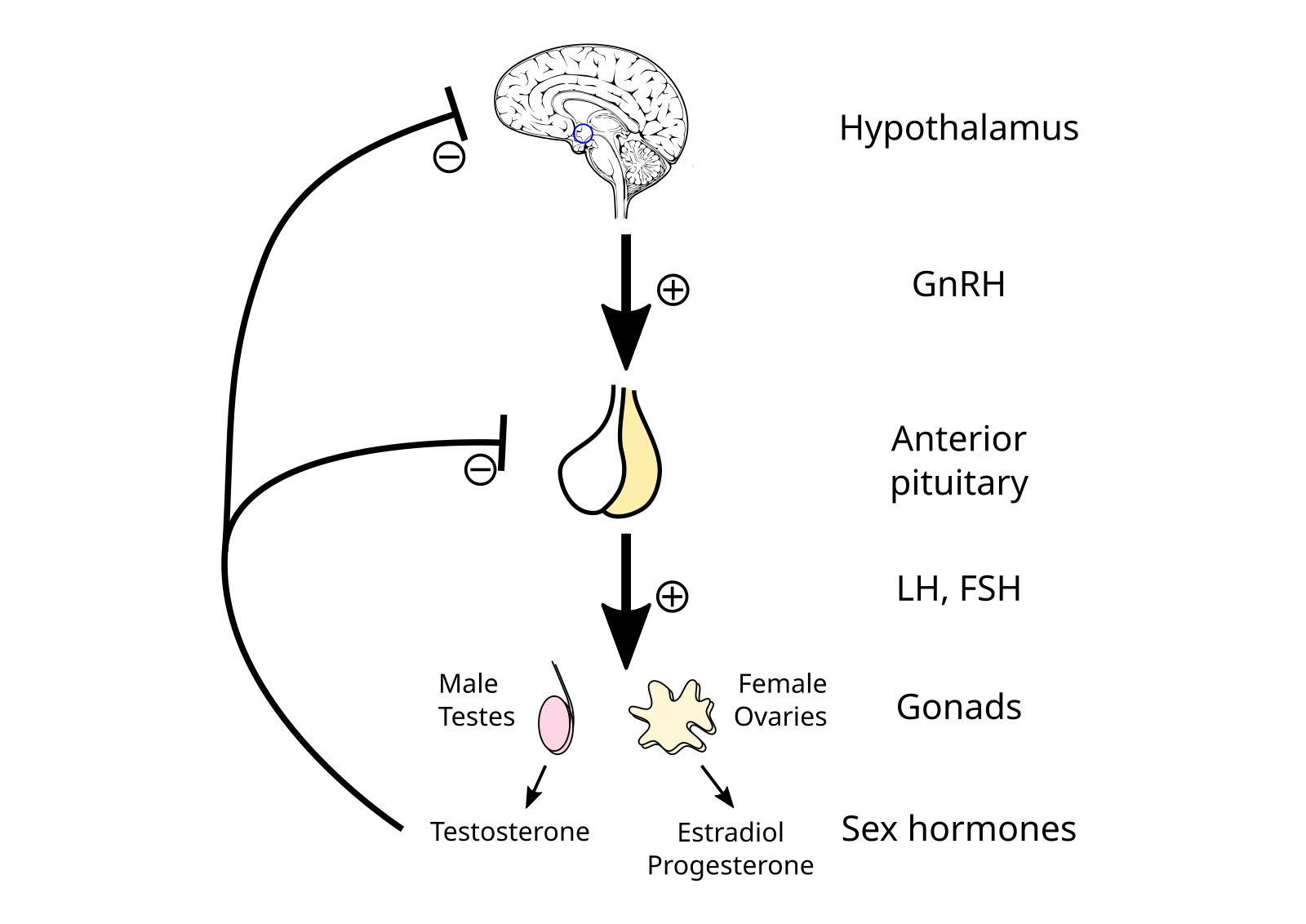
The hypothalamic-pituitary-gonadal (HPG) axis. GnRH → FSH + LH → gonads → sex hormones (testosterone or estradiol/progesterone), with negative feedback to hypothalamus and pituitary. Credit: Wikimedia Commons, CC BY 3.0
The HPG Axis: Chain of Command
The regulation of gonadal hormones follows the same hierarchical pattern as the thyroid and adrenal axes:
Hypothalamus releases GnRH (gonadotropin-releasing hormone) in a pulsatile pattern
GnRH stimulates the anterior pituitary to release FSH and LH
FSH and LH act on the gonads to stimulate gamete production and hormone secretion
Sex hormones feed back to inhibit the hypothalamus and pituitary (negative feedback)
The pulsatile nature of GnRH is critical. Constant, non-pulsatile GnRH actually downregulates LH and FSH receptors and suppresses gonadotropin release. This is the pharmacological basis for GnRH agonists used in certain treatments - continuous administration paradoxically shuts down the axis.
Testes: Testosterone and Spermatogenesis
The testes are located in the scrotum, which maintains a temperature 2-3 degrees C below core body temperature - optimal for sperm production. Two key cell types produce the two main testicular outputs:
Leydig cells (interstitial cells) - located between the seminiferous tubules. LH stimulates Leydig cells to produce testosterone. Testosterone is a steroid hormone derived from cholesterol.
Sertoli cells (sustentacular cells) - located inside the seminiferous tubules. FSH acts on Sertoli cells to support spermatogenesis. Sertoli cells also produce inhibin, which selectively feeds back to inhibit FSH release from the anterior pituitary (without significantly affecting LH).
Testosterone effects:
Development of male reproductive organs
Secondary sexual characteristics (facial hair, deepened voice, muscle mass, body hair)
Maintains spermatogenesis (works alongside FSH)
Anabolic effects on muscle and bone
Influences libido
Negative feedback on GnRH and LH
Ovaries: Estrogen, Progesterone, and the Menstrual Cycle
The ovaries contain follicles at various stages of development, each housing an oocyte (egg). Two key cell types in the follicle produce ovarian hormones:
Theca cells - the outer layer of the follicle. LH stimulates theca cells to produce androgens (androstenedione), which are then passed to granulosa cells.
Granulosa cells - the inner layer surrounding the oocyte. FSH stimulates granulosa cells to convert the androgens from theca cells into estrogen (primarily estradiol) via the enzyme aromatase. After ovulation, granulosa cells become part of the corpus luteum, which produces progesterone.
This two-cell model (theca + granulosa) is an important MCAT concept: neither cell type alone can produce estrogen efficiently. They must cooperate.
Estrogen effects:
Development of female secondary sexual characteristics (breast development, wider hips, fat distribution)
Stimulates endometrial growth during the follicular phase
Stimulates LH surge at mid-cycle (positive feedback - a rare exception to the usual negative feedback)
Maintains bone density (stimulates osteoblast activity)
Negative feedback on FSH and GnRH (at moderate levels)
Progesterone effects:
Maintains and stabilizes the endometrial lining for implantation
Inhibits uterine contractions during pregnancy
Raises body temperature slightly (basal body temperature rises after ovulation - used to detect ovulation)
Negative feedback on GnRH, FSH, and LH during the luteal phase
Key Feedback Patterns
In males, testosterone provides straightforward negative feedback - high testosterone suppresses GnRH and LH, keeping the system stable.
In females, the feedback is more complex:
Low-to-moderate estrogen (follicular phase) - negative feedback on FSH and LH
High estrogen (just before ovulation) - switches to positive feedback on LH, triggering the massive LH surge that causes ovulation
After ovulation - the corpus luteum produces progesterone, which returns the system to negative feedback on GnRH, FSH, and LH
This estrogen-mediated switch from negative to positive feedback at mid-cycle is one of the few examples of positive feedback in the endocrine system, and it is a high-yield MCAT concept.
Feature
Testes
Ovaries
Gamete-producing cells
Spermatogonia (in seminiferous tubules)
Oocytes (in follicles)
Hormone-producing cells
Leydig (testosterone), Sertoli (inhibin)
Theca (androgens), Granulosa (estrogen, inhibin)
LH target
Leydig cells
Theca cells, triggers ovulation
FSH target
Sertoli cells
Granulosa cells
Primary hormones
Testosterone
Estrogen, progesterone
Negative feedback to pituitary
Testosterone, inhibin
Estrogen, progesterone, inhibin
In the ovarian two-cell model, what does each cell type contribute and which gonadotropin stimulates each?
Click to reveal answer
Theca cells (stimulated by LH) produce androgen precursors (androstenedione). Granulosa cells (stimulated by FSH) convert these androgens to estrogen via the enzyme aromatase. Both cell types and both gonadotropins are required for estrogen synthesis.
Estrogen usually inhibits LH release via negative feedback. Under what specific condition does estrogen switch to positive feedback, and what is the physiological result?
Click to reveal answer
When estrogen reaches a high sustained level (from the dominant follicle in the late follicular phase), it switches to positive feedback on the anterior pituitary, triggering a massive LH surge. This LH surge causes ovulation - the release of the mature oocyte from the follicle. After ovulation, progesterone from the corpus luteum restores negative feedback.
The classic endocrine glands - pituitary, thyroid, adrenals, pancreas, and gonads - get most of the attention. But several other organs also produce hormones that are fair game on the MCAT. These “part-time” endocrine organs have primary functions unrelated to hormone production, yet the hormones they release are critical for homeostasis.
The Pineal Gland: Your Internal Clock
The pineal gland is a tiny, pea-sized structure deep in the brain between the two hemispheres. Its primary product is melatonin, synthesized from the amino acid tryptophan (via serotonin as an intermediate).
Melatonin regulates circadian rhythms - your body’s internal clock for sleep-wake cycles. Production is controlled by light:
Darkness stimulates melatonin synthesis (signals from the retina travel to the suprachiasmatic nucleus of the hypothalamus, which signals the pineal gland)
Light inhibits melatonin production
Melatonin promotes sleepiness, and its levels peak in the middle of the night. It also influences seasonal reproductive patterns in some animals and may have antioxidant properties.
The Thymus: Immune Training Center
The thymus is located in the upper chest behind the sternum. It is largest during childhood and gradually shrinks (involutes) after puberty. The thymus produces thymosin and other thymic hormones that are essential for the maturation and differentiation of T lymphocytes (T cells).
Immature T cells migrate from the bone marrow to the thymus, where they undergo selection - learning to distinguish self from non-self. Without a functional thymus, the immune system cannot mount effective adaptive immune responses. This is why the thymus is most active in childhood, when the immune system is being “educated.”
The Kidneys: Three Endocrine Functions
Beyond filtering blood, the kidneys produce three hormones:
1. Renin - an enzyme-hormone released by the juxtaglomerular apparatus (JGA) in response to low blood pressure, low sodium, or sympathetic stimulation. Renin initiates the RAAS cascade: renin converts angiotensinogen (from the liver) to angiotensin I, which ACE (in the lungs) converts to angiotensin II. Angiotensin II causes vasoconstriction and stimulates aldosterone release, both raising blood pressure.
2. Erythropoietin (EPO) - released by interstitial fibroblasts in the kidney cortex in response to hypoxia (low blood oxygen). EPO stimulates the bone marrow to produce more red blood cells, increasing the blood’s oxygen-carrying capacity. This is why chronic kidney disease often leads to anemia - the kidneys cannot produce adequate EPO.
3. Calcitriol (active vitamin D) - the kidneys convert 25-hydroxyvitamin D to 1,25-dihydroxyvitamin D (calcitriol) in the proximal convoluted tubule. This conversion is stimulated by PTH. Calcitriol increases calcium and phosphate absorption from the GI tract and supports bone mineralization.
The Heart: ANP and BNP
The heart is not just a pump - it is also an endocrine organ. When the atria are stretched by increased blood volume, atrial cardiomyocytes release ANP (atrial natriuretic peptide). The ventricles release a related peptide, BNP (brain natriuretic peptide), when stretched.
ANP’s effects are essentially the opposite of aldosterone:
Increases sodium excretion (natriuresis)
Increases water excretion (diuresis)
Inhibits aldosterone secretion
Inhibits ADH secretion
Dilates blood vessels
The net effect: blood volume decreases, blood pressure drops. ANP is the body’s defense against volume overload.
Hormone Pair
Raises Blood Pressure
Lowers Blood Pressure
Aldosterone vs. ANP
Aldosterone (Na+ retention)
ANP (Na+ excretion)
ADH vs. ANP
ADH (water retention)
ANP (water excretion)
The GI Tract: Digestive Hormones
The gastrointestinal tract is the largest endocrine organ in the body by cell count. Key GI hormones for the MCAT:
Gastrin - released by G cells in the stomach in response to food. Stimulates gastric acid (HCl) secretion and gastric motility.
Secretin - released by S cells in the duodenum in response to acidic chyme. Stimulates bicarbonate secretion from the pancreas to neutralize acid.
CCK (cholecystokinin) - released by I cells in the duodenum in response to fats and proteins. Stimulates bile release from the gallbladder and digestive enzyme secretion from the pancreas. Also promotes satiety (feeling full).
GIP (glucose-dependent insulinotropic peptide) - released in response to glucose and fats. Stimulates insulin release from beta cells (an “incretin” effect - oral glucose stimulates more insulin than IV glucose because of GI hormones).
Appetite Hormones
Leptin - produced by adipose tissue (fat cells). Leptin signals satiety to the hypothalamus - “you have enough fat stored, reduce appetite.” Leptin levels correlate with body fat mass. In obesity, leptin levels are high, but the brain becomes resistant to the signal (similar to insulin resistance in Type 2 diabetes).
Ghrelin - produced primarily by the stomach. Ghrelin stimulates appetite - “your stomach is empty, time to eat.” Ghrelin levels rise before meals and drop after eating.
A patient at high altitude develops increased red blood cell production over several weeks. What hormone mediates this response, where is it produced, and what stimulus triggers its release?
Click to reveal answer
Erythropoietin (EPO), produced by interstitial fibroblasts in the kidney cortex, is released in response to hypoxia (low blood oxygen levels). At high altitude, reduced atmospheric oxygen causes chronic hypoxia, stimulating EPO release, which stimulates bone marrow to increase red blood cell production over days to weeks.
ANP and aldosterone have opposing effects on sodium balance. Which hormone promotes sodium retention and which promotes sodium excretion? What triggers the release of each?
Click to reveal answer
Aldosterone (from adrenal cortex zona glomerulosa) promotes sodium retention - triggered by angiotensin II and high potassium levels. ANP (from atrial cardiomyocytes) promotes sodium excretion - triggered by atrial stretch from increased blood volume. They balance blood pressure from opposite directions.
Feedback loops are the operating system of the endocrine system. Without them, hormone levels would spiral out of control - a little too much cortisol would trigger more cortisol, which would trigger even more, until the system self-destructed. Instead, the body uses feedback to keep every hormone within a precise range.
The MCAT tests feedback loops constantly. You need to be able to predict what happens at every level of a hormonal axis when one component is blocked, destroyed, or overproduced.
Negative Feedback: The Thermostat Model
Negative feedback is the dominant control mechanism in the endocrine system. The principle is simple: the output of a process inhibits the process itself. When the product builds up, production slows down. When the product drops, production speeds up.
Major Negative Feedback Loops
The classic negative feedback loop: the effector's output reverses the original stimulus, maintaining homeostasis. This pattern applies to cortisol, thyroid hormones, blood glucose, calcium, and body temperature regulation. Credit: Lumen Learning / OpenStax Anatomy and Physiology, CC BY 4.0
HPA Axis (Cortisol Regulation)
Stress triggers the hypothalamus to release CRH
CRH stimulates the anterior pituitary to release ACTH
ACTH stimulates the adrenal cortex to produce cortisol
Cortisol feeds back to inhibit both CRH (hypothalamus) and ACTH (anterior pituitary)
Result: cortisol levels rise during stress, then return to baseline as the feedback kicks in
HPT Axis (Thyroid Regulation)
Low thyroid hormones prompt the hypothalamus to release TRH
TRH stimulates TSH from the anterior pituitary
TSH stimulates T3/T4 production from the thyroid
Rising T3/T4 inhibit TRH and TSH
Result: thyroid hormones are maintained in a tight range
Blood Glucose Regulation
High glucose after a meal triggers insulin from beta cells
Insulin drives glucose into cells, lowering blood glucose
Falling glucose reduces the stimulus for insulin secretion
Low glucose triggers glucagon from alpha cells
Glucagon raises blood glucose by stimulating the liver
Rising glucose reduces the stimulus for glucagon
Calcium Homeostasis
Low blood Ca2+ triggers PTH release from parathyroid glands
PTH raises Ca2+ (bone resorption, kidney reabsorption, vitamin D activation)
Rising Ca2+ inhibits further PTH secretion
High blood Ca2+ triggers calcitonin from thyroid C cells
Calcitonin deposits Ca2+ into bone, lowering blood levels
Water Balance (ADH)
High blood osmolarity (dehydration) triggers ADH release from posterior pituitary
ADH increases water reabsorption in collecting ducts, diluting the blood
As osmolarity normalizes, ADH secretion decreases
Predicting Disruptions: The MCAT Strategy
The MCAT loves to present scenarios where one component of a feedback loop is damaged and asks you to predict the consequences. Here is the systematic approach:
Step 1: Identify the axis (HPA, HPT, HPG, etc.) Step 2: Determine which level is affected (hypothalamus, pituitary, or target gland) Step 3: Trace the consequences up and down the axis
Example: Primary hypothyroidism (thyroid gland is damaged)
Thyroid produces less T3/T4
Less T3/T4 means less negative feedback on the pituitary and hypothalamus
Without feedback inhibition, TRH and TSH increase
Result: Low T3/T4, HIGH TSH (“primary” = the problem is at the gland level)
Example: Secondary hypothyroidism (pituitary is damaged)
Pituitary produces less TSH
Less TSH means less thyroid stimulation
Thyroid produces less T3/T4
But TRH from hypothalamus increases (trying to compensate)
Result: Low T3/T4, LOW TSH (“secondary” = the problem is at the pituitary level)
Positive Feedback: The Rare Amplifier
Positive feedback is the opposite of negative feedback: the output amplifies the process rather than inhibiting it. This creates an escalating cycle that continues until an external event breaks the loop. Positive feedback is rare in the endocrine system because runaway amplification is inherently dangerous.
Key positive feedback examples for the MCAT:
1. Oxytocin during labor
Baby’s head pushes against the cervix
Cervical stretch signals the hypothalamus to release more oxytocin
Oxytocin causes stronger uterine contractions
Stronger contractions push the baby harder against the cervix
More cervical stretch triggers even more oxytocin
Loop breaks when the baby is delivered (stimulus removed)
2. Estrogen and the LH surge
Rising estrogen from the dominant follicle reaches a threshold
High sustained estrogen switches from negative to positive feedback on the anterior pituitary
LH surge occurs
LH surge triggers ovulation
After ovulation, the corpus luteum produces progesterone, which restores negative feedback
Loop breaks when ovulation occurs
3. Prolactin during breastfeeding
Suckling stimulates nerve endings in the nipple
Signals to the hypothalamus reduce dopamine release
Less dopamine means less inhibition of prolactin
More prolactin means more milk production
More milk encourages more suckling
Loop breaks when the infant stops nursing
4. Blood clotting cascade (not endocrine, but a classic positive feedback example)
Activated platelets release signals that activate more platelets
The clot grows until the vessel is sealed
Negative vs. Positive Feedback Summary
Feature
Negative Feedback
Positive Feedback
Effect
Output inhibits the stimulus
Output amplifies the stimulus
Goal
Maintain homeostasis (stability)
Drive a process to completion
Frequency
Extremely common
Rare
Duration
Continuous
Self-limiting (needs external break)
Examples
Cortisol, thyroid, insulin/glucagon, PTH/Ca2+
Oxytocin in labor, LH surge, prolactin in lactation
A patient has low cortisol and HIGH ACTH. Is this a primary or secondary adrenal problem? Explain the feedback logic.
Click to reveal answer
This is PRIMARY adrenal insufficiency (Addison's disease). The adrenal cortex cannot produce cortisol. Low cortisol means no negative feedback on the pituitary, so ACTH rises as the pituitary tries (unsuccessfully) to stimulate the damaged adrenals. Primary = gland is broken, tropic hormone is high.
Name three examples of positive feedback in the endocrine system and identify what breaks each loop.
Click to reveal answer
(1) Oxytocin during labor - broken by delivery of the baby (cervical stretch stimulus removed). (2) Estrogen-driven LH surge - broken by ovulation (dominant follicle ruptures, progesterone restores negative feedback). (3) Prolactin during breastfeeding - broken when the infant stops nursing (suckling stimulus removed, dopamine inhibition resumes).
The MCAT does not ask you to diagnose patients, but it regularly presents endocrine disorders in passage-based questions to test whether you understand the underlying physiology. The strategy is always the same: identify which hormone is too high or too low, then reason through the feedback loop to predict the consequences.
Every disorder in this section can be understood by asking three questions:
Which hormone is abnormal?
Is it too much (hyper) or too little (hypo)?
Where in the axis is the problem - gland, pituitary, or hypothalamus?
Growth Hormone Disorders
Gigantism - GH excess before epiphyseal plate closure (in children). Excess GH/IGF-1 drives excessive linear growth.
Acromegaly - GH excess after plate closure (in adults). Long bones can no longer lengthen, so bones thicken instead (hands, feet, face).
Dwarfism - GH deficiency (or GH-receptor insensitivity) in childhood. Produces proportional short stature, unlike the disproportionate short stature of achondroplasia.
Adrenal Disorders
Cushing Syndrome - cortisol excess. Predictable from cortisol’s actions: hyperglycemia (gluconeogenesis), central fat deposition, muscle wasting (protein catabolism), hypertension, and immunosuppression. Causes include a pituitary adenoma (high ACTH), an autonomous adrenal tumor (low ACTH), or exogenous steroids.
Addison Disease - primary adrenal insufficiency. The adrenal cortex is destroyed, so both cortisol and aldosterone drop. Loss of cortisol feedback drives ACTH very high, which also drives hyperpigmentation (ACTH and MSH share a precursor). Expect low Na⁺, high K⁺ (no aldosterone), low glucose, and low blood pressure.
Thyroid Disorders (Detailed)
The HPT axis. Understanding this feedback loop is key to predicting lab values in thyroid disorders: Graves disease (high T3/T4, low TSH) vs. Hashimoto's (low T3/T4, high TSH). Credit: Wikimedia Commons, CC0 Public Domain
Graves Disease - autoimmune hyperthyroidism. Antibodies mimic TSH and continuously stimulate the thyroid. Labs: high T3/T4, LOW TSH (suppressed by feedback, but the antibodies bypass that control).
Hashimoto Thyroiditis - autoimmune destruction of the thyroid. Labs: low T3/T4, HIGH TSH (pituitary trying to rescue a damaged gland).
Iodine Deficiency Goiter - no iodine means no T3/T4. TSH stays elevated, chronically stimulating the thyroid, which enlarges into a visible goiter.
Water Balance Disorders
SIADH (Syndrome of Inappropriate ADH Secretion) - too much ADH. The kidneys reabsorb too much water, diluting the blood. Result: hyponatremia (dangerously low sodium from dilution), concentrated urine, water retention. Can be caused by certain tumors, drugs, or CNS disorders.
Diabetes Insipidus - too little ADH effect. Two types:
Central DI: hypothalamus/posterior pituitary does not produce enough ADH
Nephrogenic DI: kidneys do not respond to ADH (receptor or aquaporin defect)
Both produce the same symptoms: enormous volumes of very dilute urine (the patient “insipidly” wastes water), extreme thirst, and risk of dehydration. Note: “diabetes” here means “excessive urination” (from Greek “to pass through”) - this has nothing to do with blood sugar or insulin.
Aldosterone Disorders
Hypoaldosteronism - aldosterone deficiency. Causes sodium loss, potassium retention (hyperkalemia), low blood volume, and hypotension. Often seen as part of Addison disease.
Catecholamine Disorder
Master Disorder Table
Disorder
Hormone Problem
Key Lab Findings
Key Symptoms
Cushing Syndrome
Cortisol excess
High cortisol, variable ACTH
Moon face, central obesity, hyperglycemia
Addison Disease
Cortisol + aldosterone deficiency
Low cortisol, HIGH ACTH
Hypotension, hyperpigmentation, fatigue
Graves Disease
T3/T4 excess
High T3/T4, LOW TSH
Weight loss, tachycardia, exophthalmos
Hashimoto Thyroiditis
T3/T4 deficiency
Low T3/T4, HIGH TSH
Weight gain, fatigue, cold intolerance
Acromegaly/Gigantism
GH excess
High GH, high IGF-1
Enlarged extremities (acromegaly) or tall stature (gigantism)
Type 1 Diabetes
Insulin deficiency
High glucose, low insulin, low C-peptide
Polyuria, polydipsia, weight loss
Type 2 Diabetes
Insulin resistance
High glucose, high insulin (early)
Polyuria, polydipsia, obesity
SIADH
ADH excess
Low serum Na+, concentrated urine
Water retention, hyponatremia
Diabetes Insipidus
ADH deficiency/resistance
High serum Na+, dilute urine
Massive urine output, extreme thirst
Conn Syndrome
Aldosterone excess
Low K+, high Na+
Hypertension, hypokalemia
Pheochromocytoma
Catecholamine excess
High catecholamines/metabolites
Episodic hypertension, tachycardia
A patient has high cortisol, low ACTH, and a mass on the adrenal gland. What is the diagnosis and why is ACTH low?
Click to reveal answer
This is Cushing syndrome caused by an adrenal tumor autonomously producing cortisol. ACTH is low because the excess cortisol suppresses CRH and ACTH via negative feedback. The pituitary is functioning normally - it is responding appropriately to high cortisol by reducing ACTH. The problem is at the adrenal gland level.
A patient produces enormous volumes of dilute urine. Administration of exogenous ADH concentrates the urine normally. Is this central or nephrogenic diabetes insipidus?
Click to reveal answer
Central diabetes insipidus. The kidneys responded normally to exogenous ADH, meaning the receptors and aquaporin channels work fine - the problem is that the body is not producing enough ADH. In nephrogenic DI, the kidneys would NOT respond to exogenous ADH because the defect is in the kidney's ADH receptors or aquaporin channels.
We have covered what hormones do and where they come from. Now we need to address a question students often overlook: how do hormones actually get from their gland of origin to their target cell? And once they arrive, how does a single hormone molecule produce a massive cellular response?
The answers - carrier protein transport and signal amplification cascades - explain some of the most elegant and heavily tested physiology on the MCAT.
Hormone Transport in the Blood
How a hormone travels in the blood depends entirely on its solubility:
Water-soluble hormones (peptide hormones and catecholamines) dissolve freely in blood plasma. They do not need carrier proteins. They travel quickly, are rapidly degraded by plasma enzymes, and have short half-lives (minutes). This is why their effects are fast but brief.
Lipid-soluble hormones (steroid hormones and thyroid hormones) cannot dissolve in the aqueous blood. They must bind to carrier proteins for transport:
Albumin - a general-purpose carrier that binds many hormones loosely and nonspecifically
Specific binding globulins - highly specific carriers:
Cortisol-binding globulin (CBG/transcortin) for cortisol
Sex hormone-binding globulin (SHBG) for testosterone and estrogen
Thyroxine-binding globulin (TBG) for T3 and T4
Free vs. Bound Hormone: A Critical Distinction
Only free (unbound) hormone is biologically active. The hormone bound to a carrier protein is essentially in storage - it cannot bind to receptors or enter target cells. The carrier protein serves as a reservoir and a protective shell that prevents the hormone from being degraded or excreted.
At any moment, only a small percentage of total hormone is free - typically 1-10%. But as free hormone is used up or degraded, more bound hormone dissociates from carriers to replenish the free pool. This buffer system smooths out fluctuations and extends the effective half-life of lipid-soluble hormones.
Signal Amplification: From One Molecule to Thousands
The GPCR-cAMP signal amplification cascade: each step activates multiple downstream molecules, producing a massive amplification from a single hormone binding event. Credit: OpenStax Biology 2e, CC BY 4.0
One of the most remarkable features of peptide hormone signaling is the amplification cascade. A single hormone molecule binding to a single receptor can ultimately produce millions of product molecules inside the cell. Here is how:
Step 1: Hormone binds receptor - one hormone molecule activates one receptor
Step 2: Receptor activates G-protein - one activated receptor can activate ~100 G-proteins (the receptor acts catalytically, activating one G-protein after another before the hormone dissociates)
Step 3: G-protein activates adenylyl cyclase - each G-protein activates one adenylyl cyclase enzyme
Step 4: Adenylyl cyclase produces cAMP - each adenylyl cyclase produces ~100 cAMP molecules per second
Step 5: cAMP activates Protein Kinase A (PKA) - each cAMP activates PKA
Step 6: PKA phosphorylates target proteins - each PKA molecule can phosphorylate many substrate proteins
At each step, one molecule activates many downstream molecules. The amplification is multiplicative:
This is why epinephrine works at picomolar concentrations (10−12 M) - you need vanishingly small amounts of hormone because each molecule generates a massive intracellular response.
Other Second Messenger Systems
The cAMP pathway is the most common, but other second messenger systems exist:
IP3/DAG pathway - Some GPCRs activate phospholipase C (PLC), which cleaves PIP2 into IP3 and DAG. IP3 triggers Ca2+ release from the ER. DAG activates protein kinase C (PKC). This pathway is used by some hormones like GnRH and oxytocin.
Receptor tyrosine kinase (RTK) pathway - Used by insulin, growth factors, and IGF-1. The receptor itself has kinase activity - hormone binding causes the receptor to autophosphorylate, creating docking sites for intracellular signaling proteins (like IRS for insulin). This activates the Ras-MAPK pathway (cell growth) and the PI3K-Akt pathway (metabolic effects like GLUT4 translocation).
Intracellular receptor signaling: steroid hormones bypass surface receptors and directly modulate gene expression in the nucleus. Credit: OpenStax Biology 2e, CC BY 4.0
Intracellular receptor pathway - Steroid hormones and thyroid hormones use this pathway. No second messengers needed - the hormone directly binds a nuclear receptor that acts as a transcription factor. The “amplification” here comes from gene transcription: one activated gene can produce hundreds of mRNA copies, each translated into hundreds of protein copies.
Hormone Half-Life and Clinical Implications
A hormone’s half-life determines how quickly its effects can change:
Hormone Type
Typical Half-Life
Why
Catecholamines
1-2 minutes
Free in plasma, rapidly degraded by COMT and MAO
Peptide hormones
4-40 minutes
Free in plasma, degraded by plasma proteases
Thyroid hormones
1-7 days
Bound to TBG, protected from degradation
Steroid hormones
60-90 minutes (cortisol)
Bound to carriers, but still cleared by liver
Master Hormone Table
Gland
Hormone
Type
Target
Key Actions
Anterior pituitary
GH
Peptide
Liver, bone
Growth via IGF-1, raises blood glucose
Anterior pituitary
TSH
Peptide
Thyroid
Stimulates T3/T4 production
Anterior pituitary
ACTH
Peptide
Adrenal cortex
Stimulates cortisol production
Anterior pituitary
FSH
Peptide
Gonads
Follicle growth / spermatogenesis
Anterior pituitary
LH
Peptide
Gonads
Ovulation, testosterone production
Anterior pituitary
Prolactin
Peptide
Mammary glands
Milk production
Posterior pituitary
ADH
Peptide
Kidneys
Water reabsorption
Posterior pituitary
Oxytocin
Peptide
Uterus, breast
Contractions, milk letdown
Thyroid
T3/T4
Amino acid-derived
Most cells
Increase metabolic rate
Thyroid C cells
Calcitonin
Peptide
Bone, kidney
Lowers blood calcium
Parathyroid
PTH
Peptide
Bone, kidney, gut
Raises blood calcium
Adrenal cortex
Aldosterone
Steroid
Kidney
Na+ retention, K+ excretion
Adrenal cortex
Cortisol
Steroid
Most cells
Raises glucose, anti-inflammatory
Adrenal cortex
DHEA
Steroid
Peripheral tissues
Sex hormone precursor
Adrenal medulla
Epinephrine
Amino acid-derived
Heart, lungs, liver
Fight-or-flight response
Pancreas (beta)
Insulin
Peptide
Liver, muscle, fat
Lowers blood glucose
Pancreas (alpha)
Glucagon
Peptide
Liver
Raises blood glucose
Testes
Testosterone
Steroid
Many tissues
Male development, anabolic
Ovaries
Estrogen
Steroid
Many tissues
Female development, endometrial growth
Ovaries
Progesterone
Steroid
Uterus
Maintains endometrium, pregnancy
Pineal
Melatonin
Amino acid-derived
Brain
Circadian rhythm, sleep
Kidney
EPO
Peptide
Bone marrow
Red blood cell production
Kidney
Renin
Enzyme
Angiotensinogen
Initiates RAAS cascade
Heart
ANP
Peptide
Kidney, vessels
Lowers blood pressure, Na+ excretion
Why do steroid hormones have longer-lasting effects than peptide hormones, even though their plasma half-lives may not be dramatically different?
Click to reveal answer
Steroid hormones alter gene transcription, producing new mRNA and new proteins. Even after the steroid hormone is degraded, the proteins it induced continue to function - the effects outlast the hormone itself. Peptide hormones activate second messenger cascades that are quickly deactivated by phosphatases once the hormone dissociates, so effects stop shortly after the signal stops.
If a drug blocks adenylyl cyclase, which type of hormones would be most affected and why?
Click to reveal answer
Peptide hormones and catecholamines that use the cAMP second messenger pathway (via GPCRs) would be most affected. Blocking adenylyl cyclase prevents cAMP production, cutting off the entire amplification cascade. Steroid hormones would be unaffected because they bypass surface receptors entirely and act through intracellular nuclear receptors.