
The Pancreas
You just finished a massive pasta dinner. Your blood glucose is spiking. Without intervention, that sugar would damage your blood vessels, nerves, and organs. But within minutes, your pancreas has detected the rise, released insulin, and directed cells throughout your body to absorb the excess glucose. By the time you feel sleepy on the couch, your blood sugar is already heading back to normal.
Now imagine it is 6 a.m. and you have not eaten in 10 hours. Your blood glucose is dropping. Your brain - which runs almost entirely on glucose - is at risk. Your pancreas detects the drop, releases glucagon, and your liver begins converting stored glycogen back into glucose, pushing your blood sugar back up.
This insulin-glucagon seesaw is one of the most elegant regulatory systems in the body - and one of the most heavily tested topics on the MCAT.
Dual Function: Exocrine and Endocrine
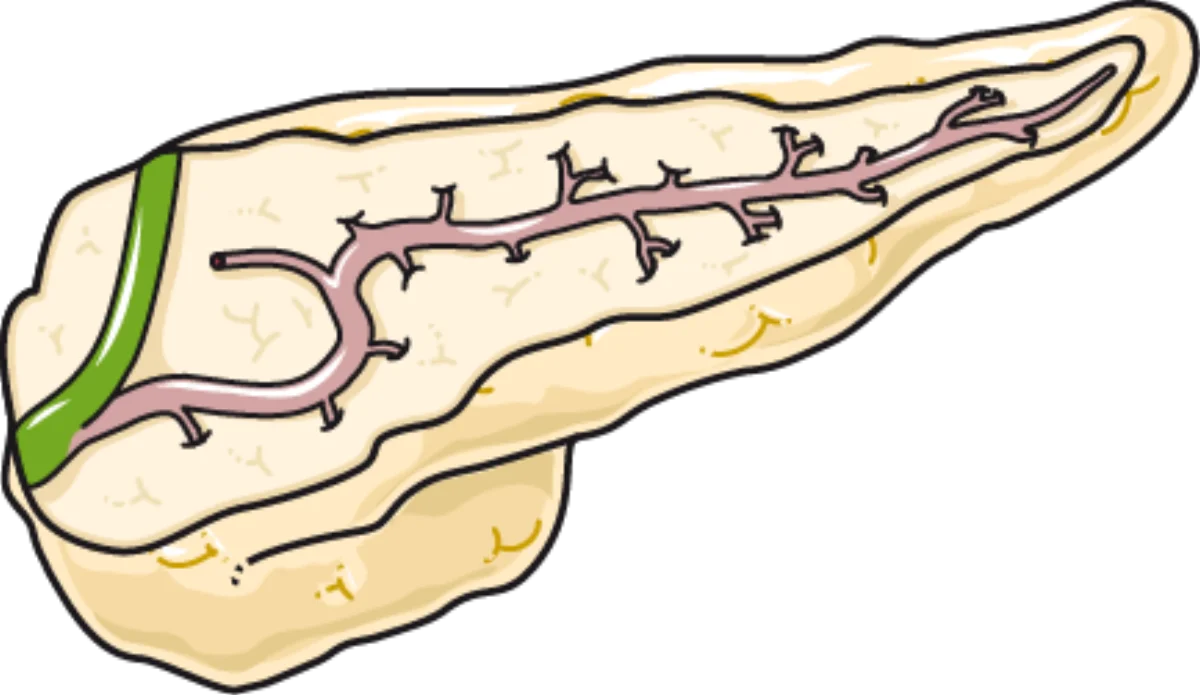
The pancreas is unique because it functions as both an exocrine and an endocrine gland. The exocrine portion (about 99% of the pancreas) consists of acinar cells that produce digestive enzymes (amylase, lipase, trypsin, chymotrypsin) and secrete them through the pancreatic duct into the duodenum. The digestive role of these enzymes is covered in detail in the pancreas section of the digestive system chapter.
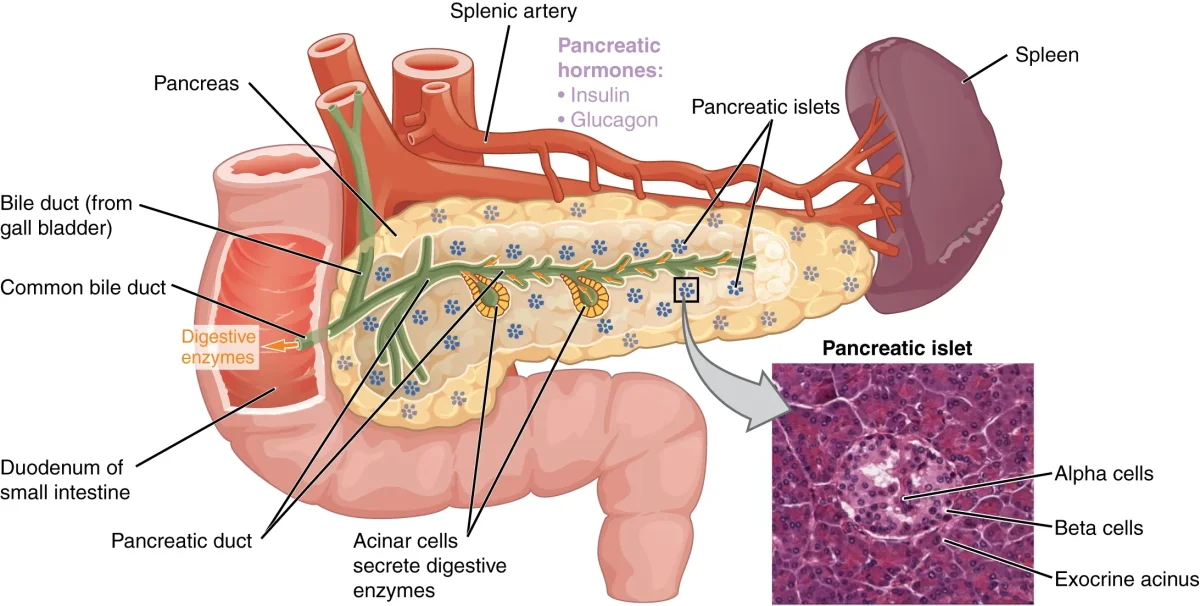
The endocrine portion consists of small clusters of hormone-producing cells called the islets of Langerhans (about 1% of pancreatic mass, but critical for metabolic regulation). Each islet contains several cell types:
| Cell Type | Hormone | % of Islet | Function |
|---|---|---|---|
| Beta (B) cells | Insulin | ~60-70% | Lowers blood glucose |
| Alpha (A) cells | Glucagon | ~20-30% | Raises blood glucose |
| Delta (D) cells | Somatostatin | ~5-10% | Inhibits insulin AND glucagon |
| PP (F) cells | Pancreatic polypeptide | ~1-2% | Regulates pancreatic secretions |
| Epsilon cells | Ghrelin | <1% | Stimulates appetite |
Insulin: The Storage Hormone
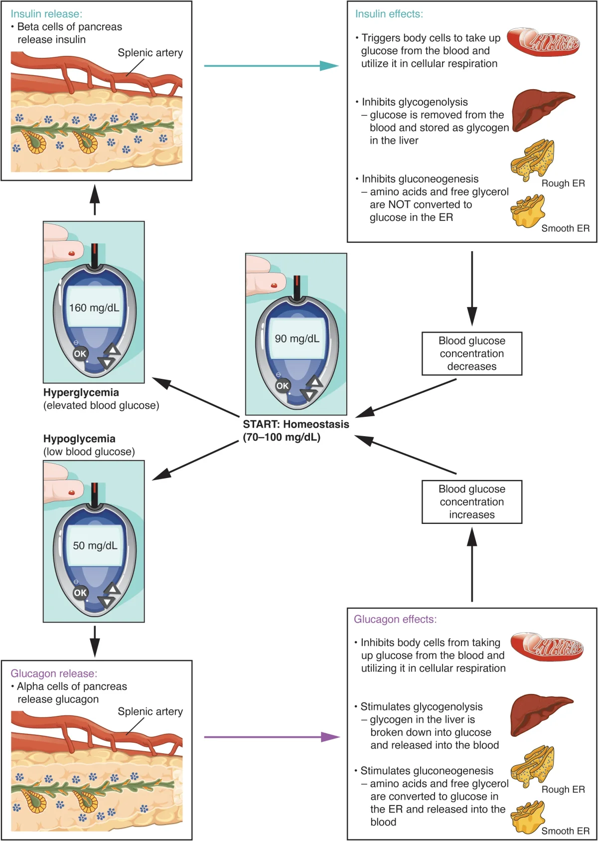
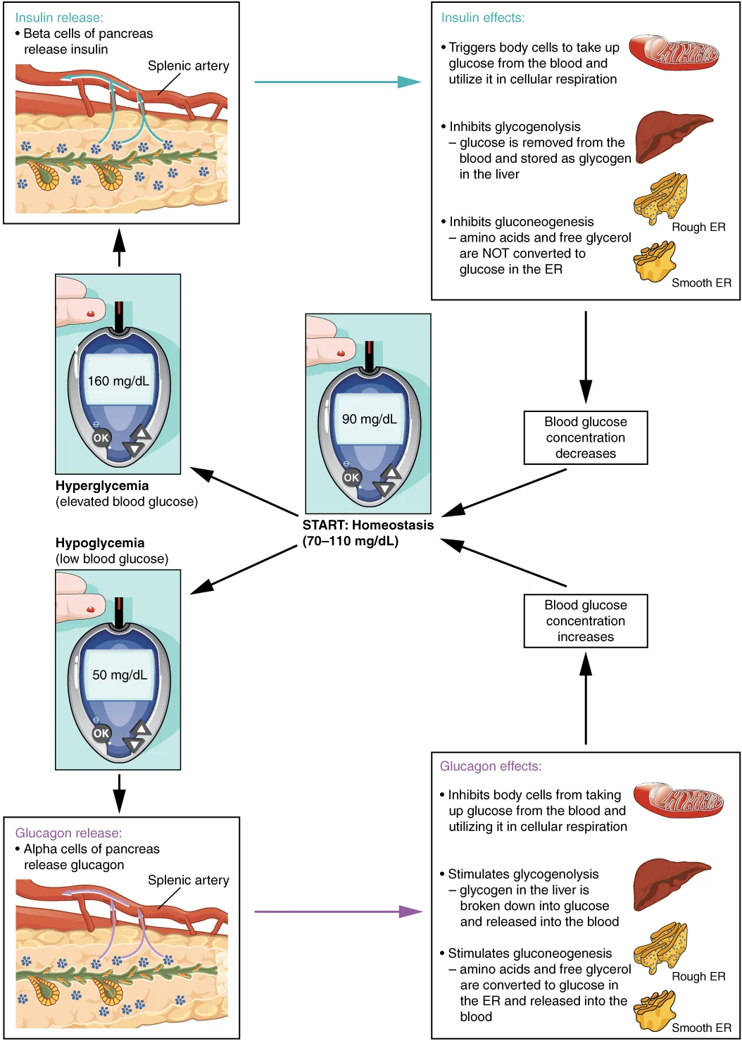
Insulin is a peptide hormone released by beta cells in response to elevated blood glucose (the primary stimulus). It is also stimulated by elevated amino acids, certain GI hormones (incretins like GLP-1), and parasympathetic stimulation (via the vagus nerve during eating).
Insulin’s key metabolic effects:
In the liver:
- Stimulates glycogenesis (glucose to glycogen storage)
- Inhibits glycogenolysis (prevents glycogen breakdown)
- Inhibits gluconeogenesis (prevents new glucose production)
- Promotes lipogenesis (fatty acid synthesis)
In muscle:
- Increases glucose uptake by promoting GLUT4 transporter insertion into the cell membrane
- Stimulates glycogenesis and protein synthesis
In adipose tissue:
- Increases glucose uptake via GLUT4
- Stimulates lipogenesis (glucose to triglycerides)
- Inhibits lipolysis (prevents fat breakdown)
The net effect: blood glucose drops as glucose is taken up by cells and stored as glycogen or fat.
Glucagon: The Mobilization Hormone
Glucagon is a peptide hormone released by alpha cells in response to low blood glucose (the primary stimulus). It is also stimulated by elevated amino acids (which makes physiological sense - a high-protein, low-carb meal needs glucagon to prevent hypoglycemia) and by epinephrine during stress.
Glucagon’s effects are essentially the opposite of insulin, and its primary target is the liver:
- Stimulates glycogenolysis (glycogen breakdown to glucose)
- Stimulates gluconeogenesis (production of new glucose from amino acids, lactate, and glycerol)
- Stimulates lipolysis (fat breakdown, releasing fatty acids and glycerol)
- Stimulates ketogenesis (conversion of fatty acids to ketone bodies during prolonged fasting)
The net effect: blood glucose rises as the liver dumps glucose into the bloodstream.
Somatostatin: The Brake Pedal
Somatostatin from delta cells inhibits both insulin and glucagon secretion. It also inhibits the release of growth hormone from the anterior pituitary (where it is called Growth Hormone-Inhibiting Hormone, or GHIH). In the GI tract, somatostatin slows digestion by reducing gut motility, enzyme secretion, and nutrient absorption.
Somatostatin prevents wild swings in blood glucose by dampening both sides of the seesaw simultaneously. It is released after meals when GI hormone levels and nutrient concentrations are high.
Blood Glucose Regulation: The Complete Picture
| State | Blood Glucose | Dominant Hormone | Liver Response | Muscle/Adipose Response |
|---|---|---|---|---|
| Fed (after meal) | High | Insulin | Glycogenesis, lipogenesis, inhibit gluconeogenesis | Glucose uptake (GLUT4), storage |
| Fasting (hours) | Low | Glucagon | Glycogenolysis, gluconeogenesis | Reduced glucose uptake, lipolysis |
| Prolonged fast | Very low | Glucagon + cortisol | Gluconeogenesis, ketogenesis | Fatty acid oxidation, ketone use |
| Acute stress | Rising | Epinephrine + cortisol | Glycogenolysis, gluconeogenesis | Reduced glucose uptake |
Diabetes Mellitus
Type 1 Diabetes - autoimmune destruction of beta cells. The body produces little to no insulin. Blood glucose stays high because cells cannot take it up. Patients require exogenous insulin injections. Onset typically in childhood/adolescence.
Type 2 Diabetes - target cells become resistant to insulin. The pancreas initially compensates by producing more insulin, but over time the beta cells become exhausted and insulin production declines. Associated with obesity, physical inactivity, and genetic factors. Most common form (~90-95% of diabetes cases). Managed with lifestyle changes, oral medications that improve insulin sensitivity, and sometimes insulin.