
Innate Immunity
Before your body ever manufactures a single antibody or trains a single T cell, it has an entire defense system already in place - one that was ready the day you were born. This is innate immunity: fast, nonspecific, and always on duty.
Think of it like a building’s security system. The locked doors, security cameras, and fire alarms do not need to know who the intruder is. They just detect “something is wrong” and respond immediately. Innate immunity works the same way - it recognizes general danger signals rather than specific invaders.
First Line of Defense: Physical and Chemical Barriers
Your body’s outermost defenses prevent pathogens from ever entering in the first place.
Skin is the largest organ and the most important barrier. The epidermis is a layer of tightly packed dead cells that most pathogens simply cannot penetrate. The skin also actively fights microbes by secreting:
- Lysozyme (in sweat) - an enzyme that breaks down bacterial cell walls
- Sebum - an oily secretion from sebaceous glands with antimicrobial properties
- Antimicrobial peptides (in sweat) - small proteins that inhibit pathogen growth
Mucous membranes line the respiratory, gastrointestinal, and urogenital tracts. Mucus is a thick, sticky fluid that physically traps pathogens and contains lysozyme to destroy bacterial cell walls.
Stomach acid (HCl, pH ~1.5-2) destroys most ingested bacteria and viruses by denaturing their proteins. This is why food poisoning requires a large enough dose of bacteria to overwhelm the acid.
Cilia in the respiratory tract beat in coordinated waves to sweep mucus (and trapped pathogens) upward toward the throat, where it is swallowed or expelled. Smoking paralyzes cilia - which is why smokers are more susceptible to respiratory infections.
Normal microbiota - the trillions of beneficial bacteria on your skin and in your gut - outcompete pathogenic microbes for nutrients and binding sites. Some even produce toxins that kill pathogens. Think of them as the “regulars” at a bar who take up all the seats so troublemakers cannot sit down.
Pathogen Recognition: PAMPs and PRRs
How does the innate immune system know what to attack? It recognizes pathogen-associated molecular patterns (PAMPs) - molecular features found on microbes but never on human cells. Examples include:
- Lipopolysaccharide (LPS) on gram-negative bacterial cell walls
- Peptidoglycan on gram-positive bacterial cell walls
- Double-stranded RNA (dsRNA) from replicating viruses
- Flagellin from bacterial flagella
Immune cells detect PAMPs using pattern recognition receptors (PRRs), the most important being Toll-like receptors (TLRs) on the surface of macrophages and dendritic cells. When a TLR binds a PAMP, it triggers an immediate immune response - no prior exposure required.
Second Line of Defense: The Inflammatory Response
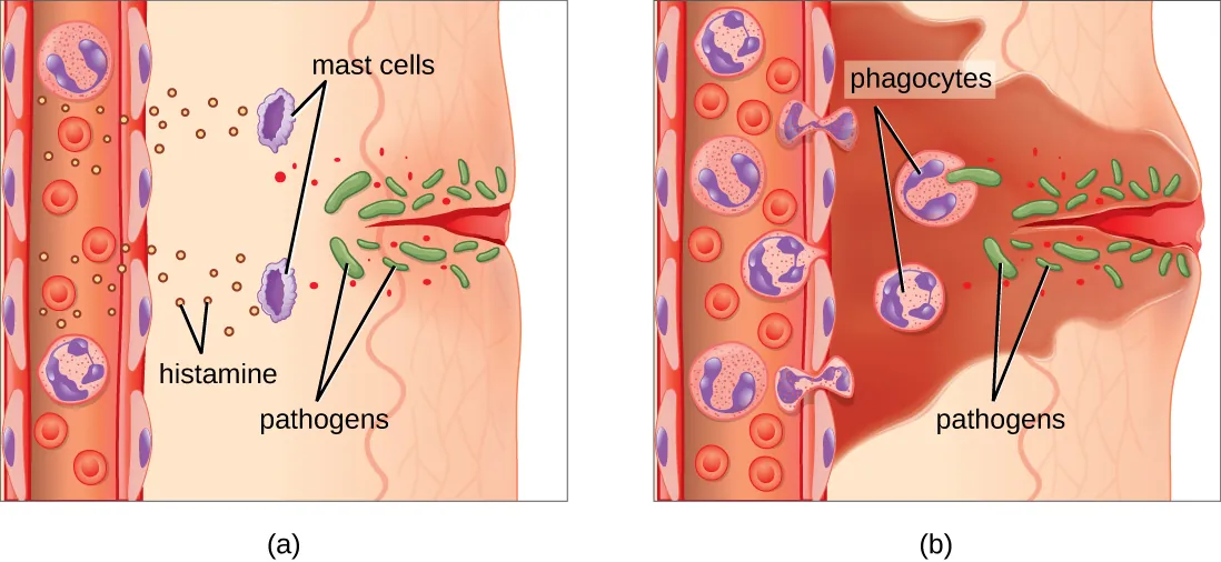
When pathogens breach the physical barriers, the body launches inflammation - a rapid, localized response designed to contain the threat and recruit immune cells. Here is what happens step by step:
-
Tissue damage or pathogen detection - Damaged cells release chemical signals. Macrophages in the tissue detect PAMPs and release cytokines.
-
Mast cell activation - Mast cells stationed in tissues release histamine, which causes nearby blood vessels to dilate (widen).
-
Vasodilation and increased permeability - Dilated vessels bring more blood to the area. Vessel walls become “leaky,” allowing fluid and immune cells to exit the blood and enter the tissue.
-
Immune cell recruitment - White blood cells such as neutrophils are the first responders, arriving within minutes. They squeeze between endothelial cells (diapedesis) and follow chemical gradients (chemotaxis) to the infection site.
-
Phagocytosis and pathogen destruction - Neutrophils and macrophages engulf and destroy pathogens using enzymes and reactive oxygen species.
The four classic signs of inflammation are easy to remember:
| Sign | Cause |
|---|---|
| Redness | Vasodilation brings more blood |
| Heat | Increased blood flow raises local temperature |
| Swelling | Fluid leaks from permeable vessels into tissue |
| Pain | Swelling puts pressure on nerve endings |
Fever is a systemic extension of inflammation. Pyrogens (fever-inducing molecules) released by immune cells or pathogens reset the hypothalamic thermostat to a higher temperature. Fever helps fight infection by inhibiting bacterial and viral reproduction and by increasing the metabolic rate of immune cells.
Interferons are antiviral cytokines. A virus-infected cell secretes interferons that bind neighbors and switch them into an antiviral state (slowing protein synthesis, degrading viral RNA). The infected cell often sacrifices itself, but the interferon signal stops the virus from spreading. Think of it as a burning building warning the whole block.
The Complement System
The complement system is a set of ~30 proteins circulating in the blood in inactive form. When activated, they work in a cascade (each protein activates the next) to destroy pathogens through three mechanisms:
Three activation pathways all converge on the same endpoint:
| Pathway | Trigger | Key Detail |
|---|---|---|
| Classical | Antibody bound to pathogen (antigen-antibody complex) | Links adaptive immunity to complement; starts with C1 |
| Alternative | Spontaneous C3 cleavage on pathogen surfaces | No antibody needed; truly innate |
| Lectin | Mannose-binding lectin binds pathogen surface sugars | Pattern recognition of carbohydrates |
Three functions of complement:
-
Opsonization - C3b coats the pathogen surface, making it much easier for phagocytes to recognize and engulf it. Think of opsonization as “painting a target” on the enemy.
-
Inflammation - Small complement fragments (C3a, C5a) attract immune cells to the site and promote inflammation by triggering mast cell degranulation and chemotaxis.
-
Membrane Attack Complex (MAC) - Proteins C5b, C6, C7, C8, and multiple C9s (the C5b-9 complex) assemble into a ring-shaped pore in the pathogen’s membrane, causing lysis and death. Gram-negative bacteria are especially vulnerable; gram-positive bacteria are usually protected by their thick peptidoglycan wall.