Every time you scrape your knee, your body launches a construction project. Cells around the wound start duplicating themselves to patch the gap. But a cell cannot just split in half whenever it feels like it. It has to copy all 6 billion base pairs of DNA, duplicate its organelles, stockpile enough energy and raw materials, and verify everything is correct - all before it divides. That preparation process is the cell cycle.
The Big Picture
The cell cycle has two main parts:
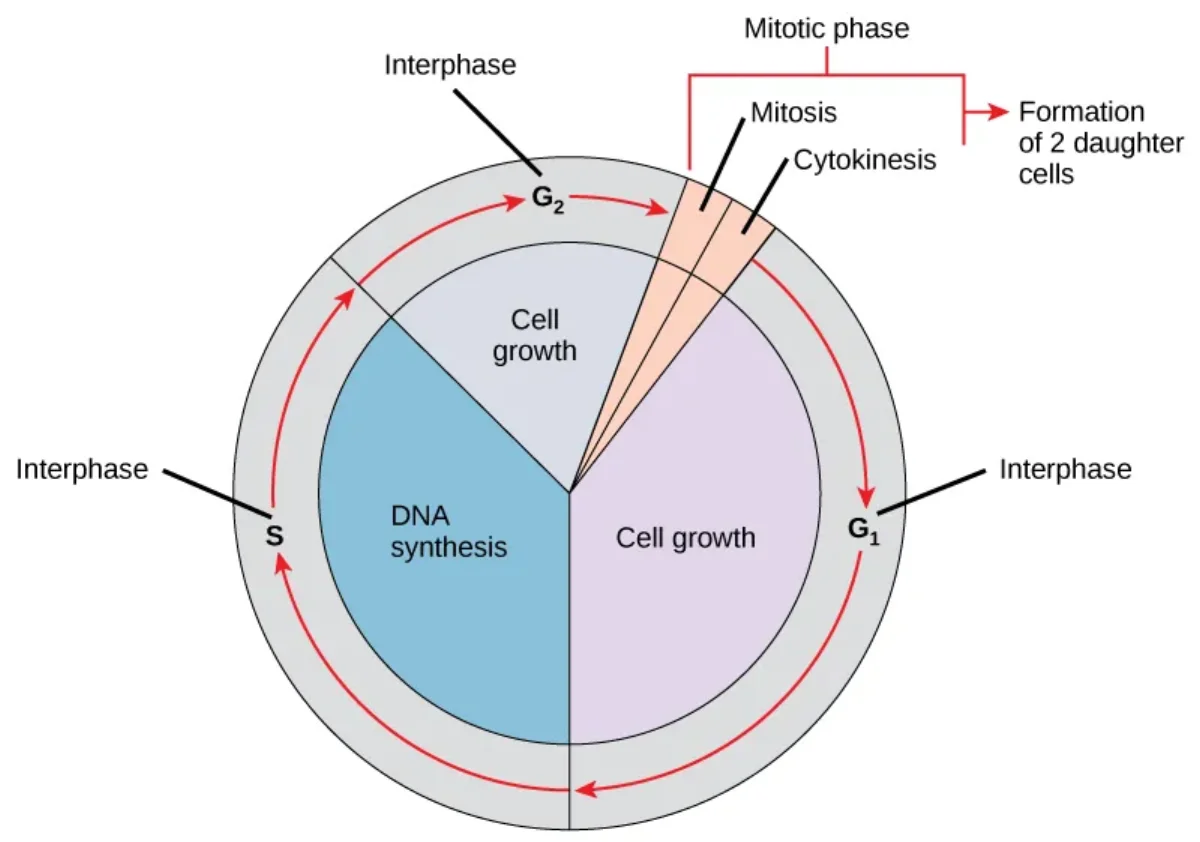
Interphase - the long “preparation” period (G1 + S + G2). The cell spends about 90% of its life here.
M phase (Mitotic phase) - the actual division. This is the shortest part.
Most students make the mistake of thinking “cell division” is the whole cycle. It is not. Division is the grand finale. The bulk of the work happens in interphase.
The cell cycle shown as a circular diagram. Interphase (G1, S, G2) dominates the cycle, with the M phase (mitosis + cytokinesis) occupying the shortest segment. Credit: OpenStax Biology 2e, CC BY 4.0
G1 Phase (First Gap) - “Get Ready”
G1 is the cell’s growth phase. The cell:
Increases in size
Synthesizes proteins and ribosomes
Produces the building blocks (nucleotides) it will need to copy its DNA
Accumulates energy reserves (ATP)
This is also when the cell receives external signals (growth factors) telling it whether to proceed with division or stop. If the cell does not receive the right signals, or if conditions are unfavorable, it can exit the cycle and enter G0.
G1 is the longest and most variable phase. Fast-dividing cells (like gut epithelium) blow through G1 quickly. Slow-dividing cells (like liver cells) linger in G1 for a long time.
S Phase (Synthesis) - “Copy the Blueprint”
This is when DNA replication occurs. Every chromosome in the nucleus is duplicated, producing two identical copies called sister chromatids, joined at a structure called the centromere.
Before S phase: 46 chromosomes, each made of 1 chromatid.
After S phase: still 46 chromosomes, but each now made of 2 sister chromatids (92 chromatids total).
The centrosome (the cell’s microtubule organizing center) also duplicates during S phase.
G2 Phase (Second Gap) - “Final Check”
G2 is the final preparation before division. The cell:
Continues to grow and produce proteins
Synthesizes proteins specifically needed for mitosis (like tubulin for the mitotic spindle)
Checks that DNA replication in S phase was completed accurately
Verifies there is no DNA damage
If errors are detected, the cell pauses here to repair them before committing to division.
M Phase (Mitosis) - “Divide”
The cell physically splits into two daughter cells. This involves:
Mitosis - division of the nucleus (covered in detail in Section 2.2)
Cytokinesis - division of the cytoplasm
G0 Phase - “Off the Clock”
Not every cell is constantly dividing. Cells that have exited the cell cycle enter G0, a quiescent (resting) state. G0 cells are alive and metabolically active - they are just not preparing to divide.
Some cells enter G0 temporarily and can be called back into the cycle by the right signals:
Liver cells (hepatocytes) - normally in G0, but re-enter the cycle if the liver is damaged
Lymphocytes - re-enter the cycle when activated by an antigen
Other cells enter G0 permanently and never divide again:
Neurons - once mature, they do not divide (this is why brain damage is often irreversible)
Cardiac muscle cells (cardiomyocytes) - heart cells cannot regenerate after a heart attack, which is why damaged heart tissue is replaced by scar tissue
Chromosome Counting Across the Cell Cycle
This table is critical for the MCAT. Know these numbers cold:
Phase
Chromosomes
Chromatids
DNA Content
Ploidy
G1
46
46
2n
Diploid
After S phase
46
92
4n
Diploid
After G2
46
92
4n
Diploid
After Mitosis
46
46
2n
Diploid
After Meiosis I
23
46
2n
Haploid
After Meiosis II
23
23
n
Haploid
A cell in G2 has how many chromosomes and how many chromatids?
Click to reveal answer
46 chromosomes and 92 chromatids. DNA was replicated in S phase, so each of the 46 chromosomes now consists of 2 sister chromatids joined at the centromere. The chromosome number does not change until the homologs separate in meiosis I.
Name two cell types that permanently remain in G0 and never re-enter the cell cycle.
Click to reveal answer
Neurons and cardiac muscle cells (cardiomyocytes). Both are terminally differentiated and do not divide. This is why brain damage and heart attacks result in permanent tissue loss rather than regeneration.
Imagine photocopying a book. You put the original on the glass, press the button, and get an identical copy. The original is unchanged, the copy is perfect, and now you have two identical books. That is mitosis - one cell becomes two genetically identical daughter cells.
Mitosis is the “factory” side of cell division. It is how your body grows from a single fertilized egg into trillions of cells, how it replaces the skin cells you shed every day, and how it repairs a wound. The goal is always the same: make a perfect copy.
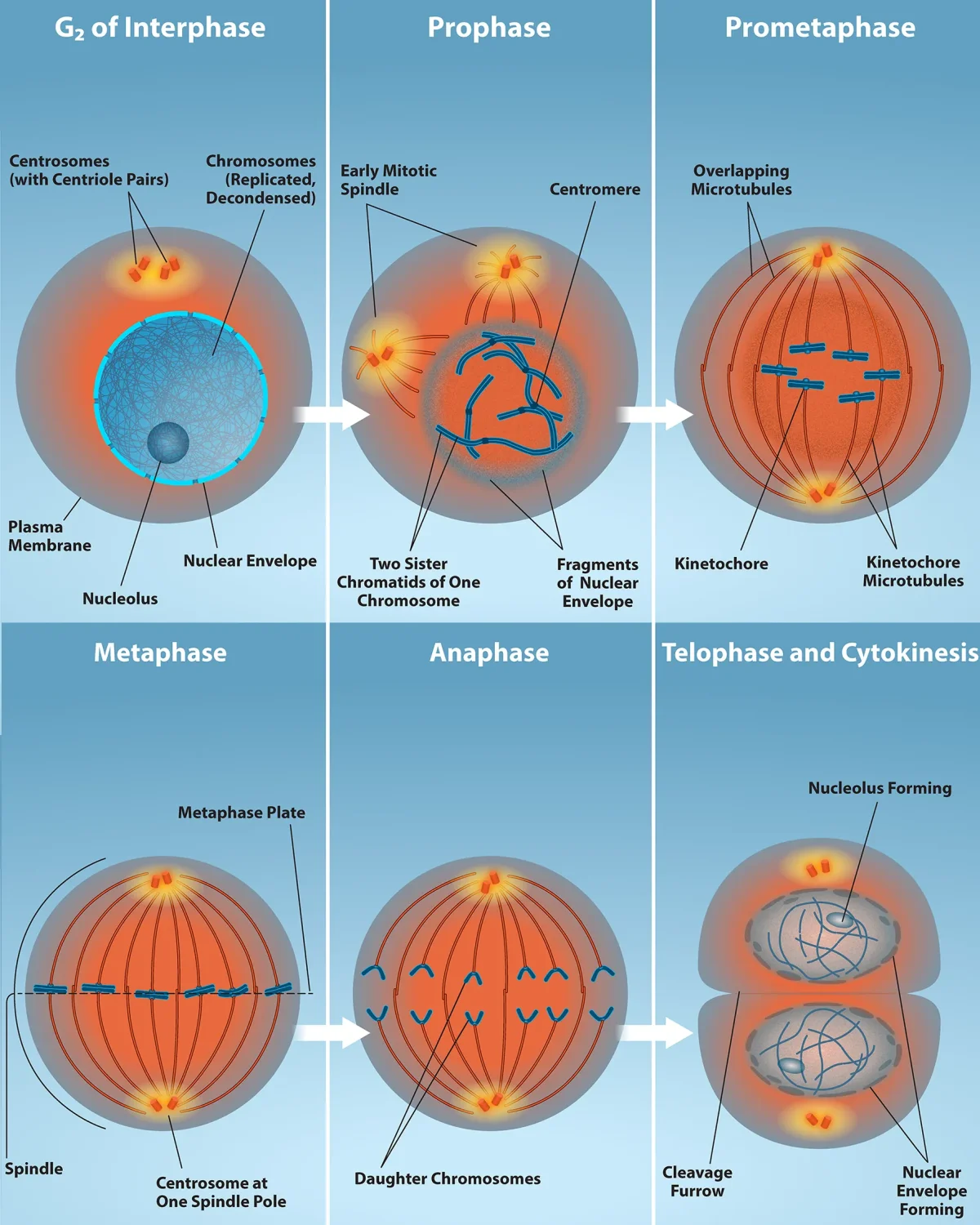
The stages of mitosis from prophase through telophase and cytokinesis, showing chromosome behavior at each stage. Credit: OpenStax Biology 2e, CC BY 4.0Real microscopy of onion root tip cells in various stages of mitosis. Chromosomes are stained dark blue-purple, making it easy to spot cells in prophase (condensing), metaphase (aligned at center), and anaphase (pulling apart). Credit: Wikimedia Commons (ELaurent), CC BY-SA 4.0
Prophase - “Prepare for War”
Prophase is the cell gearing up for division. Several things happen simultaneously:
Chromatin condenses into visible chromosomes. The loose DNA threads tighten and compact so they can be moved without tangling. Think of winding up earbuds before putting them in your pocket.
Each chromosome is visible as two sister chromatids joined at the centromere.
The nuclear envelope begins to break down. This must happen so the spindle fibers can reach the chromosomes.
The nucleolus disappears.
Centrosomes migrate to opposite poles of the cell. As they move, they begin assembling the mitotic spindle - a network of microtubules that will pull the chromosomes apart.
Aster fibers radiate from each centrosome and anchor it to the cell membrane for stability.
Some textbooks split this into prophase and prometaphase. In prometaphase, the nuclear envelope is fully dissolved and spindle fibers attach to the chromosomes at their kinetochores - protein structures on the centromere that serve as attachment points for microtubules.
Metaphase - “Middle Lineup”
The chromosomes are dragged to the center of the cell and align along the metaphase plate (an imaginary equator halfway between the two poles).
Each chromosome is attached to spindle fibers from both poles via its kinetochores.
Tension from opposite spindle fibers holds each chromosome at the midline.
The cell runs its spindle assembly checkpoint (M checkpoint) here: are all chromosomes properly attached to spindle fibers from both poles? If not, division stalls until the problem is fixed.
Memory trick:Metaphase = Middle. Chromosomes line up in the middle.
Anaphase - “Apart”
This is the shortest and most dramatic stage:
Cohesin proteins holding sister chromatids together are cleaved by the enzyme separase.
Sister chromatids are pulled to opposite poles by the shortening of kinetochore microtubules.
Simultaneously, non-kinetochore microtubules lengthen, pushing the poles further apart and elongating the cell.
By the end of anaphase, each pole has a complete set of 46 chromosomes.
Memory trick:Anaphase = Apart. Chromatids are pulled apart.
Telophase - “Two Nuclei”
Telophase is essentially prophase in reverse:
Nuclear envelopes reform around each set of chromosomes.
Chromosomes decondense back into loose chromatin.
Nucleoli reappear.
The spindle apparatus disassembles.
At this point, you have a single cell with two complete nuclei.
Memory trick:Telophase = Two nuclei.
Cytokinesis - “Cut the Cell”
Cytokinesis divides the cytoplasm, completing the physical separation into two daughter cells. It usually begins during late anaphase or telophase.
In animal cells: A ring of actin and myosin filaments (the contractile ring) pinches the cell membrane inward, forming a cleavage furrow that deepens until the cell splits in two. Think of tightening a drawstring on a bag.
In plant cells: A rigid cell wall prevents pinching. Instead, vesicles from the Golgi apparatus line up at the cell’s equator and fuse to form a cell plate, which grows outward until it separates the two cells completely.
Mitosis Summary Table
Stage
Key Events
Memory Cue
Prophase
Chromatin condenses, nuclear envelope breaks down, spindle forms
”Prepare”
Metaphase
Chromosomes align at metaphase plate, spindle checkpoint
”Middle”
Anaphase
Sister chromatids separate and move to opposite poles
”Apart”
Telophase
Nuclear envelopes reform, chromosomes decondense
”Two nuclei”
Cytokinesis
Cytoplasm divides (cleavage furrow in animals, cell plate in plants)
“Cut”
Key Outcome
Mitosis produces 2 daughter cells that are genetically identical to the parent cell and to each other. Each has 46 chromosomes (2n).
During which stage of mitosis do sister chromatids separate?
Click to reveal answer
Anaphase. The enzyme separase cleaves cohesin proteins at the centromere, and kinetochore microtubules pull the sister chromatids (now individual chromosomes) to opposite poles of the cell.
What structure on the chromosome serves as the attachment point for spindle fibers?
Click to reveal answer
The kinetochore - a protein complex assembled on the centromere of each chromatid. Kinetochore microtubules from the spindle attach here to pull chromosomes during anaphase. Do not confuse the kinetochore (protein structure) with the centromere (DNA region).
How does cytokinesis differ between animal and plant cells?
Click to reveal answer
Animal cells use a contractile ring of actin and myosin that pinches inward, forming a cleavage furrow. Plant cells cannot pinch because of their rigid cell wall, so they build a cell plate from Golgi vesicles that grows outward to divide the cell.
Imagine an airport with three security checkpoints. Before you board, you must pass through all three. At each one, guards check your passport, your ticket, and your bags. If anything is wrong, you are stopped until the problem is resolved. If the problem cannot be fixed, you are removed from the airport entirely.
The cell cycle works the same way. There are three major checkpoints where the cell pauses to verify that everything is in order before proceeding to the next phase. If conditions are not right, the cell stops. If the damage is unrepairable, the cell destroys itself (apoptosis). Cancer is what happens when someone sneaks past every checkpoint.
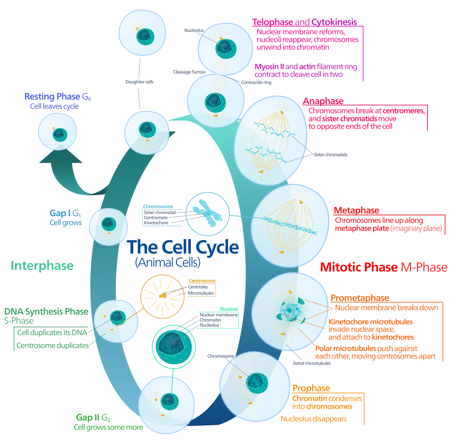
The cell cycle end to end: G1, S and G2 make up interphase, prophase through telophase and cytokinesis divide the cell, and cells that stop dividing exit to G0. The diagram labels the phases, not the control points. The three checkpoints sit at boundaries you can see here: late G1 (is the cell ready to replicate DNA?), the G2 to M transition (was replication error-free?), and within metaphase (are all chromosomes attached to the spindle?). Cyclin-CDK complexes drive progression across each of those boundaries. Credit: Wikimedia Commons, CC0 Public Domain
The Three Major Checkpoints
G1 Checkpoint (Restriction Point) - The most important decision point. The cell asks:
Is the cell large enough to divide?
Are there sufficient nutrients and energy?
Is the DNA undamaged?
Are growth factors present?
If conditions are favorable, the cell commits to division and enters S phase. If not, it enters G0 (quiescence). Once past the G1 checkpoint, the cell is committed - it will complete the cycle without needing additional external signals.
G2 Checkpoint - Before entering mitosis, the cell verifies:
Was DNA replication completed successfully?
Is there any remaining DNA damage?
Are mitosis-promoting factors (MPF) present?
M Checkpoint (Spindle Assembly Checkpoint) - During metaphase, the cell checks:
Are all chromosomes attached to spindle fibers from both poles?
Is there proper tension on each kinetochore?
If even one chromosome is unattached, the checkpoint halts division. Failure at this checkpoint leads to nondisjunction - unequal chromosome distribution that causes conditions like Down syndrome (trisomy 21).
Cyclins and CDKs - The Molecular Engine
The cell cycle is driven forward by a partnership between two types of proteins:
Cyclins - regulatory proteins whose concentrations rise and fall in a predictable pattern as the cell progresses through the cycle (hence the name “cyclin”)
CDKs (cyclin-dependent kinases) - enzyme proteins that are always present in the cell but are inactive on their own
Here is the key: CDKs only become active when a cyclin binds to them. The cyclin-CDK complex then phosphorylates (adds phosphate groups to) target proteins that push the cell into the next phase.
Think of it this way: the CDK is the car engine (always there, always ready) and the cyclin is the ignition key (comes and goes). No key, no ignition. No cyclin, no cell cycle progression.
Phase Transition
Cyclin
CDK Partner
Function
G1 to S
Cyclin D
CDK64
Passes the restriction point
S phase entry
Cyclin E
CDK2
Initiates DNA replication
S to G2
Cyclin A
CDK2
Completes DNA replication
G2 to M
Cyclin B
CDK1
Triggers mitosis (MPF)
The Cyclin B-CDK1 complex is also known as MPF (Maturation Promoting Factor) or M-phase promoting factor. It triggers the cascade of events that initiates mitosis: chromosome condensation, nuclear envelope breakdown, and spindle formation.
Growth Factors and External Signals
Cells do not just decide to divide on their own. They need permission from the body in the form of growth factors - signaling molecules that bind to cell surface receptors and activate signal transduction pathways leading to the production of cyclins.
Without growth factors, most normal cells remain in G0. This is why:
Wound healing is localized (growth factors are released at the injury site)
Cells in a dish stop dividing when they run out of growth factor in the media
Two additional controls on cell growth:
Density-dependent inhibition (contact inhibition) - cells stop dividing when they become too crowded and physically touch neighboring cells
Anchorage dependence - normal cells must be attached to a surface (extracellular matrix) to divide
Cancer cells ignore both of these signals.
Tumor Suppressors - The Brakes
Tumor suppressor genes encode proteins that slow down or stop cell division. They are the brakes on the cell cycle.
p53 - “The Guardian of the Genome”:
Monitors DNA integrity
If damage is detected, p53 halts the cell cycle at the G1 checkpoint to allow time for repair
If repair fails, p53 triggers apoptosis (programmed cell death)
p53 is the most commonly mutated gene in human cancers - when p53 is lost, damaged cells keep dividing instead of self-destructing
Rb (Retinoblastoma protein):
Controls the G1 checkpoint by binding to and inhibiting the transcription factor E2F
When Rb is phosphorylated by Cyclin D-CDK64, it releases E2F, allowing the cell to enter S phase
Loss of Rb removes the G1 brake, leading to uncontrolled entry into S phase
Proto-Oncogenes and Oncogenes - The Gas Pedal
Proto-oncogenes are normal genes that encode proteins promoting cell division (growth factors, receptors, signal transduction proteins, transcription factors). They are the gas pedal.
When a proto-oncogene is mutated into an oncogene, it becomes a stuck gas pedal - always on, driving cell division even without the proper signals. Unlike tumor suppressors, only one copy of a proto-oncogene needs to be mutated to cause problems (it is a gain-of-function mutation).
Feature
Tumor Suppressors
Oncogenes
Normal function
Inhibit cell division (brakes)
Promote cell division (gas pedal)
Mutation type
Loss-of-function
Gain-of-function
Copies needed to cause cancer
Both (two-hit)
One (dominant)
Examples
p53, Rb
Ras
How Cancer Develops
Cancer is not a single mutation - it is an accumulation of mutations in multiple regulatory genes. A typical cancer cell might have:
Activated oncogenes (stuck gas pedal)
Inactivated tumor suppressors (broken brakes)
Loss of contact inhibition
Loss of anchorage dependence
Telomerase reactivation (immortality - can divide indefinitely)
Ability to stimulate angiogenesis (growing new blood vessels to feed the tumor)
What happens when cyclin B binds to CDK1?
Click to reveal answer
It forms the MPF (Maturation/M-phase Promoting Factor) complex, which triggers entry into mitosis. MPF phosphorylates proteins that cause chromosome condensation, nuclear envelope breakdown, and spindle assembly. Without this complex, the cell cannot enter M phase.
Why does a mutation in p53 increase cancer risk?
Click to reveal answer
p53 is the "guardian of the genome." It monitors DNA for damage, halts the cell cycle at G1 to allow repairs, and triggers apoptosis if damage is irreparable. Without functional p53, cells with damaged DNA continue to divide and accumulate further mutations, leading to cancer. It is the most commonly mutated gene in human cancers.
Mitosis is the photocopier - it makes identical copies. Meiosis is the card shuffler. It takes a full deck (46 chromosomes), shuffles it in ways that guarantee every hand is unique, and deals out four half-decks (23 chromosomes each). That is how your body produces sperm and egg cells that are all genetically different from each other - and from you.
Why Meiosis Exists
The fundamental problem: if sperm and egg both had 46 chromosomes, the resulting embryo would have 92. Next generation: 184. This would double every generation. Meiosis solves this by halving the chromosome number, so that when sperm (23) meets egg (23), the result is a normal 46.
This halving is why Meiosis I is called the reduction division.
Key Vocabulary Before We Start
Homologous chromosomes (homologs) - a matching pair of chromosomes, one from mom and one from dad. They carry the same genes at the same positions (loci) but may have different versions (alleles).
Sister chromatids - the two identical copies of a chromosome produced by DNA replication, joined at the centromere.
Tetrad (bivalent) - the structure formed when a pair of homologs lines up during meiosis I. Each homolog has two sister chromatids, so a tetrad has 4 chromatids total.
Synapsis - the physical pairing of homologous chromosomes, held together by the synaptonemal complex.
Chiasma (plural: chiasmata) - the X-shaped site where crossing over occurs between homologs.
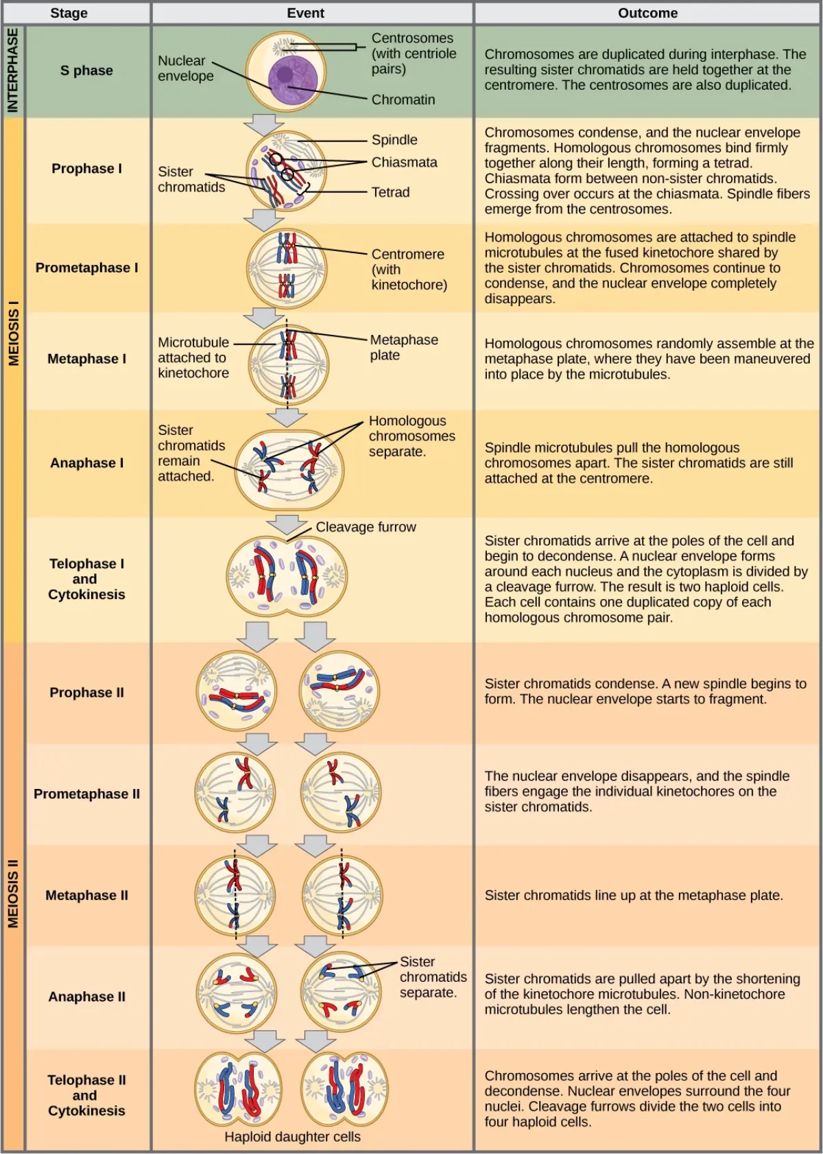
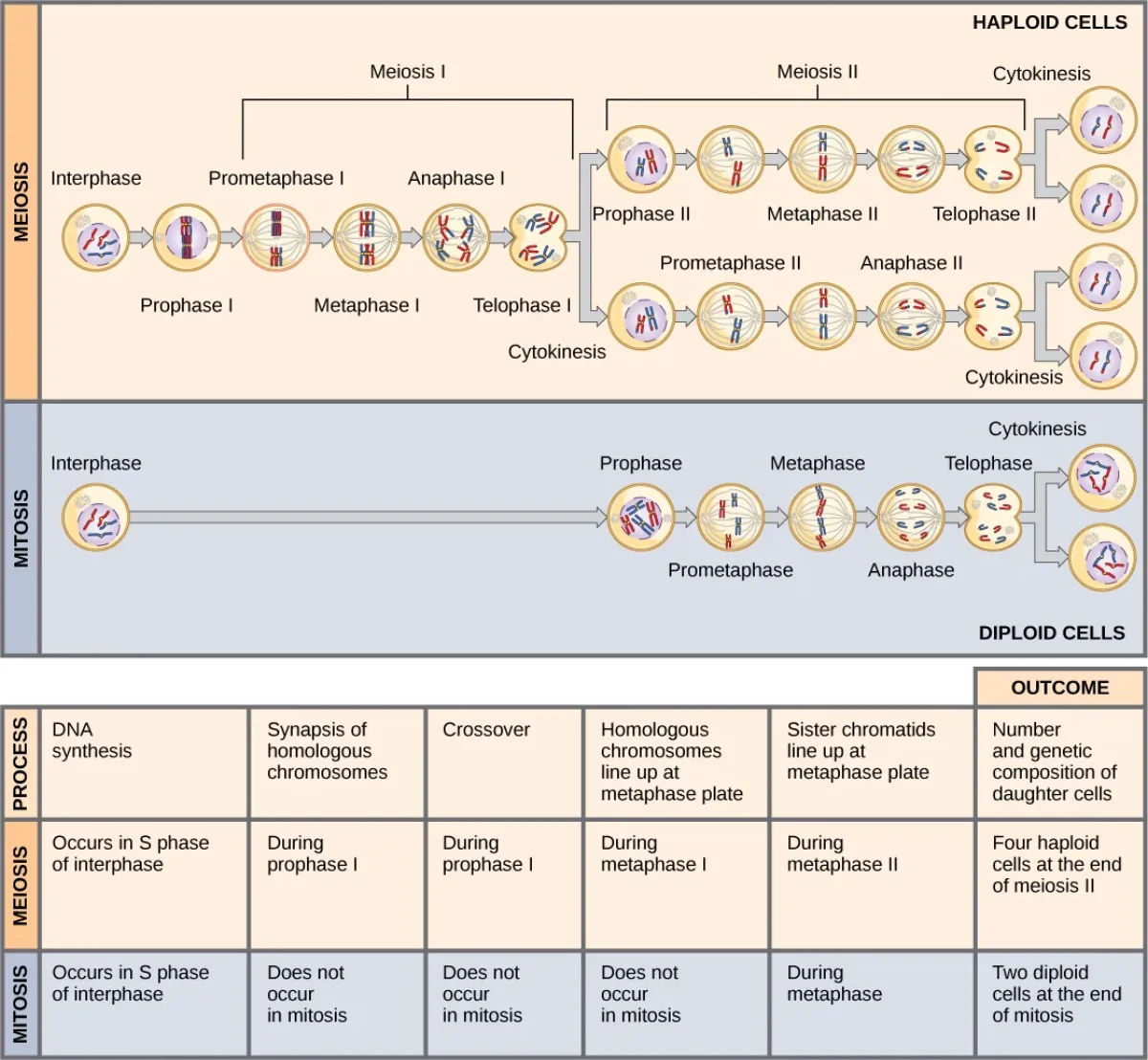
The complete process of meiosis, showing both divisions. Meiosis I separates homologous chromosomes (reduction division). Meiosis II separates sister chromatids (equational division). Credit: OpenStax Biology 2e, CC BY 4.0
Meiosis I - The Reduction Division
Meiosis I is where the real action happens. This is the division that is fundamentally different from mitosis.
Prophase I - The longest and most complex phase of meiosis:
Chromosomes condense.
Synapsis occurs: homologous chromosomes pair up, forming tetrads held together by the synaptonemal complex.
Crossing over happens at chiasmata - homologs swap segments of DNA. This is the major source of genetic recombination (covered in detail in Section 2.5).
Nuclear envelope breaks down, spindle forms.
Metaphase I - Tetrads (paired homologs) align at the metaphase plate.
Critical difference from mitosis: in mitosis, individual chromosomes line up. In meiosis I, pairs of homologs line up together.
The orientation of each pair is random - mom’s chromosome could face either pole. This is independent assortment (Mendel’s Second Law), and it is a second major source of genetic variation.
Anaphase I - Homologous chromosomes are pulled to opposite poles.
Critical difference from mitosis: in mitosis, sister chromatids separate. In meiosis I, homologs separate but sister chromatids stay together.
This separation (disjunction) is what reduces the chromosome number from 2n to n.
Telophase I and Cytokinesis - Nuclear envelopes may reform. The cell divides into two cells, each with 23 chromosomes (but each chromosome still consists of 2 sister chromatids).
Meiosis II - The Equational Division
Meiosis II is essentially mitosis performed on haploid cells. There is no additional DNA replication between Meiosis I and Meiosis II (the brief pause is called interkinesis, not a full interphase).
Metaphase II - Individual chromosomes (each still consisting of 2 sister chromatids) align at the metaphase plate.
Anaphase II - Sister chromatids finally separate and move to opposite poles.
Telophase II and Cytokinesis - Nuclear envelopes reform, cells divide. The end result is 4 haploid cells, each with 23 chromosomes (single chromatids).
Meiosis vs. Mitosis - Side by Side
Feature
Mitosis
Meiosis
Number of divisions
1
2
Daughter cells produced
2
4
Genetic result
Identical to parent
Genetically unique
Chromosome number in daughters
2n (diploid)
n (haploid)
Crossing over?
No
Yes (Prophase I)
Synapsis/tetrads?
No
Yes (Prophase I)
Independent assortment?
No
Yes (Metaphase I)
What separates in division I?
Sister chromatids
Homologs
Purpose
Growth, repair
Gamete production
Where it occurs
Somatic cells
Germ cells (gonads)
Direct comparison of mitosis and meiosis. Mitosis produces two identical diploid cells. Meiosis produces four unique haploid cells. Credit: OpenStax Biology 2e, CC BY 4.0
Sources of Genetic Variation in Meiosis
Meiosis generates genetic diversity through three mechanisms:
Crossing over (Prophase I) - homologs swap DNA segments, creating new allele combinations on each chromosome.
Independent assortment (Metaphase I) - each homologous pair orients randomly, giving 223 = 8,388,608 possible chromosome combinations per gamete.
Random fertilization - any one of millions of genetically unique sperm can fertilize any one of millions of genetically unique eggs.
Together, these three mechanisms ensure that no two gametes - and no two offspring (except identical twins) - are ever genetically identical.
Nondisjunction - When Meiosis Goes Wrong
Nondisjunction is the failure of chromosomes to separate properly during meiosis. It can occur in:
Meiosis I - homologs fail to separate. Both members of a homologous pair go to the same pole. Result: two gametes with an extra chromosome (n+1) and two gametes missing a chromosome (n-1).
Meiosis II - sister chromatids fail to separate. Result: one gamete with n+1, one with n-1, and two normal (n) gametes.
When an n+1 gamete is fertilized by a normal gamete, the resulting embryo has trisomy (2n+1 = 47 chromosomes). The most commonly tested example is Trisomy 21 (Down syndrome) - three copies of chromosome 21. Nondisjunction can also affect sex chromosomes, such as XXY (Klinefelter syndrome) or XO (Turner syndrome).
What is the key difference between what separates in Anaphase I vs. Anaphase II?
Click to reveal answer
In Anaphase I, homologous chromosomes separate (one homolog to each pole). In Anaphase II, sister chromatids separate (identical to mitotic anaphase). This is the fundamental distinction: Meiosis I = reduction (homologs split, 2n to n), Meiosis II = equational (sisters split, n stays n).
How many genetically unique gametes can a human produce from independent assortment alone (without considering crossing over)?
Click to reveal answer
223 = 8,388,608 possible combinations per gamete. Each of the 23 homologous pairs independently orients at the metaphase plate during Meiosis I, and each can face either pole. With crossing over included, the number of unique gametes is virtually unlimited.
Imagine you have two decks of cards - one red (from your mother) and one blue (from your father). If you just randomly assigned whole decks to your gametes, each gamete would get either the full red deck or the full blue deck. But that is not what happens. During meiosis, individual cards get swapped between the decks. A red 7 of hearts trades places with a blue 7 of hearts. A red queen of spades swaps with a blue queen of spades. Now each deck is a unique mosaic of red and blue cards. That swap is crossing over.
How Crossing Over Works
Crossing over occurs during Prophase I of meiosis, after homologous chromosomes have paired up through synapsis.
Homologous chromosomes align precisely, gene for gene, forming a tetrad (4 chromatids total).
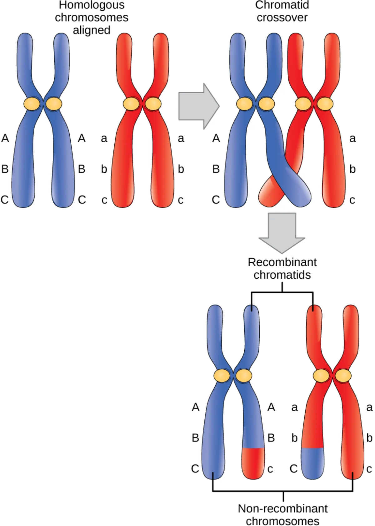
Non-sister chromatids (one from each homolog) physically overlap at points called chiasmata.
At each chiasma, the DNA strands are cut by enzymes and reconnected to the other homolog.
The result: each chromatid now carries a new combination of alleles - some originally from the maternal chromosome, some from the paternal chromosome.
Crossing over during Prophase I. Homologous chromosomes (blue and red) exchange segments at chiasmata, producing recombinant chromatids with new allele combinations. Credit: Lumen Learning / OpenStax Biology, CC BY 4.0
What Gets Swapped
Crossing over swaps corresponding segments between non-sister chromatids of homologous chromosomes. It does NOT occur between:
Sister chromatids of the same chromosome (they are identical, so swapping would change nothing)
Non-homologous chromosomes (chromosome 1 does not swap with chromosome 15)
The exchange is always reciprocal - if chromatid A gives a segment to chromatid B, chromatid B gives the equivalent segment back to chromatid A.
Recombinant vs. Parental Chromatids
After crossing over in a tetrad:
2 chromatids are recombinant - they carry new allele combinations that did not exist in either parent
2 chromatids are parental - they retain the original allele combinations
This means that of the four gametes produced by a single meiosis, some will be recombinant and some will be parental.
Gene Linkage
Genes located on the same chromosome are said to be linked. Linked genes tend to be inherited together because they travel on the same physical piece of DNA. However, crossing over can break this linkage.
The key principle: the further apart two genes are on a chromosome, the more likely crossing over will occur between them. If two genes are very close together, the chance that a chiasma will form between them is low, so they stay linked. If they are far apart, there are many potential chiasma sites between them, so they are frequently separated by crossing over.
Genes on the same chromosome but far apart behave almost as if they are on different chromosomes (they assort nearly independently). Genes very close together are tightly linked and rarely separated.
Recombination Frequency
Recombination frequency is the percentage of offspring that show recombinant (non-parental) phenotypes. It directly reflects the distance between two genes on a chromosome.
0% recombination = genes are so close they are never separated = completely linked
50% recombination = genes behave as if they are on different chromosomes = unlinked (either very far apart on the same chromosome or on different chromosomes entirely)
Anything between 0% and 50% = partially linked
Recombination frequency is measured in centimorgans (cM) or map units. 1 cM = 1% recombination frequency.
Recombination Frequency
Interpretation
0%
Completely linked (always inherited together)
1-49%
Partially linked (on same chromosome, some crossing over)
50%
Unlinked (different chromosomes or very far apart)
Independent Assortment and Crossing Over Together
Mendel’s Law of Independent Assortment states that alleles of different genes sort into gametes independently. This law is perfectly true for genes on different chromosomes. For genes on the same chromosome, it only holds if they are far enough apart that crossing over separates them frequently (recombination frequency approaching 50%).
The combination of independent assortment (random orientation of homologs in Metaphase I) and crossing over (physical swapping of DNA segments) produces a staggering number of unique gametes:
With crossing over: the number is effectively infinite
This genetic diversity is the raw material for natural selection and evolution.
Why Genetic Diversity Matters
Crossing over is not just a molecular curiosity - it is an evolutionary necessity. Populations with greater genetic variation are better equipped to adapt to changing environments, resist new pathogens, and survive selective pressures. Without recombination, every individual would carry the exact same combination of alleles found in their parents, and evolution would be far slower.
Genes A and B have a recombination frequency of 8%. What does this tell you about their physical relationship?
Click to reveal answer
They are on the same chromosome and are 8 centimorgans (map units) apart. A recombination frequency between 0% and 50% indicates the genes are linked (on the same chromosome). The 8% frequency means crossing over separates them in 8% of meioses. If they were on different chromosomes, the frequency would be 50%.
Between which types of chromatids does crossing over occur?
Click to reveal answer
Non-sister chromatids of homologous chromosomes. Crossing over occurs between one chromatid from the maternal homolog and one chromatid from the paternal homolog. It does not occur between sister chromatids (which are identical) or between non-homologous chromosomes.
The body needs to produce sex cells (gametes) that have half the normal chromosome number. That is the job of meiosis. But meiosis does not happen the same way in males and females. The male version is a mass production line. The female version is an artisan operation. Understanding the differences between spermatogenesis and oogenesis is one of the highest-yield topics for the MCAT.
Spermatogenesis - The Assembly Line
Spermatogenesis occurs in the seminiferous tubules of the testes, beginning at puberty and continuing throughout life. The entire process takes about 65-75 days and produces approximately 200-300 million sperm per day.
The pathway:
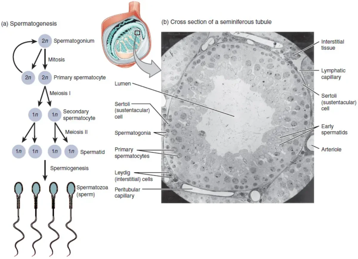
Spermatogonia (2n) - diploid stem cells lining the outer wall of the seminiferous tubule. They divide by mitosis to maintain the stem cell population and produce cells committed to becoming sperm.
Primary spermatocyte (2n) - the spermatogonium that commits to meiosis. Still diploid, with 46 chromosomes (92 chromatids after DNA replication). Undergoes Meiosis I.
Secondary spermatocyte (n) - two haploid cells result from Meiosis I. Each has 23 chromosomes (46 chromatids). Undergoes Meiosis II.
Spermatid (n) - four haploid cells result from Meiosis II. Each has 23 chromosomes (23 chromatids). These are round, immature cells.
Spermatozoon (mature sperm) - spermatids undergo spermiogenesis, a maturation process (NOT a division) that transforms them into streamlined sperm cells.
Net result: 1 spermatogonium produces 4 functional sperm cells.
Spermatogenesis in the seminiferous tubule. Cells progress from the outer wall (spermatogonia) toward the lumen (mature sperm), passing through meiotic divisions along the way. Credit: OpenStax Anatomy & Physiology 2e, CC BY 4.0
Supporting Cells in Spermatogenesis
Sertoli cells (sustentacular cells):
Located within the seminiferous tubules
Nourish and support developing sperm cells
Form the blood-testis barrier - a tight junction barrier that protects developing germ cells from the immune system (sperm are “foreign” to the body’s immune cells because they have a unique haploid genome)
Secrete androgen-binding protein (ABP) to concentrate testosterone near developing sperm
Secrete inhibin, which provides negative feedback on FSH
Activated by FSH from the anterior pituitary
Leydig cells (interstitial cells):
Located in the spaces between seminiferous tubules
Produce testosterone and other androgens
Activated by LH from the anterior pituitary
Spermiogenesis and Sperm Structure
Spermiogenesis transforms round spermatids into specialized sperm cells. No cell division occurs - it is purely a structural remodeling:
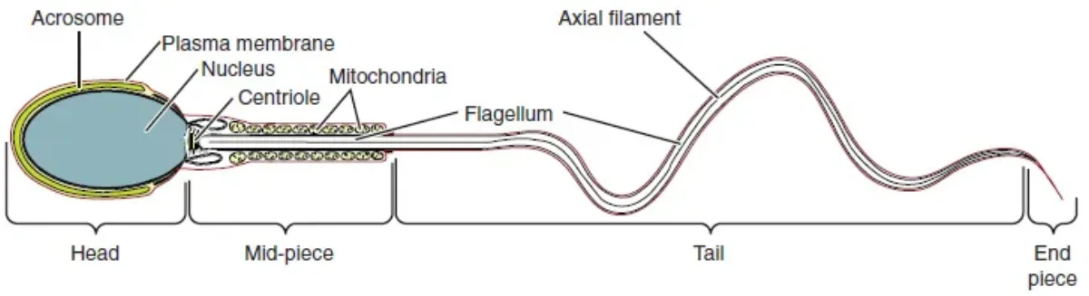
Head - contains highly condensed DNA and is capped by the acrosome, a vesicle filled with hydrolytic enzymes (derived from the Golgi apparatus) needed to penetrate the egg
Midpiece - packed with mitochondria wrapped in a spiral, providing ATP for the flagellum
Tail (flagellum) - a long whip-like structure for motility, built from microtubules in a 9+2 arrangement
Structure of a mature sperm cell: the acrosome-capped head carries DNA, the midpiece provides energy, and the flagellar tail drives motility. Credit: OpenStax Anatomy & Physiology 2e, CC BY 4.0
Oogenesis - The Artisan Process
Oogenesis is dramatically different from spermatogenesis. It begins before birth, pauses for years, produces only one functional gamete per cycle, and involves two arrest points.
The pathway:
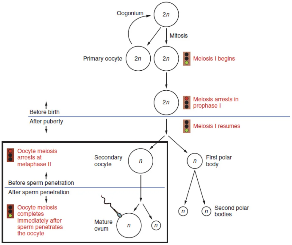
Oogonia (2n) - diploid germ cells in the fetal ovary that multiply by mitosis. By week 20 of fetal development, the female fetus has approximately 7 million oogonia - this is the maximum. No new oogonia are produced after birth.
Primary oocyte (2n) - oogonia enter meiosis I and arrest at Prophase I before birth. They remain frozen at this stage, enclosed in primordial follicles, from before birth until they are recruited for ovulation (potentially decades later). By puberty, only about 400,000 primary oocytes remain.
Secondary oocyte (n) + First polar body - each month after puberty, one primary oocyte completes Meiosis I. The division is unequal: one large cell (the secondary oocyte) gets almost all the cytoplasm, and one tiny cell (the first polar body) gets almost none. The secondary oocyte then arrests at Metaphase II.
Ovulation releases the secondary oocyte (arrested at Metaphase II).
Mature ovum (n) + Second polar body - Meiosis II is only completed if fertilization occurs. The sperm triggers the completion of Meiosis II, producing a mature ovum and a second polar body.
Oogenesis: the pathway from oogonium to mature ovum, showing the two arrest points and unequal divisions that produce polar bodies. Credit: Lumen Learning / OpenStax Anatomy & Physiology, CC BY 4.0
Spermatogenesis vs. Oogenesis - Comparison
Feature
Spermatogenesis
Oogenesis
Location
Seminiferous tubules (testes)
Ovaries
Begins
Puberty
Before birth (fetal development)
Ends
Continues throughout life
Menopause (~age 50)
Functional gametes per meiosis
4 sperm
1 ovum (+ 3 polar bodies)
Daily production
~200-300 million
1 per month
Total lifetime gametes
Trillions
~400 ovulated
Arrest points
None
Prophase I (years) and Metaphase II (until fertilization)
Cytoplasm division
Equal
Unequal (egg gets most)
Cell size
Tiny (60 µm with tail)
Large (120 µm - largest human cell)
Why Unequal Division?
The egg must supply all the cytoplasm, organelles, mRNA, ribosomes, and nutrients needed to sustain the early embryo until it implants and the placenta takes over. Splitting these resources into four equal cells would leave each one too small to support development. By concentrating everything into one large egg and discarding the rest as polar bodies, oogenesis ensures the embryo starts with everything it needs.
At what stages is oogenesis arrested, and what triggers resumption at each point?
Click to reveal answer
First arrest: Prophase I (from before birth until the LH surge triggers ovulation). Second arrest: Metaphase II (from ovulation until fertilization by a sperm). If fertilization does not occur, the oocyte degenerates without completing Meiosis II.
Which cells produce testosterone in the testes, and which hormone stimulates them?
Click to reveal answer
Leydig cells (interstitial cells), stimulated by LH from the anterior pituitary. Leydig cells are located between the seminiferous tubules. Sertoli cells, on the other hand, are inside the tubules and are stimulated by FSH.
What is the function of the acrosome?
Click to reveal answer
The acrosome is a cap-like vesicle at the tip of the sperm head, derived from the Golgi apparatus, containing hydrolytic enzymes. During fertilization, the acrosome reaction releases these enzymes to digest through the zona pellucida (glycoprotein layer) surrounding the egg, allowing the sperm to reach and fuse with the oocyte membrane.
The male reproductive system has one job: produce sperm, mix them with protective fluid, and deliver them to the female reproductive tract. Every structure exists to support some part of that pipeline. If you understand the pathway sperm follow from production to delivery, you understand the entire system.
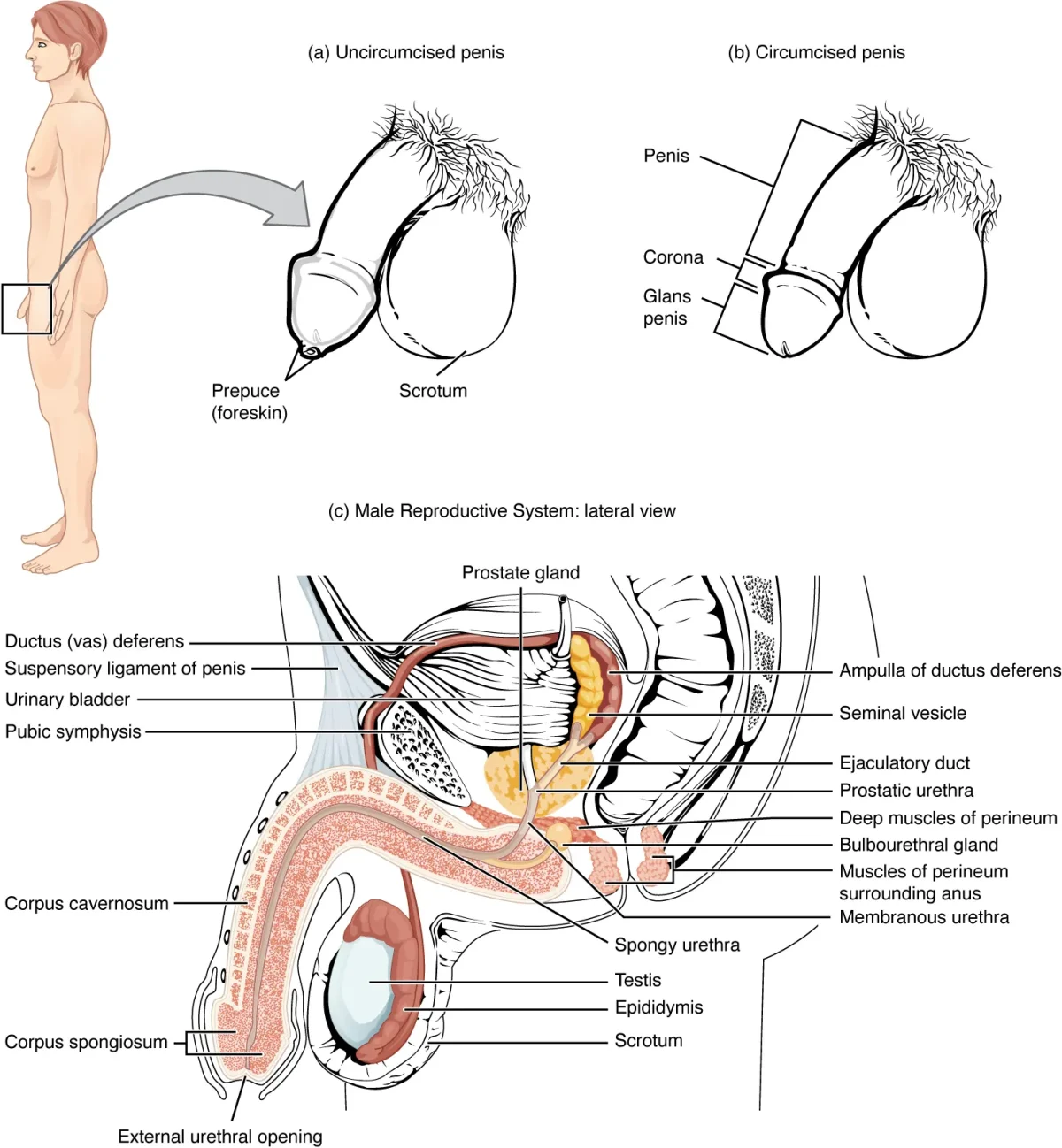
Lateral cross-section of the male reproductive system, showing the pathway from testes to urethra. Credit: OpenStax Anatomy & Physiology 2e, CC BY 4.0
The Testes
The testes are the male gonads - they produce both sperm and testosterone. They are located outside the body in the scrotum, which keeps them 2-4 degrees C cooler than core body temperature. This lower temperature is essential for spermatogenesis - heat kills developing sperm.
The cremaster muscle adjusts the position of the testes: it contracts to pull them closer to the body in cold temperatures (conserve heat) and relaxes to let them hang further away in warm temperatures (cool down).
Inside each testis:
Seminiferous tubules - highly coiled tubes where spermatogenesis occurs. Sertoli cells and developing sperm line the walls; mature sperm are released into the lumen.
Interstitial space - the area between tubules, containing Leydig cells that produce testosterone in response to LH.
The Sperm Pathway
After being produced in the seminiferous tubules, sperm follow this route:
1. Epididymis - A long, coiled tube sitting on top of each testis. Sperm spend 2-3 weeks here, during which they:
Gain motility (learn to swim)
Complete maturation
Are stored until ejaculation
2. Vas deferens (ductus deferens) - A muscular tube that carries sperm from the epididymis up through the spermatic cord, over the bladder, and into the pelvic cavity. Its thick muscular wall produces peristaltic contractions that propel sperm during ejaculation.
This is the structure cut during a vasectomy - a male sterilization procedure. After a vasectomy, sperm are still produced but cannot reach the ejaculatory duct. Ejaculation still occurs (seminal fluid is still produced by the accessory glands), but the ejaculate contains no sperm.
3. Ejaculatory duct - Formed where the vas deferens merges with the duct from the seminal vesicle. Passes through the prostate gland and empties into the urethra.
4. Urethra - The final shared pathway for both urine and semen (but never simultaneously - a sphincter prevents urine from mixing with semen during ejaculation). The urethra runs through the prostate gland and then through the length of the penis.
The Three Accessory Glands
Sperm make up only about 2-5% of semen. The rest is seminal fluid contributed by three glands:
Gland
Contribution to Semen
Key Secretions
Seminal vesicles
~60-70% of volume
Fructose (energy for sperm), prostaglandins, alkaline fluid
Prostate gland
~25-30% of volume
Alkaline fluid, enzymes (PSA), citric acid
Bulbourethral (Cowper’s) glands
~5% of volume
Mucus for lubrication, clears urethra of residual urine
The alkaline nature of seminal fluid is critical - it neutralizes the acidic environment of both the male urethra (residual urine) and the female vagina (pH ~3.5-4.5), protecting sperm from acid damage.
Male Sexual Response
The male sexual response is controlled by the autonomic nervous system:
Arousal and erection - controlled by the parasympathetic nervous system. Parasympathetic fibers cause vasodilation of arteries supplying the erectile tissue (corpora cavernosa and corpus spongiosum), increasing blood flow and producing an erection.
Emission and ejaculation - controlled by the sympathetic nervous system. Sympathetic stimulation causes contractions of the vas deferens, seminal vesicles, and prostate (emission), followed by rhythmic contractions of muscles at the base of the penis (ejaculation).
Resolution - also sympathetic. Arteries constrict, blood drains, penis returns to flaccid state.
Embryological Development
The sex of the embryo is determined at fertilization by the 23rd chromosome pair (XX = female, XY = male), but the reproductive organs do not differentiate until about week 7 of development.
The SRY gene on the Y chromosome activates a genetic cascade that causes undifferentiated gonads to develop into testes.
The testes produce testosterone, which stimulates development of the Wolffian ducts into male internal structures (epididymis, vas deferens, seminal vesicles, ejaculatory ducts).
The testes also produce Mullerian Inhibiting Factor (MIF), which causes the Mullerian ducts to degenerate (preventing female internal structures from forming).
Dihydrotestosterone (DHT) - a more potent androgen converted from testosterone by 5-alpha-reductase - drives the development of external male genitalia (penis, scrotum).
Without the SRY gene (XX embryos), the default pathway is female development.
Name the three accessory glands that contribute to semen and the primary secretion of each.
What is the role of the SRY gene, and what happens if it is absent?
Click to reveal answer
The SRY gene (Sex-determining Region Y) on the Y chromosome triggers the development of testes from undifferentiated gonads. The testes then produce testosterone (Wolffian duct development) and MIF (Mullerian duct degeneration). Without SRY, the gonads develop into ovaries and the default female pathway proceeds - Mullerian ducts form the uterus, fallopian tubes, and upper vagina.
The female reproductive system does something no other organ system attempts: it creates a brand-new environment inside the body every month, optimized for receiving and nourishing a fertilized egg. If no egg implants, the entire setup is torn down and rebuilt from scratch the next month. This monthly construction-and-demolition cycle is the menstrual cycle, and understanding it is essential for the MCAT.
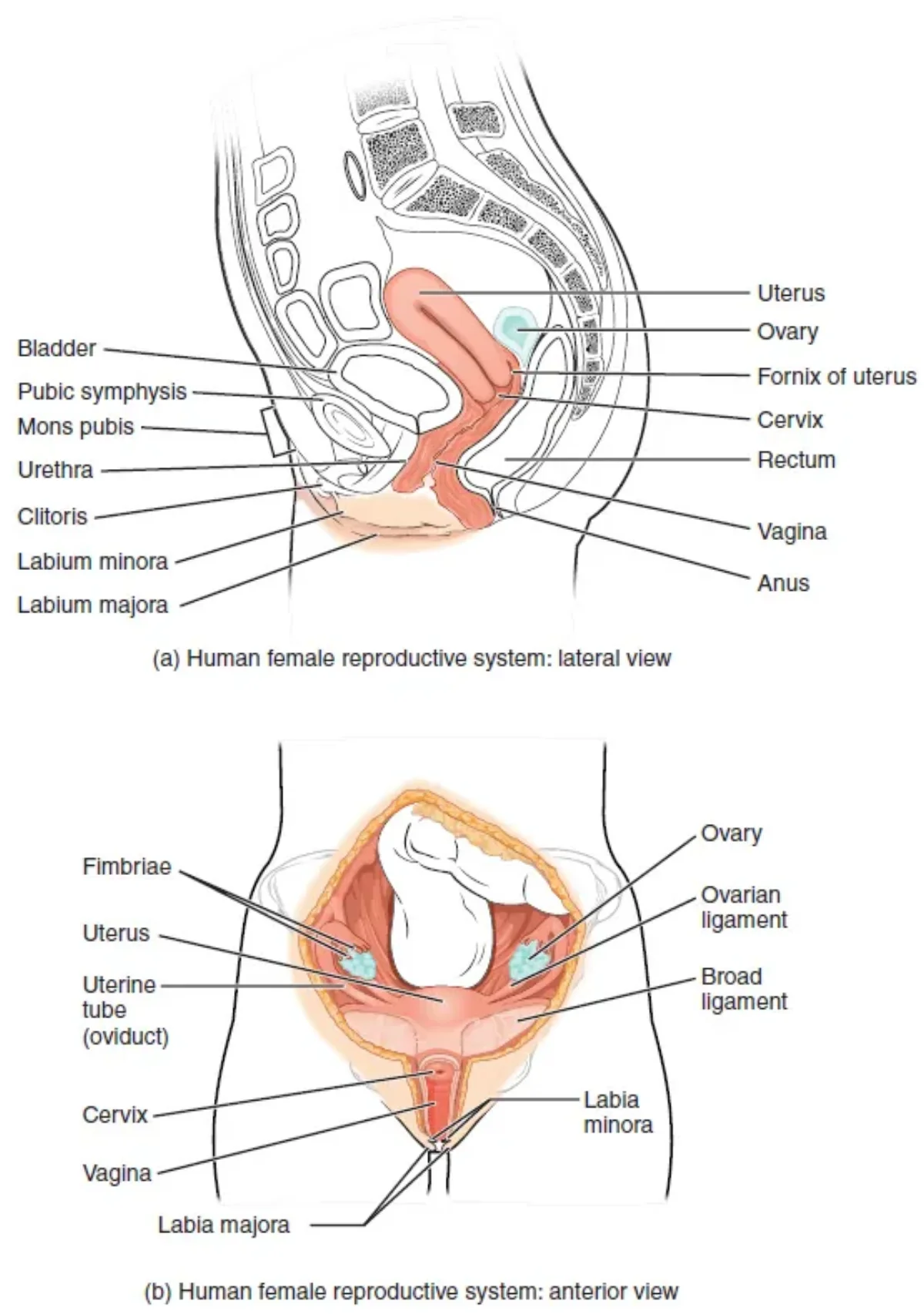
The female reproductive system, showing the ovaries, fallopian tubes (oviducts), uterus, cervix, and vagina. Credit: OpenStax Anatomy & Physiology 2e, CC BY 4.0
The Ovaries
The ovaries are the female gonads - they produce both ova (eggs) and the hormones estrogen and progesterone. Each ovary is about the size and shape of an almond, located on either side of the uterus.
Inside each ovary are follicles - structures that contain and support developing oocytes:
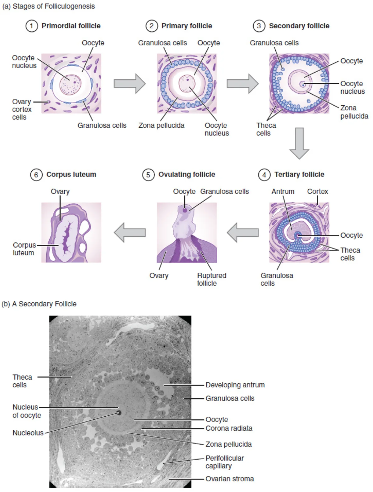
Primordial follicles - the most immature stage, containing a primary oocyte surrounded by a single layer of flat granulosa cells. Present from before birth.
Primary follicles - granulosa cells become cuboidal and begin to multiply. The zona pellucida (a glycoprotein layer) forms around the oocyte.
Secondary follicles - multiple layers of granulosa cells, plus an outer layer of theca cells. A fluid-filled cavity (antrum) begins to form.
Graafian (tertiary) follicle - the fully mature follicle with a large antrum. Contains the secondary oocyte ready for ovulation. Only one follicle per cycle typically reaches this stage (the dominant follicle); the rest undergo atresia (degeneration).
Stages of folliculogenesis from primordial follicle to corpus luteum, with an electron micrograph showing internal follicle structure. Credit: Lumen Learning / OpenStax Anatomy & Physiology, CC BY 4.0
The Egg’s Journey
After ovulation, the egg follows this path:
Ovary - the Graafian follicle ruptures and releases the secondary oocyte into the peritoneal cavity.
Fimbriae - finger-like projections at the end of the fallopian tube sweep the oocyte into the tube. The egg does not physically “jump” from the ovary into the tube - it is captured by these waving fimbriae.
Fallopian tube (oviduct) - the oocyte travels down the tube, propelled by cilia lining the tube’s inner surface and by peristaltic contractions. Fertilization normally occurs here, specifically in the ampulla (the widest section, closest to the ovary).
Uterus - if fertilized, the embryo arrives and implants in the endometrium about 6-7 days after fertilization. If unfertilized, the oocyte degenerates.
The Uterus
The uterus is a pear-shaped, muscular organ where the embryo implants and develops during pregnancy. It has three layers:
Endometrium (inner lining) - the layer that thickens each month and is shed during menstruation. Contains glands and blood vessels.
Myometrium (middle layer) - thick smooth muscle that contracts during labor to push the baby out. Also responsible for menstrual cramps.
Perimetrium (outer layer) - the serous membrane covering the uterus.
The Cervix and Vagina
Cervix - the narrow, lower portion of the uterus that opens into the vagina. It produces mucus that changes consistency throughout the menstrual cycle (thin and watery at ovulation to help sperm pass; thick and sticky at other times to block sperm and pathogens). The cervix dilates to approximately 10 cm during labor.
Vagina - a muscular canal connecting the cervix to the outside of the body. It serves as the birth canal, the passage for menstrual blood, and the receptacle for the penis during intercourse.
The external female genitalia (collectively called the vulva) include the labia majora, labia minora, clitoris, and the openings of the urethra and vagina. In females, the urinary and reproductive tracts are completely separate (unlike in males, where the urethra carries both urine and semen).
The Ovarian Cycle
The ovarian cycle describes what happens in the ovaries over a typical 28-day period:
Follicular Phase (Days 1-14):
Multiple follicles begin developing under the influence of FSH.
Granulosa cells in the follicles produce increasing amounts of estrogen.
One dominant follicle emerges; the rest degenerate (atresia).
Rising estrogen causes the endometrium to thicken (proliferative phase of the uterine cycle).
At the end of this phase, estrogen levels peak and trigger the LH surge.
Ovulation (Day ~14):
The LH surge causes the Graafian follicle to rupture, releasing the secondary oocyte.
The oocyte completes Meiosis I just before ovulation, producing the secondary oocyte and a polar body.
This is the most fertile window of the cycle.
Luteal Phase (Days 15-28):
The ruptured follicle transforms into the corpus luteum, which secretes progesterone (primarily) and some estrogen.
Progesterone prepares the endometrium for implantation (secretory phase of the uterine cycle).
High progesterone exerts negative feedback on GnRH, FSH, and LH, preventing additional ovulation.
If no fertilization occurs, the corpus luteum degenerates after about 12 days, becoming the corpus albicans.
The drop in progesterone and estrogen triggers menstruation.
The Uterine Cycle
The uterine cycle describes what happens to the endometrium, driven by the hormones of the ovarian cycle:
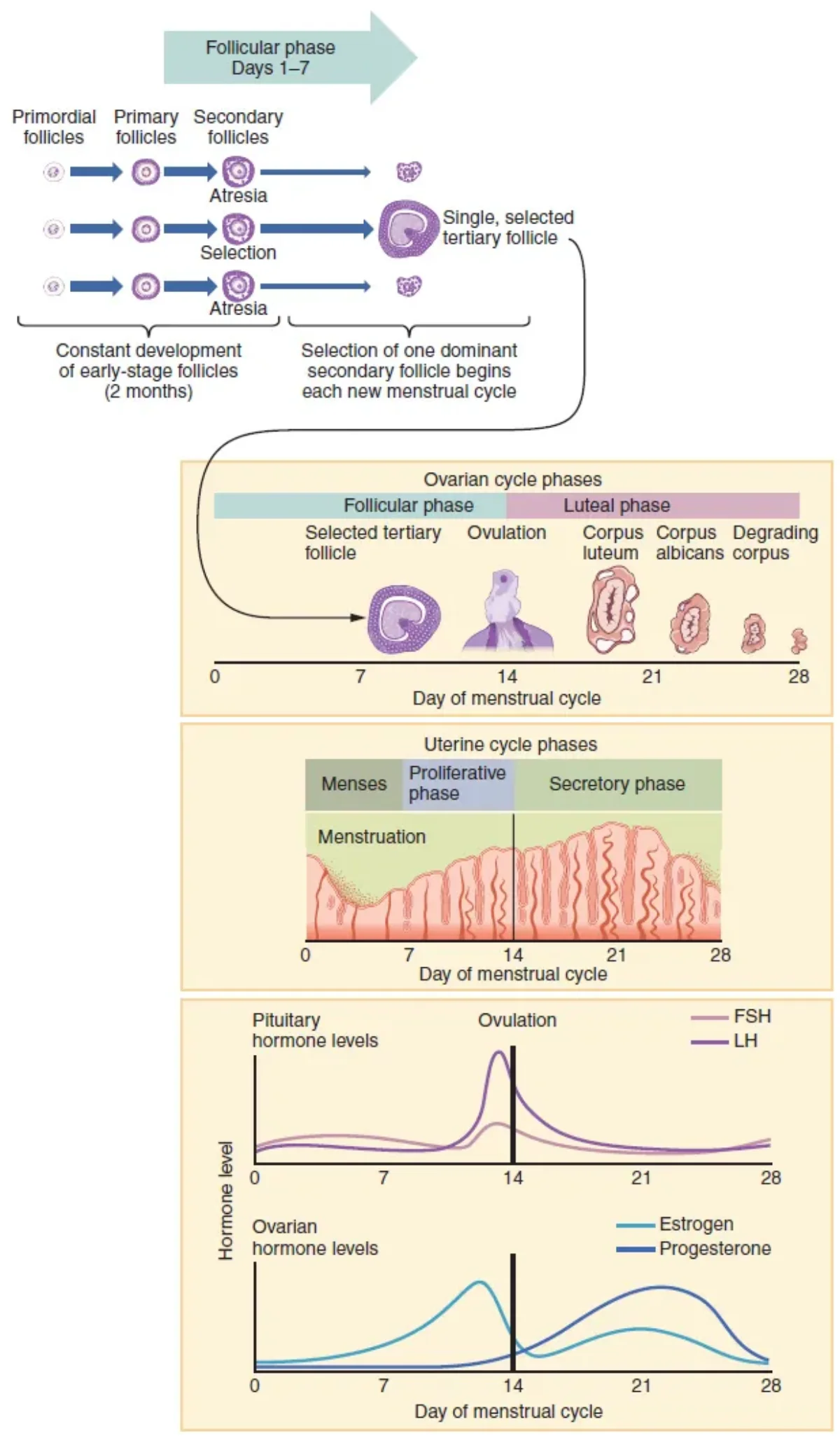
The correlation between hormone levels, ovarian events, and uterine changes across a 28-day cycle. The LH surge triggers ovulation around day 14. Credit: OpenStax Anatomy & Physiology 2e, CC BY 4.0
How to Read the Menstrual Hormone Graph
MCAT passages almost always show some version of this four-line graph. Here is how to identify each curve without memorizing axes:
FSH - rises early (days 1-5) to recruit follicles, drops as estrogen feeds back, then a small mid-cycle bump at day ~14.
Estrogen - low at menses, climbs steeply during the follicular phase, peaks just before ovulation, dips, then a smaller luteal peak.
LH - flat baseline, then a sharp narrow spike around day 14 (the LH surge). This spike is the easiest landmark on the whole graph.
Progesterone - flat and low until after ovulation, then climbs to a broad luteal peak (day ~21), then drops before menses.
If Pregnancy Occurs
If fertilization and implantation occur, the embryo’s developing placenta secretes human chorionic gonadotropin (hCG). hCG “rescues” the corpus luteum from degeneration, keeping progesterone levels high and maintaining the endometrium. This is the hormone detected by pregnancy tests. After about 10-12 weeks, the placenta takes over progesterone production, and the corpus luteum is no longer needed.
What happens to the corpus luteum if fertilization does NOT occur?
Click to reveal answer
Without hCG from an implanting embryo, the corpus luteum degenerates after about 12 days, becoming the corpus albicans (a scar-like remnant). The resulting drop in progesterone and estrogen triggers shedding of the endometrium (menstruation) and allows FSH/LH to rise again, starting a new cycle.
Where in the female reproductive tract does fertilization normally occur?
Click to reveal answer
In the ampulla of the fallopian tube (oviduct) - the widest section closest to the ovary. The egg is captured by the fimbriae after ovulation and travels through the tube. If sperm are present, fertilization occurs here, and the embryo then continues to the uterus for implantation.
The reproductive system runs on a chain of command that starts in the brain and ends in the gonads. If you understand this hierarchy - who gives the orders, who carries them out, and who reports back - you can answer almost any MCAT question about reproductive hormones.
The HPG Axis
The hypothalamic-pituitary-gonadal (HPG) axis is the master control system for reproduction in both males and females:
Hypothalamus releases GnRH (gonadotropin-releasing hormone) in pulsatile bursts.
Anterior pituitary responds to GnRH by releasing FSH (follicle-stimulating hormone) and LH (luteinizing hormone).
Gonads respond to FSH and LH by producing sex hormones and gametes.
Sex hormones feed back to the hypothalamus and pituitary to regulate the system.
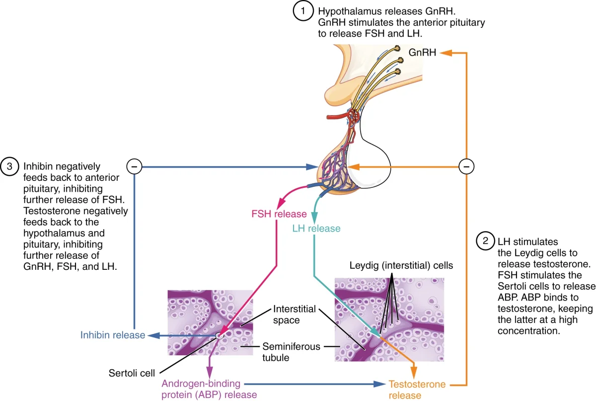
The HPG axis in males. GnRH from the hypothalamus stimulates FSH and LH release. LH drives testosterone production from Leydig cells; FSH supports spermatogenesis via Sertoli cells. Testosterone and inhibin provide negative feedback. Credit: OpenStax Anatomy & Physiology 2e, CC BY 4.0
HPG Axis in Males
Hormone
Source
Target
Action
GnRH
Hypothalamus
Anterior pituitary
Stimulates FSH and LH release
FSH
Anterior pituitary
Sertoli cells
Supports spermatogenesis, stimulates ABP production
LH
Anterior pituitary
Leydig cells
Stimulates testosterone production
Testosterone
Leydig cells
Whole body + hypothalamus/pituitary
Male development, secondary sex characteristics; negative feedback on GnRH and LH
Inhibin
Sertoli cells
Anterior pituitary
Negative feedback specifically on FSH
DHT
Target tissues (from testosterone)
External genitalia, prostate, hair follicles
More potent androgen for specific targets
The male system is relatively straightforward: it is a simple negative feedback loop. Testosterone and inhibin keep GnRH, LH, and FSH in check. There is no positive feedback in the male system.
HPG Axis in Females
The female system is more complex because it involves cycling hormone levels and a switch between negative and positive feedback.
Hormone
Source
Target
Action
GnRH
Hypothalamus
Anterior pituitary
Stimulates FSH and LH release
FSH
Anterior pituitary
Granulosa cells
Stimulates follicle growth and estrogen production
LH
Anterior pituitary
Theca cells, corpus luteum
Stimulates androgen production (theca), triggers ovulation, maintains corpus luteum
Estrogen
Granulosa cells
Whole body + hypothalamus/pituitary
Female development, endometrial growth; negative feedback at low levels, POSITIVE feedback at high levels (triggers LH surge)
Progesterone
Corpus luteum (then placenta)
Uterus + hypothalamus/pituitary
Maintains endometrium, negative feedback on GnRH/LH/FSH
Inhibin
Granulosa cells
Anterior pituitary
Negative feedback on FSH
hCG
Embryonic trophoblast/placenta
Corpus luteum
Maintains corpus luteum during early pregnancy
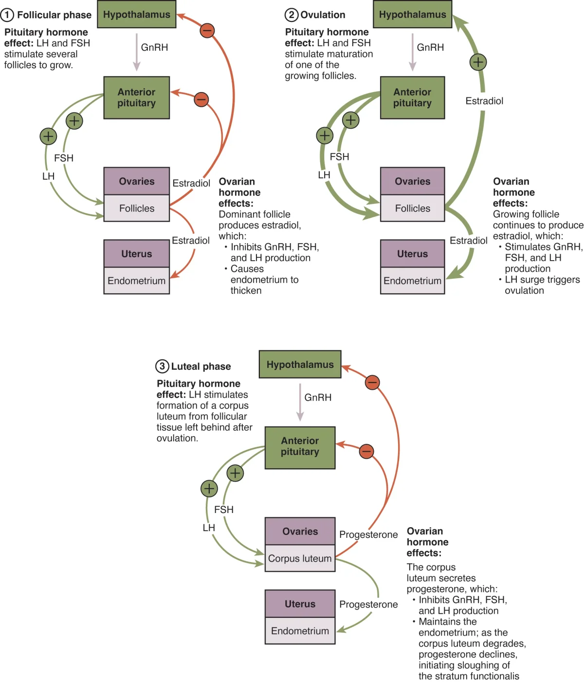
Hormonal regulation of the female reproductive system, showing the interplay between GnRH, FSH, LH, estrogen, and progesterone. Credit: OpenStax Anatomy & Physiology 2e, CC BY 4.0
The Estrogen Feedback Switch
This is the single most important concept in reproductive endocrinology:
Low to moderate estrogen → negative feedback → suppresses GnRH, FSH, and LH
Sustained high estrogen (from the dominant follicle in late follicular phase) → positive feedback → stimulates a surge of GnRH and LH → triggers ovulation
After ovulation, the corpus luteum produces progesterone, which exerts strong negative feedback, preventing another LH surge and another ovulation for the rest of the cycle.
Estrogen Cooperation: Theca and Granulosa Cells
Estrogen production in the ovary requires teamwork between two cell types:
Theca cells (outer layer of the follicle) - stimulated by LH to produce androgens (androstenedione)
Granulosa cells (inner layer) - stimulated by FSH, they express the enzyme aromatase, which converts the androgens into estrogen
This is called the two-cell, two-gonadotropin model. Neither cell type can make estrogen alone - theca cells make the precursor, granulosa cells convert it.
Puberty
Puberty is triggered by activation of the HPG axis - the hypothalamus begins pulsatile GnRH secretion that was suppressed during childhood. Rising sex hormones then drive secondary sex characteristics.
Males (testosterone): testis/penis growth, spermatogenesis, deeper voice, facial and body hair, muscle mass, growth spurt.
Females (estrogen): breast development (thelarche, usually first), widened hips, body fat redistribution, menarche, growth spurt.
Both testosterone and estrogen ultimately cause fusion of the epiphyseal (growth) plates, ending height growth. Children who enter puberty early tend to be shorter as adults because their plates close sooner.
Hormonal Contraception as a Feedback Demonstration
The main MCAT point about contraception is that it exploits negative feedback. Combined oral contraceptives (synthetic estrogen + progestin) hold estrogen and progesterone steadily high, which suppresses GnRH, FSH, and LH. Without the FSH rise and LH surge, follicle development and ovulation cannot occur.
The body essentially “thinks” it is already in the luteal phase or pregnant. This is the same mechanism the corpus luteum uses to prevent a second ovulation within one cycle.
Menopause
As a woman ages, her ovaries run out of viable follicles and stop responding to FSH and LH. Estrogen and progesterone fall, and follicle development ceases. Menopause typically occurs between ages 45 and 55.
The MCAT-testable signature is a paired hormone shift:
Estrogen and progesterone drop because the ovaries no longer produce functional follicles or corpora lutea.
FSH and LH rise sharply because their negative feedback has been removed. The pituitary keeps shouting at ovaries that cannot answer.
Elevated FSH is the clinical marker used to confirm menopause. Secondary effects include endometrial atrophy, loss of bone density (estrogen normally supports bone), and vasomotor symptoms like hot flashes.
hCG - The LH Mimic
hCG (human chorionic gonadotropin) is secreted by the trophoblast once the blastocyst implants. Structurally it looks like LH, so it binds the LH receptor on the corpus luteum and keeps it producing progesterone past its normal 12-day lifespan. That sustained progesterone prevents menstruation and protects the embryo.
After ~10-12 weeks, the placenta takes over progesterone production and hCG declines. hCG in urine or blood is the molecule detected by pregnancy tests.
Key Hormone Summary
Hormone
Primary Source
Key Functions
GnRH
Hypothalamus
Stimulates FSH and LH release; pulsatile secretion
FSH
Anterior pituitary
Follicle growth (females), spermatogenesis support (males)
LH
Anterior pituitary
Ovulation trigger, corpus luteum formation (females); testosterone production (males)
Endometrial maintenance, negative feedback, pregnancy support
Testosterone
Leydig cells, adrenal cortex
Male development, spermatogenesis, secondary sex characteristics
Inhibin
Sertoli cells (males), granulosa cells (females)
Negative feedback specifically on FSH
hCG
Trophoblast/placenta
Maintains corpus luteum in early pregnancy
When does estrogen switch from negative to positive feedback, and what is the result?
Click to reveal answer
When estrogen reaches a sustained high level in the late follicular phase (from the dominant follicle), it switches to positive feedback on the hypothalamus and anterior pituitary. This triggers a massive LH surge (and a smaller FSH surge), which causes the Graafian follicle to rupture and release the egg (ovulation). This is the only example of positive feedback in the HPG axis.
What is the "two-cell, two-gonadotropin model" of estrogen synthesis?
Click to reveal answer
Theca cells (stimulated by LH) produce androgens (androstenedione). Granulosa cells (stimulated by FSH) use the enzyme aromatase to convert those androgens into estrogen. Neither cell type can make estrogen alone - it requires both cell types and both gonadotropins working together.
Of the 200-300 million sperm released during ejaculation, only a few hundred reach the egg in the fallopian tube. Of those, only one will fertilize it. The journey from vagina to ampulla of the fallopian tube is a brutal obstacle course - acidic pH, cervical mucus, immune cells, wrong turns, and sheer distance all eliminate the vast majority. The sperm that makes it is not the “strongest” - it is the luckiest, with help from some clever molecular biology at the finish line.
Capacitation
Freshly ejaculated sperm cannot fertilize an egg. They must first undergo capacitation - a process that occurs in the female reproductive tract over several hours. During capacitation:
Cholesterol is removed from the sperm membrane, making it more fluid and reactive
Intracellular calcium levels increase
The sperm becomes hyperactive (vigorous, whip-like tail movement)
The acrosomal membrane becomes primed for the acrosome reaction
Only capacitated sperm can undergo the acrosome reaction and bind to the zona pellucida.
Penetrating the Egg - Two Barriers
Barrier 1: The Corona Radiata
The corona radiata is a layer of granulosa cells surrounding the egg. Sperm must physically push through these cells, aided by hyaluronidase and other enzymes released from the acrosome and from the collective action of many sperm (though only one will ultimately fertilize the egg).
Barrier 2: The Zona Pellucida
The zona pellucida is a thick glycoprotein shell around the oocyte. It contains specific receptor proteins:
ZP3 - the sperm receptor. It binds species-specific sperm and triggers the acrosome reaction.
ZP2 - helps maintain sperm binding after the acrosome reaction.
The Acrosome Reaction
When a capacitated sperm binds to ZP3 on the zona pellucida:
The acrosomal membrane fuses with the sperm’s plasma membrane
Hydrolytic enzymes (including acrosin) are released
These enzymes digest a path through the zona pellucida
The sperm reaches the oocyte’s plasma membrane and fuses with it
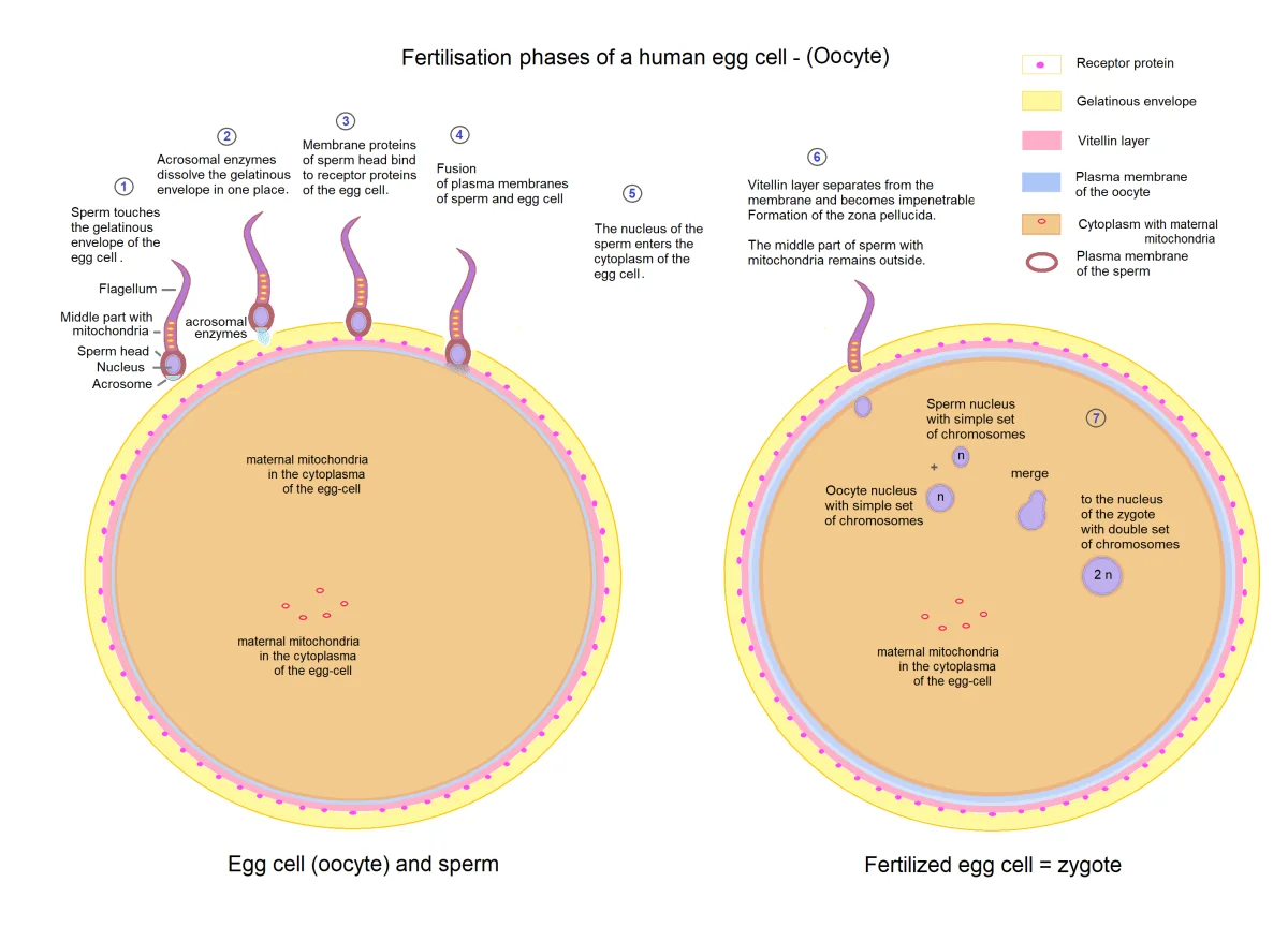
The process of fertilization: sperm penetrates the corona radiata, undergoes the acrosome reaction through the zona pellucida, fuses with the oocyte membrane, and triggers the cortical reaction to prevent polyspermy. Credit: OpenStax, CC BY 4.0
The Cortical Reaction - Blocking Polyspermy
The moment one sperm fuses with the oocyte membrane, two things happen rapidly to prevent additional sperm from entering:
Fast block (immediate):
The oocyte membrane depolarizes (membrane potential shifts from negative to positive)
This electrical change prevents other sperm from fusing with the membrane
Temporary measure, lasting only a few minutes
Slow block (permanent):
Sperm entry triggers a wave of calcium ions (Ca2+) released from the oocyte’s endoplasmic reticulum
This calcium wave causes cortical granules (vesicles just beneath the oocyte membrane) to fuse with the membrane and release their contents into the space between the membrane and the zona pellucida
Cortical granule enzymes harden the zona pellucida into the fertilization membrane, destroying ZP3 receptors and making it impenetrable to additional sperm
Polyspermy (fertilization by more than one sperm) would give the zygote too many chromosomes, which is lethal. The fast and slow blocks ensure this does not happen.
Completing Meiosis and Forming the Zygote
Remember that the “egg” released at ovulation is actually a secondary oocyte arrested at Metaphase II. Sperm entry triggers the completion of Meiosis II:
The secondary oocyte completes Meiosis II, producing a mature ovum and a second polar body (which degenerates).
The sperm nucleus (now called the male pronucleus) and the ovum nucleus (the female pronucleus) each form within the cell.
The two pronuclei migrate toward each other and their membranes dissolve, combining their chromosomes.
The resulting cell - the zygote - has the full diploid complement of 46 chromosomes (23 from sperm + 23 from egg).
The zygote is the first cell of the new organism.
Early Cleavage
The zygote immediately begins dividing as it travels down the fallopian tube toward the uterus:
Cleavage - rapid mitotic divisions that increase cell number without increasing overall size. Each division produces smaller and smaller cells called blastomeres.
2-cell stage → 4-cell → 8-cell → 16-cell
By the 16-32 cell stage, the embryo is a solid ball of cells called the morula (Latin for “mulberry” - it looks like one)
The morula then develops a fluid-filled cavity (the blastocoel), becoming the blastocyst (around day 5)
The Blastocyst and Implantation
The blastocyst has two distinct cell populations:
Inner cell mass (ICM) - a cluster of cells on one side that will become the embryo proper. These cells are pluripotent.
Trophoblast - the outer layer of cells surrounding the blastocoel. It will form the placenta and other extraembryonic membranes. The trophoblast secretes enzymes that allow the blastocyst to burrow into the endometrium.
Implantation occurs about 6-7 days after fertilization. The trophoblast invades the endometrial lining and establishes connections with the mother’s blood supply. Once implanted, the trophoblast begins secreting hCG to maintain the corpus luteum and its progesterone production. From here, the blastocyst undergoes gastrulation and organogenesis, which we cover in early embryonic development.
Indeterminate vs. Determinate Cleavage
Indeterminate cleavage (humans and other deuterostomes) - each early blastomere retains the ability to develop into a complete organism. This is why identical twins can form if the embryo splits in the early cleavage stages.
Determinate cleavage (most protostomes) - the fate of each blastomere is fixed from the first division. Separating cells would not produce complete organisms.
Extraembryonic Membranes
Four membranes develop to support the embryo:
Membrane
Function
Chorion
Outermost membrane; develops into the fetal part of the placenta; chorionic villi establish maternal-fetal exchange
Amnion
Encloses the embryo in the amniotic sac filled with amniotic fluid; cushions and protects
Allantois
Contributes to umbilical cord and bladder development; waste exchange in non-mammalian vertebrates
Yolk sac
Early blood cell formation and nutrient transfer; largely vestigial in humans (no yolk)
The umbilical cord connects the fetus to the placenta and contains two umbilical arteries (carrying deoxygenated blood from fetus to placenta) and one umbilical vein (carrying oxygenated blood from placenta to fetus).
What are the fast and slow blocks to polyspermy?
Click to reveal answer
Fast block: membrane depolarization of the oocyte (immediate, temporary, electrical). Slow block: the cortical reaction - calcium-triggered release of cortical granules that harden the zona pellucida into the fertilization membrane, destroying sperm receptors (takes minutes, permanent, chemical). Together they prevent more than one sperm from fertilizing the egg.
What is the role of hCG in early pregnancy?
Click to reveal answer
hCG (human chorionic gonadotropin) is secreted by the trophoblast after implantation. It maintains the corpus luteum, preventing it from degenerating. This keeps progesterone levels high, which maintains the endometrium and prevents menstruation. hCG is the hormone detected by pregnancy tests. After about 10-12 weeks, the placenta takes over progesterone production.