
Mitosis
Imagine photocopying a book. You put the original on the glass, press the button, and get an identical copy. The original is unchanged, the copy is perfect, and now you have two identical books. That is mitosis - one cell becomes two genetically identical daughter cells.
Mitosis is the “factory” side of cell division. It is how your body grows from a single fertilized egg into trillions of cells, how it replaces the skin cells you shed every day, and how it repairs a wound. The goal is always the same: make a perfect copy.
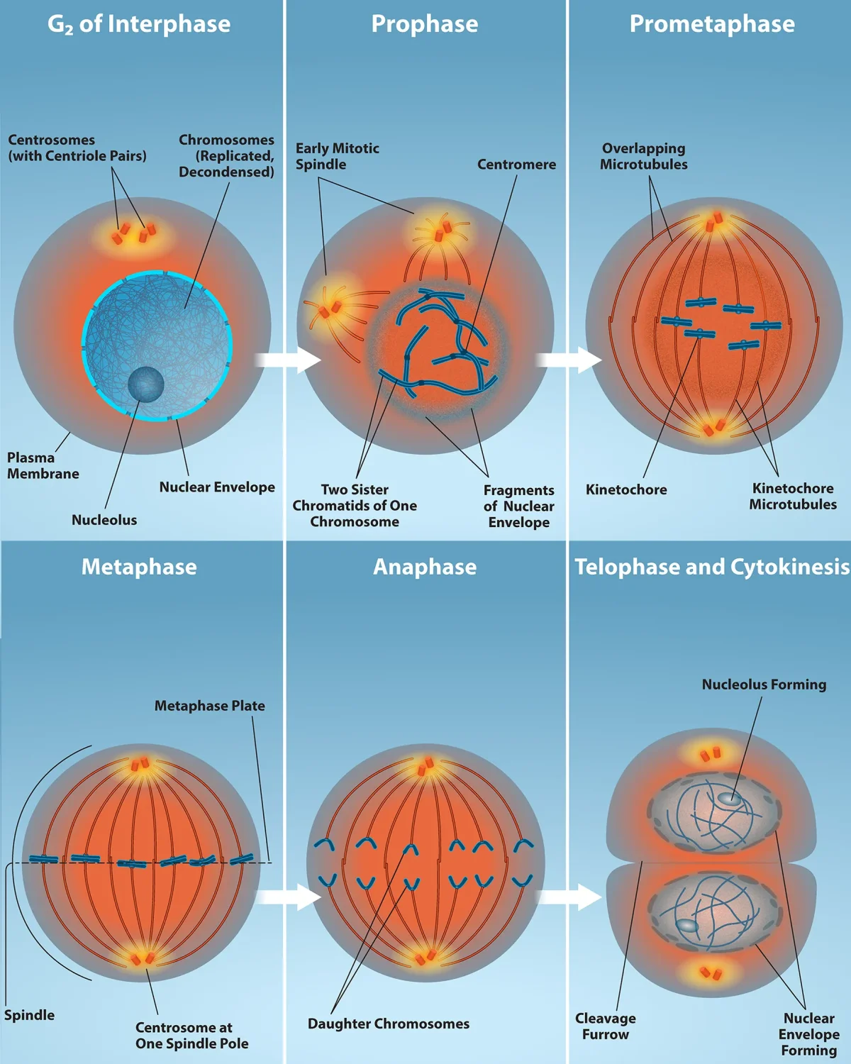

Prophase - “Prepare for War”
Prophase is the cell gearing up for division. Several things happen simultaneously:
- Chromatin condenses into visible chromosomes. The loose DNA threads tighten and compact so they can be moved without tangling. Think of winding up earbuds before putting them in your pocket.
- Each chromosome is visible as two sister chromatids joined at the centromere.
- The nuclear envelope begins to break down. This must happen so the spindle fibers can reach the chromosomes.
- The nucleolus disappears.
- Centrosomes migrate to opposite poles of the cell. As they move, they begin assembling the mitotic spindle - a network of microtubules that will pull the chromosomes apart.
- Aster fibers radiate from each centrosome and anchor it to the cell membrane for stability.
Some textbooks split this into prophase and prometaphase. In prometaphase, the nuclear envelope is fully dissolved and spindle fibers attach to the chromosomes at their kinetochores - protein structures on the centromere that serve as attachment points for microtubules.
Metaphase - “Middle Lineup”
The chromosomes are dragged to the center of the cell and align along the metaphase plate (an imaginary equator halfway between the two poles).
- Each chromosome is attached to spindle fibers from both poles via its kinetochores.
- Tension from opposite spindle fibers holds each chromosome at the midline.
- The cell runs its spindle assembly checkpoint (M checkpoint) here: are all chromosomes properly attached to spindle fibers from both poles? If not, division stalls until the problem is fixed.
Memory trick: Metaphase = Middle. Chromosomes line up in the middle.
Anaphase - “Apart”
This is the shortest and most dramatic stage:
- Cohesin proteins holding sister chromatids together are cleaved by the enzyme separase.
- Sister chromatids are pulled to opposite poles by the shortening of kinetochore microtubules.
- Simultaneously, non-kinetochore microtubules lengthen, pushing the poles further apart and elongating the cell.
- By the end of anaphase, each pole has a complete set of 46 chromosomes.
Memory trick: Anaphase = Apart. Chromatids are pulled apart.
Telophase - “Two Nuclei”
Telophase is essentially prophase in reverse:
- Nuclear envelopes reform around each set of chromosomes.
- Chromosomes decondense back into loose chromatin.
- Nucleoli reappear.
- The spindle apparatus disassembles.
At this point, you have a single cell with two complete nuclei.
Memory trick: Telophase = Two nuclei.
Cytokinesis - “Cut the Cell”
Cytokinesis divides the cytoplasm, completing the physical separation into two daughter cells. It usually begins during late anaphase or telophase.
In animal cells: A ring of actin and myosin filaments (the contractile ring) pinches the cell membrane inward, forming a cleavage furrow that deepens until the cell splits in two. Think of tightening a drawstring on a bag.
In plant cells: A rigid cell wall prevents pinching. Instead, vesicles from the Golgi apparatus line up at the cell’s equator and fuse to form a cell plate, which grows outward until it separates the two cells completely.
Mitosis Summary Table
| Stage | Key Events | Memory Cue |
|---|---|---|
| Prophase | Chromatin condenses, nuclear envelope breaks down, spindle forms | ”Prepare” |
| Metaphase | Chromosomes align at metaphase plate, spindle checkpoint | ”Middle” |
| Anaphase | Sister chromatids separate and move to opposite poles | ”Apart” |
| Telophase | Nuclear envelopes reform, chromosomes decondense | ”Two nuclei” |
| Cytokinesis | Cytoplasm divides (cleavage furrow in animals, cell plate in plants) | “Cut” |
Key Outcome
Mitosis produces 2 daughter cells that are genetically identical to the parent cell and to each other. Each has 46 chromosomes (2n).