
Gametogenesis
The body needs to produce sex cells (gametes) that have half the normal chromosome number. That is the job of meiosis. But meiosis does not happen the same way in males and females. The male version is a mass production line. The female version is an artisan operation. Understanding the differences between spermatogenesis and oogenesis is one of the highest-yield topics for the MCAT.
Spermatogenesis - The Assembly Line
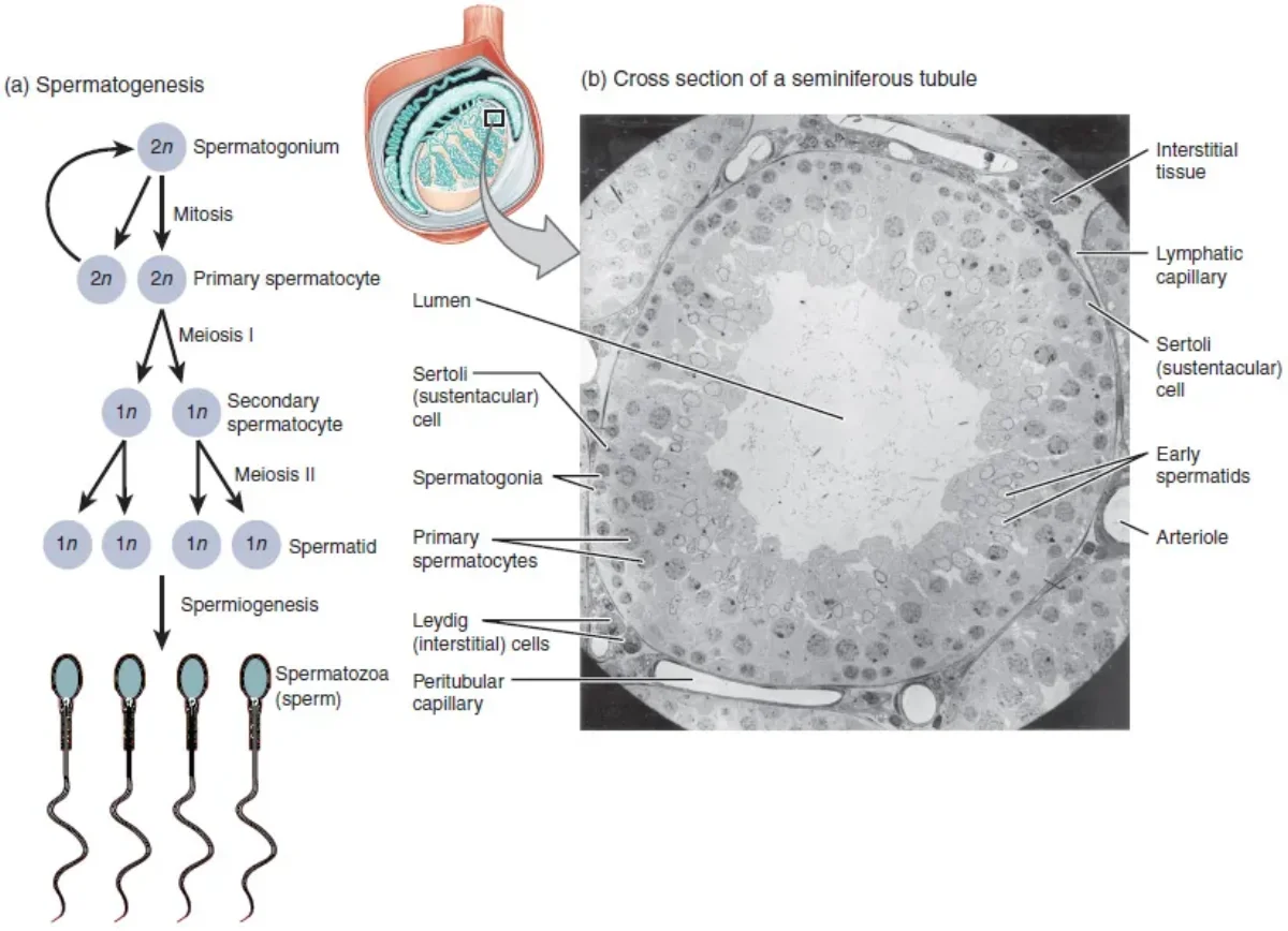
Spermatogenesis occurs in the seminiferous tubules of the testes, beginning at puberty and continuing throughout life. The entire process takes about 65-75 days and produces approximately 200-300 million sperm per day.
The pathway:
-
Spermatogonia (2n) - diploid stem cells lining the outer wall of the seminiferous tubule. They divide by mitosis to maintain the stem cell population and produce cells committed to becoming sperm.
-
Primary spermatocyte (2n) - the spermatogonium that commits to meiosis. Still diploid, with 46 chromosomes (92 chromatids after DNA replication). Undergoes Meiosis I.
-
Secondary spermatocyte (n) - two haploid cells result from Meiosis I. Each has 23 chromosomes (46 chromatids). Undergoes Meiosis II.
-
Spermatid (n) - four haploid cells result from Meiosis II. Each has 23 chromosomes (23 chromatids). These are round, immature cells.
-
Spermatozoon (mature sperm) - spermatids undergo spermiogenesis, a maturation process (NOT a division) that transforms them into streamlined sperm cells.
Net result: 1 spermatogonium produces 4 functional sperm cells.
Supporting Cells in Spermatogenesis
Sertoli cells (sustentacular cells):
- Located within the seminiferous tubules
- Nourish and support developing sperm cells
- Form the blood-testis barrier - a tight junction barrier that protects developing germ cells from the immune system (sperm are “foreign” to the body’s immune cells because they have a unique haploid genome)
- Secrete androgen-binding protein (ABP) to concentrate testosterone near developing sperm
- Secrete inhibin, which provides negative feedback on FSH
- Activated by FSH from the anterior pituitary
Leydig cells (interstitial cells):
- Located in the spaces between seminiferous tubules
- Produce testosterone and other androgens
- Activated by LH from the anterior pituitary
Spermiogenesis and Sperm Structure
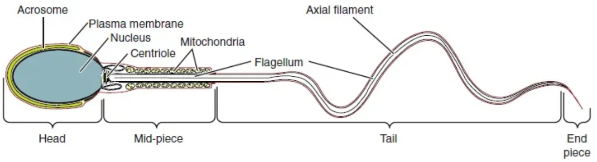
Spermiogenesis transforms round spermatids into specialized sperm cells. No cell division occurs - it is purely a structural remodeling:
- Head - contains highly condensed DNA and is capped by the acrosome, a vesicle filled with hydrolytic enzymes (derived from the Golgi apparatus) needed to penetrate the egg
- Midpiece - packed with mitochondria wrapped in a spiral, providing ATP for the flagellum
- Tail (flagellum) - a long whip-like structure for motility, built from microtubules in a 9+2 arrangement
Oogenesis - The Artisan Process
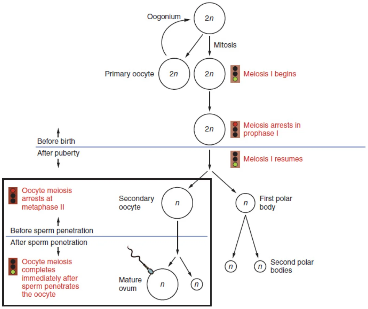
Oogenesis is dramatically different from spermatogenesis. It begins before birth, pauses for years, produces only one functional gamete per cycle, and involves two arrest points.
The pathway:
-
Oogonia (2n) - diploid germ cells in the fetal ovary that multiply by mitosis. By week 20 of fetal development, the female fetus has approximately 7 million oogonia - this is the maximum. No new oogonia are produced after birth.
-
Primary oocyte (2n) - oogonia enter meiosis I and arrest at Prophase I before birth. They remain frozen at this stage, enclosed in primordial follicles, from before birth until they are recruited for ovulation (potentially decades later). By puberty, only about 400,000 primary oocytes remain.
-
Secondary oocyte (n) + First polar body - each month after puberty, one primary oocyte completes Meiosis I. The division is unequal: one large cell (the secondary oocyte) gets almost all the cytoplasm, and one tiny cell (the first polar body) gets almost none. The secondary oocyte then arrests at Metaphase II.
-
Ovulation releases the secondary oocyte (arrested at Metaphase II).
-
Mature ovum (n) + Second polar body - Meiosis II is only completed if fertilization occurs. The sperm triggers the completion of Meiosis II, producing a mature ovum and a second polar body.
Net result: 1 oogonium produces 1 functional egg + 3 polar bodies (which degenerate).
Spermatogenesis vs. Oogenesis - Comparison
| Feature | Spermatogenesis | Oogenesis |
|---|---|---|
| Location | Seminiferous tubules (testes) | Ovaries |
| Begins | Puberty | Before birth (fetal development) |
| Ends | Continues throughout life | Menopause (~age 50) |
| Functional gametes per meiosis | 4 sperm | 1 ovum (+ 3 polar bodies) |
| Daily production | ~200-300 million | 1 per month |
| Total lifetime gametes | Trillions | ~400 ovulated |
| Arrest points | None | Prophase I (years) and Metaphase II (until fertilization) |
| Cytoplasm division | Equal | Unequal (egg gets most) |
| Cell size | Tiny (60 µm with tail) | Large (120 µm - largest human cell) |
Why Unequal Division?
The egg must supply all the cytoplasm, organelles, mRNA, ribosomes, and nutrients needed to sustain the early embryo until it implants and the placenta takes over. Splitting these resources into four equal cells would leave each one too small to support development. By concentrating everything into one large egg and discarding the rest as polar bodies, oogenesis ensures the embryo starts with everything it needs.