
Male Anatomy
The male reproductive system has one job: produce sperm, mix them with protective fluid, and deliver them to the female reproductive tract. Every structure exists to support some part of that pipeline. If you understand the pathway sperm follow from production to delivery, you understand the entire system.
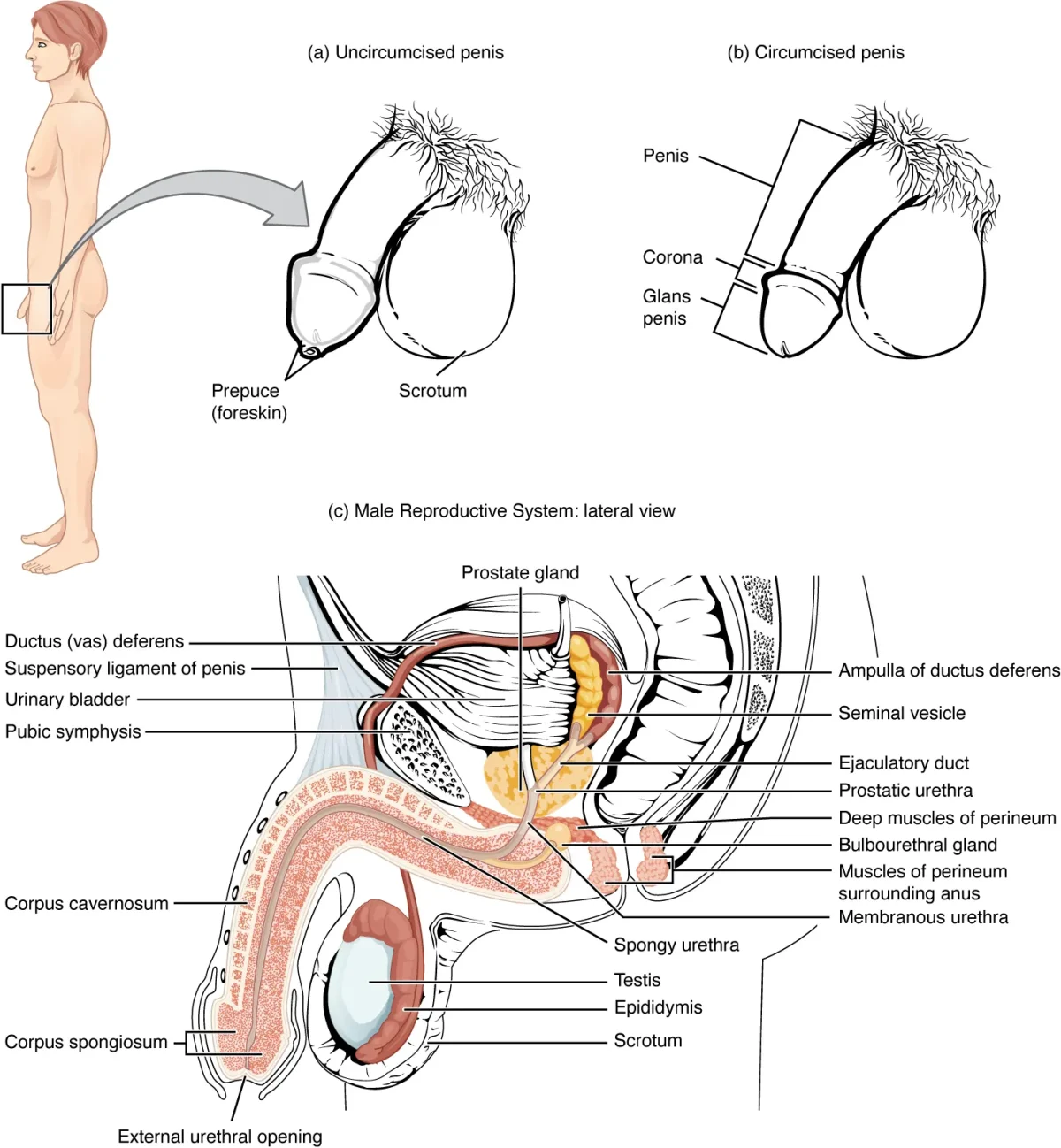
The Testes
The testes are the male gonads - they produce both sperm and testosterone. They are located outside the body in the scrotum, which keeps them 2-4 degrees C cooler than core body temperature. This lower temperature is essential for spermatogenesis - heat kills developing sperm.
The cremaster muscle adjusts the position of the testes: it contracts to pull them closer to the body in cold temperatures (conserve heat) and relaxes to let them hang further away in warm temperatures (cool down).
Inside each testis:
- Seminiferous tubules - highly coiled tubes where spermatogenesis occurs. Sertoli cells and developing sperm line the walls; mature sperm are released into the lumen.
- Interstitial space - the area between tubules, containing Leydig cells that produce testosterone in response to LH.
The Sperm Pathway
After being produced in the seminiferous tubules, sperm follow this route:
1. Epididymis - A long, coiled tube sitting on top of each testis. Sperm spend 2-3 weeks here, during which they:
- Gain motility (learn to swim)
- Complete maturation
- Are stored until ejaculation
2. Vas deferens (ductus deferens) - A muscular tube that carries sperm from the epididymis up through the spermatic cord, over the bladder, and into the pelvic cavity. Its thick muscular wall produces peristaltic contractions that propel sperm during ejaculation.
This is the structure cut during a vasectomy - a male sterilization procedure. After a vasectomy, sperm are still produced but cannot reach the ejaculatory duct. Ejaculation still occurs (seminal fluid is still produced by the accessory glands), but the ejaculate contains no sperm.
3. Ejaculatory duct - Formed where the vas deferens merges with the duct from the seminal vesicle. Passes through the prostate gland and empties into the urethra.
4. Urethra - The final shared pathway for both urine and semen (but never simultaneously - a sphincter prevents urine from mixing with semen during ejaculation). The urethra runs through the prostate gland and then through the length of the penis.
The Three Accessory Glands
Sperm make up only about 2-5% of semen. The rest is seminal fluid contributed by three glands:
| Gland | Contribution to Semen | Key Secretions |
|---|---|---|
| Seminal vesicles | ~60-70% of volume | Fructose (energy for sperm), prostaglandins, alkaline fluid |
| Prostate gland | ~25-30% of volume | Alkaline fluid, enzymes (PSA), citric acid |
| Bulbourethral (Cowper’s) glands | ~5% of volume | Mucus for lubrication, clears urethra of residual urine |
The alkaline nature of seminal fluid is critical - it neutralizes the acidic environment of both the male urethra (residual urine) and the female vagina (pH ~3.5-4.5), protecting sperm from acid damage.
Male Sexual Response
The male sexual response is controlled by the autonomic nervous system:
- Arousal and erection - controlled by the parasympathetic nervous system. Parasympathetic fibers cause vasodilation of arteries supplying the erectile tissue (corpora cavernosa and corpus spongiosum), increasing blood flow and producing an erection.
- Emission and ejaculation - controlled by the sympathetic nervous system. Sympathetic stimulation causes contractions of the vas deferens, seminal vesicles, and prostate (emission), followed by rhythmic contractions of muscles at the base of the penis (ejaculation).
- Resolution - also sympathetic. Arteries constrict, blood drains, penis returns to flaccid state.
Embryological Development
The sex of the embryo is determined at fertilization by the 23rd chromosome pair (XX = female, XY = male), but the reproductive organs do not differentiate until about week 7 of development.
- The SRY gene on the Y chromosome activates a genetic cascade that causes undifferentiated gonads to develop into testes.
- The testes produce testosterone, which stimulates development of the Wolffian ducts into male internal structures (epididymis, vas deferens, seminal vesicles, ejaculatory ducts).
- The testes also produce Mullerian Inhibiting Factor (MIF), which causes the Mullerian ducts to degenerate (preventing female internal structures from forming).
- Dihydrotestosterone (DHT) - a more potent androgen converted from testosterone by 5-alpha-reductase - drives the development of external male genitalia (penis, scrotum).
Without the SRY gene (XX embryos), the default pathway is female development.