
Female Anatomy
The female reproductive system does something no other organ system attempts: it creates a brand-new environment inside the body every month, optimized for receiving and nourishing a fertilized egg. If no egg implants, the entire setup is torn down and rebuilt from scratch the next month. This monthly construction-and-demolition cycle is the menstrual cycle, and understanding it is essential for the MCAT.
The Ovaries
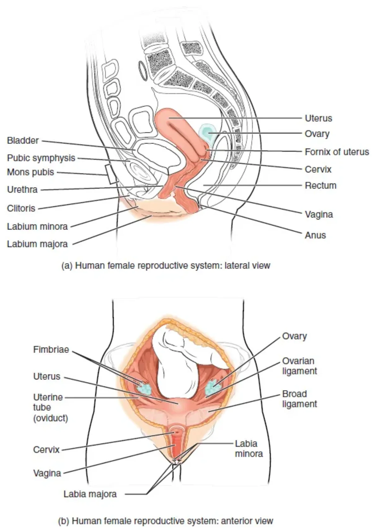
The ovaries are the female gonads - they produce both ova (eggs) and the hormones estrogen and progesterone. Each ovary is about the size and shape of an almond, located on either side of the uterus.
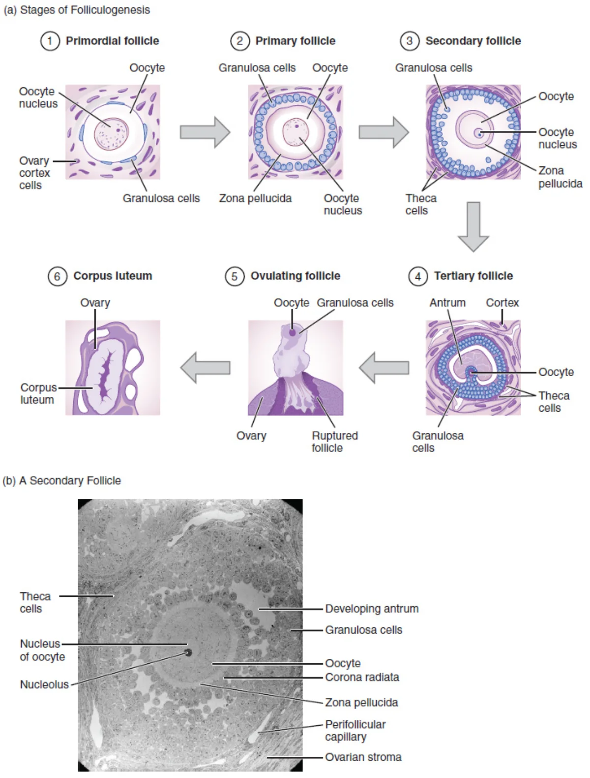
Inside each ovary are follicles - structures that contain and support developing oocytes:
- Primordial follicles - the most immature stage, containing a primary oocyte surrounded by a single layer of flat granulosa cells. Present from before birth.
- Primary follicles - granulosa cells become cuboidal and begin to multiply. The zona pellucida (a glycoprotein layer) forms around the oocyte.
- Secondary follicles - multiple layers of granulosa cells, plus an outer layer of theca cells. A fluid-filled cavity (antrum) begins to form.
- Graafian (tertiary) follicle - the fully mature follicle with a large antrum. Contains the secondary oocyte ready for ovulation. Only one follicle per cycle typically reaches this stage (the dominant follicle); the rest undergo atresia (degeneration).
The Egg’s Journey
After ovulation, the egg follows this path:
- Ovary - the Graafian follicle ruptures and releases the secondary oocyte into the peritoneal cavity.
- Fimbriae - finger-like projections at the end of the fallopian tube sweep the oocyte into the tube. The egg does not physically “jump” from the ovary into the tube - it is captured by these waving fimbriae.
- Fallopian tube (oviduct) - the oocyte travels down the tube, propelled by cilia lining the tube’s inner surface and by peristaltic contractions. Fertilization normally occurs here, specifically in the ampulla (the widest section, closest to the ovary).
- Uterus - if fertilized, the embryo arrives and implants in the endometrium about 6-7 days after fertilization. If unfertilized, the oocyte degenerates.
The Uterus
The uterus is a pear-shaped, muscular organ where the embryo implants and develops during pregnancy. It has three layers:
- Endometrium (inner lining) - the layer that thickens each month and is shed during menstruation. Contains glands and blood vessels.
- Myometrium (middle layer) - thick smooth muscle that contracts during labor to push the baby out. Also responsible for menstrual cramps.
- Perimetrium (outer layer) - the serous membrane covering the uterus.
The Cervix and Vagina
- Cervix - the narrow, lower portion of the uterus that opens into the vagina. It produces mucus that changes consistency throughout the menstrual cycle (thin and watery at ovulation to help sperm pass; thick and sticky at other times to block sperm and pathogens). The cervix dilates to approximately 10 cm during labor.
- Vagina - a muscular canal connecting the cervix to the outside of the body. It serves as the birth canal, the passage for menstrual blood, and the receptacle for the penis during intercourse.
The external female genitalia (collectively called the vulva) include the labia majora, labia minora, clitoris, and the openings of the urethra and vagina. In females, the urinary and reproductive tracts are completely separate (unlike in males, where the urethra carries both urine and semen).
The Ovarian Cycle
The ovarian cycle describes what happens in the ovaries over a typical 28-day period:
Follicular Phase (Days 1-14):
- Multiple follicles begin developing under the influence of FSH.
- Granulosa cells in the follicles produce increasing amounts of estrogen.
- One dominant follicle emerges; the rest degenerate (atresia).
- Rising estrogen causes the endometrium to thicken (proliferative phase of the uterine cycle).
- At the end of this phase, estrogen levels peak and trigger the LH surge.
Ovulation (Day ~14):
- The LH surge causes the Graafian follicle to rupture, releasing the secondary oocyte.
- The oocyte completes Meiosis I just before ovulation, producing the secondary oocyte and a polar body.
- This is the most fertile window of the cycle.
Luteal Phase (Days 15-28):
- The ruptured follicle transforms into the corpus luteum, which secretes progesterone (primarily) and some estrogen.
- Progesterone prepares the endometrium for implantation (secretory phase of the uterine cycle).
- High progesterone exerts negative feedback on GnRH, FSH, and LH, preventing additional ovulation.
- If no fertilization occurs, the corpus luteum degenerates after about 12 days, becoming the corpus albicans.
- The drop in progesterone and estrogen triggers menstruation.
The Uterine Cycle
The uterine cycle describes what happens to the endometrium, driven by the hormones of the ovarian cycle:
| Phase | Days | Driving Hormone | What Happens |
|---|---|---|---|
| Menstruation | 1-5 | Low progesterone/estrogen (trigger) | Endometrium sheds; bleeding |
| Proliferative phase | 6-14 | Estrogen (from growing follicles) | Endometrium regrows and thickens |
| Secretory phase | 15-28 | Progesterone (from corpus luteum) | Endometrium becomes glandular, vascular, secretory |
How to Read the Menstrual Hormone Graph
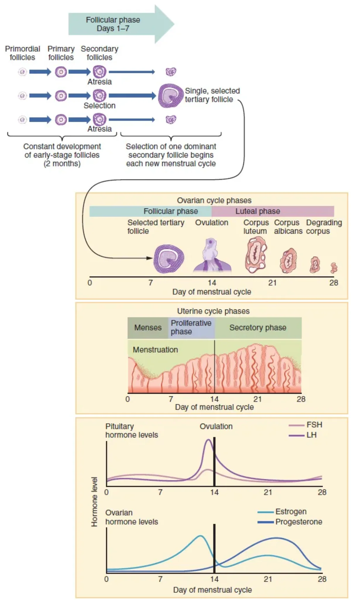
MCAT passages almost always show some version of this four-line graph. Here is how to identify each curve without memorizing axes:
- FSH - rises early (days 1-5) to recruit follicles, drops as estrogen feeds back, then a small mid-cycle bump at day ~14.
- Estrogen - low at menses, climbs steeply during the follicular phase, peaks just before ovulation, dips, then a smaller luteal peak.
- LH - flat baseline, then a sharp narrow spike around day 14 (the LH surge). This spike is the easiest landmark on the whole graph.
- Progesterone - flat and low until after ovulation, then climbs to a broad luteal peak (day ~21), then drops before menses.
If Pregnancy Occurs
If fertilization and implantation occur, the embryo’s developing placenta secretes human chorionic gonadotropin (hCG). hCG “rescues” the corpus luteum from degeneration, keeping progesterone levels high and maintaining the endometrium. This is the hormone detected by pregnancy tests. After about 10-12 weeks, the placenta takes over progesterone production, and the corpus luteum is no longer needed.