Think of the GI tract as one long conveyor belt running through a factory - except instead of building something, it tears things apart. Food enters at one end (mouth) and waste exits at the other (anus), passing through specialized stations along the way.
The technical name for this tube is the alimentary canal. It runs about 9 meters (30 feet) from mouth to anus. Here is a crucial insight: the lumen (interior) of this tube is technically outside your body. Food sitting in your stomach has not actually entered your tissues yet. It must be digested and then absorbed across the intestinal wall to truly enter the body.
The Route: Station by Station
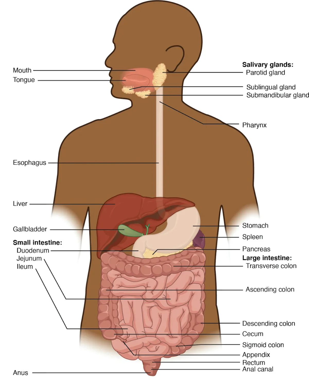
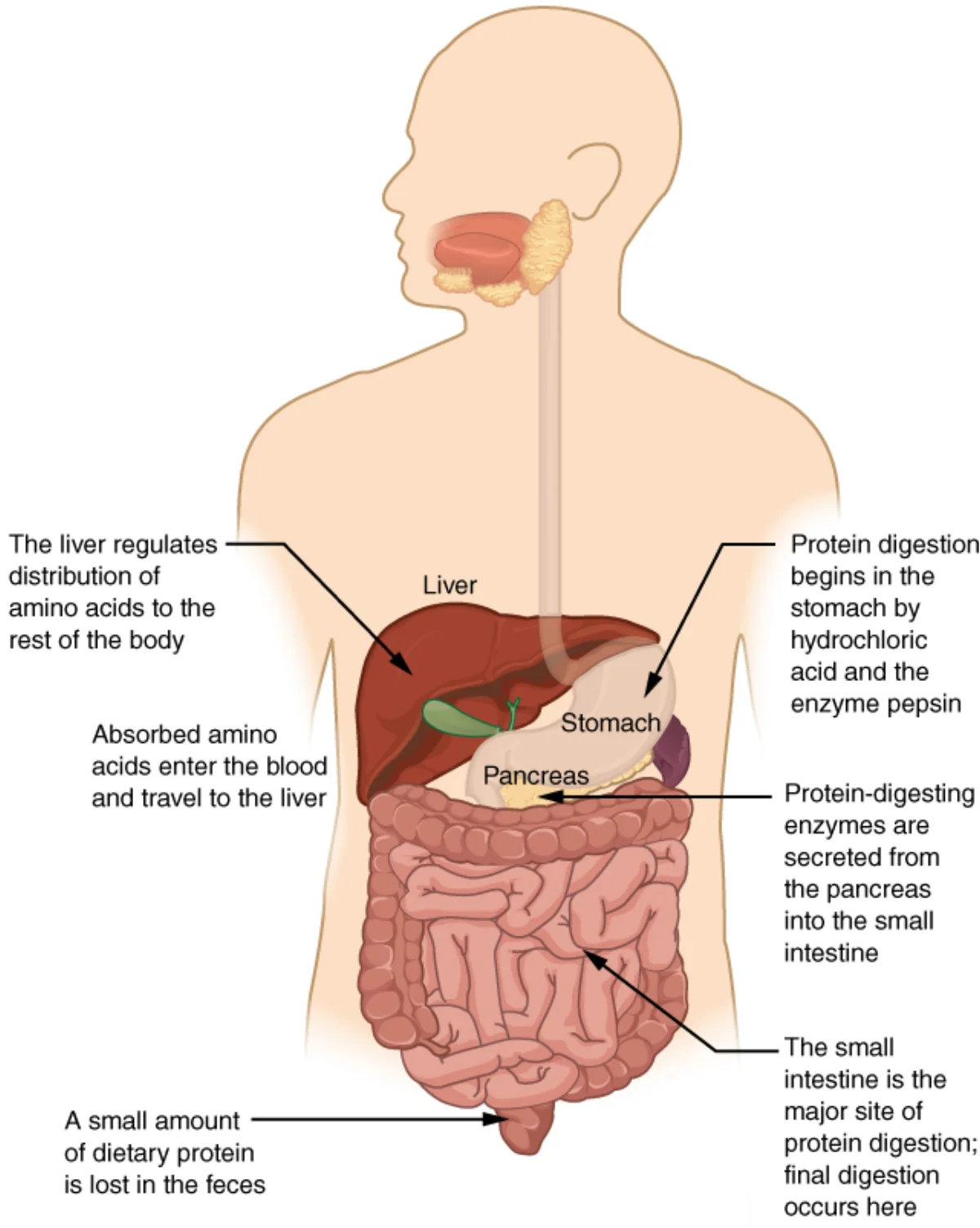
The complete GI tract and accessory organs. Food travels from mouth to anus through the alimentary canal. Credit: OpenStax Anatomy and Physiology 2e, CC BY 4.0
Interactive 3D GI Tract. Rotate to follow the alimentary canal from esophagus to rectum. Note the relative size and position of each organ.Credit: Ebers via Sketchfab, CC BY
Food follows this path, in order:
Oral cavity (mouth) - mechanical and chemical digestion begin
Pharynx - shared passage with respiratory tract
Esophagus - transport tube to stomach
Stomach - acid bath and protein digestion
Small intestine (duodenum, jejunum, ileum) - most digestion and absorption
Large intestine (cecum, colon, rectum) - water absorption, feces formation
Anus - elimination
Four accessory organs contribute secretions but food never passes through them: salivary glands, liver, gallbladder, and pancreas.
Two Types of Digestion
Mechanical digestion physically breaks food into smaller pieces without changing its chemical structure. Chewing is the most obvious example, but the stomach’s churning and the small intestine’s segmentation contractions also count. The purpose is to increase surface area so enzymes can work faster.
Chemical digestion uses enzymes to break covalent bonds in macromolecules:
Macromolecule
Broken Into
Key Enzymes (Preview)
Carbohydrates (starch)
Monosaccharides (glucose, fructose, galactose)
Amylase, disaccharidases
Proteins
Amino acids
Pepsin, trypsin, chymotrypsin
Lipids (triglycerides)
Fatty acids + monoglycerides
Lipase (with bile assistance)
Nucleic acids
Nucleotides
Nucleases
The Four Histological Layers
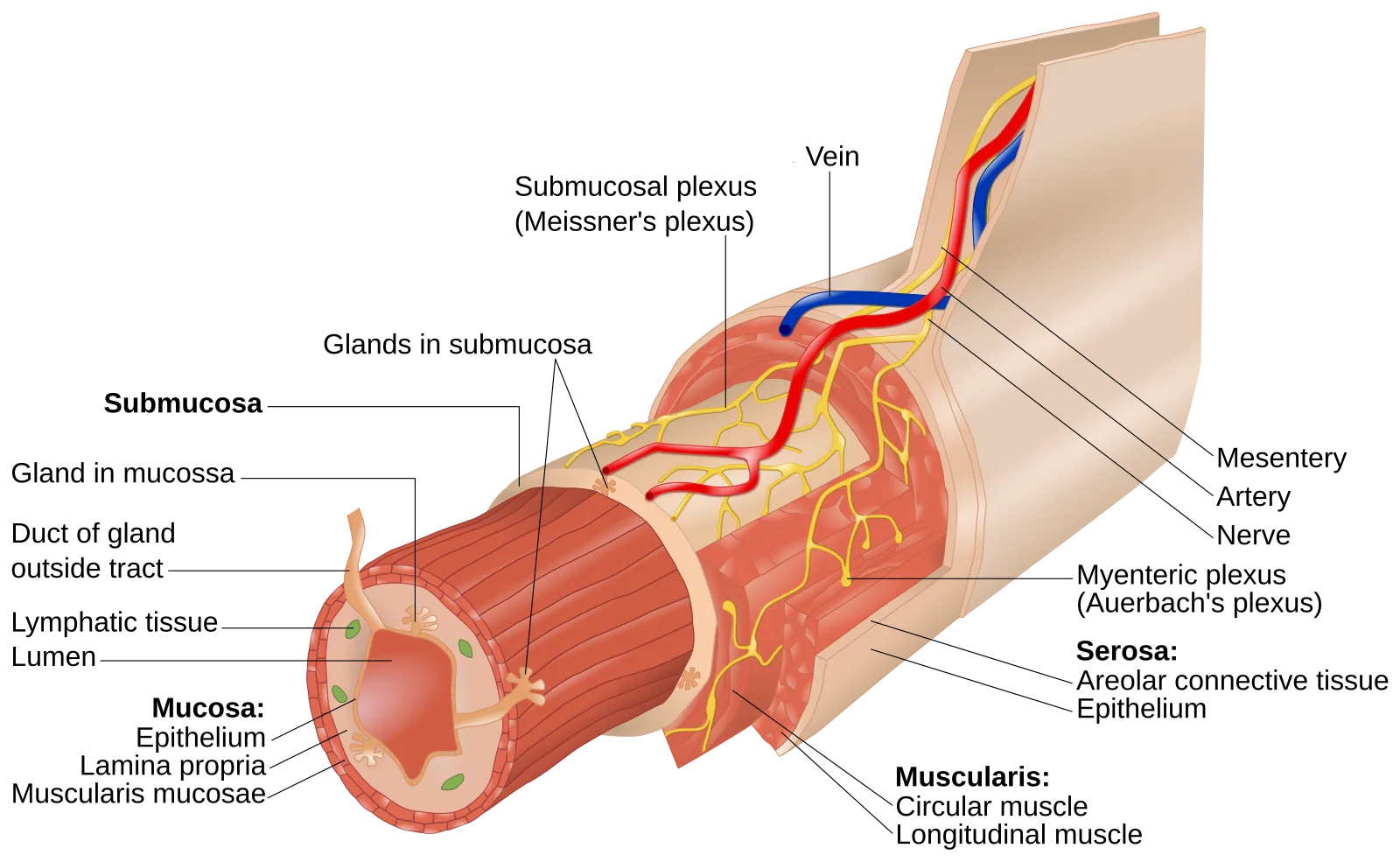
Every region of the alimentary canal shares the same four-layer wall structure, from innermost to outermost:
The four layers of the GI wall. From the lumen outward: mucosa, submucosa, muscularis externa (circular + longitudinal smooth muscle for peristalsis), and serosa. Credit: Wikimedia Commons, CC BY-SA 4.0
Mucosa - innermost layer; contains the epithelium that contacts food, plus the lamina propria (connective tissue with blood vessels and lymph) and muscularis mucosae (thin smooth muscle)
Submucosa - dense connective tissue with blood vessels, lymphatics, and the submucosal plexus (Meissner’s plexus) of the enteric nervous system
Muscularis externa - two layers of smooth muscle (inner circular, outer longitudinal) responsible for peristalsis; contains the myenteric plexus (Auerbach’s plexus) between the muscle layers
Serosa (or adventitia) - outermost connective tissue covering; serosa where the organ is surrounded by peritoneum, adventitia where it is not (esophagus)
Peristalsis and Segmentation
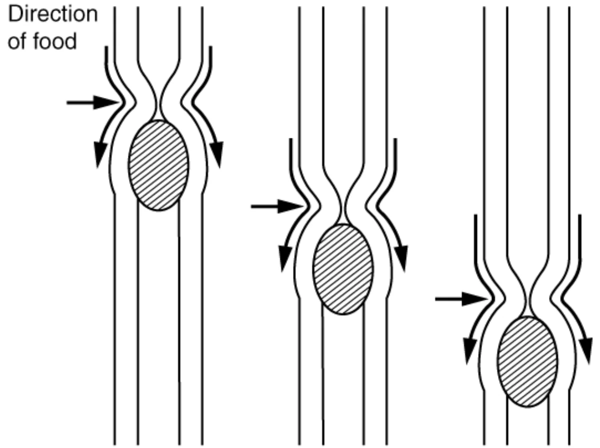
Peristalsis: coordinated contraction of circular muscle behind the bolus and longitudinal muscle ahead of it creates a wave that pushes food forward. Credit: OpenStax Anatomy and Physiology 2e, CC BY 4.0
Peristalsis is the wave-like contraction that pushes food forward through the GI tract. Behind the food bolus, circular muscle contracts (squeezing); ahead of it, longitudinal muscle contracts (shortening, which widens the lumen). This coordinated wave moves food in one direction - mouth to anus.
Segmentation is a back-and-forth contraction pattern found mainly in the small intestine. Unlike peristalsis, segmentation does not push food forward. It chops and mixes the chyme with digestive juices, ensuring maximum contact between enzymes and food.
Sphincters: The Gatekeepers
Sphincters are rings of smooth muscle that act as one-way valves between GI regions:
Sphincter
Location
Function
Upper esophageal
Top of esophagus
Prevents air from entering esophagus
Lower esophageal (cardiac)
Esophagus-stomach junction
Prevents acid reflux into esophagus
Pyloric
Stomach-duodenum junction
Controls gastric emptying rate
Ileocecal valve
Ileum-cecum junction
Prevents backflow from large intestine
Internal anal
Upper anus
Involuntary; maintains continence
External anal
Lower anus
Voluntary; controls defecation
The lumen of the GI tract is considered topologically "outside" the body. What process must occur for nutrients to actually enter the body?
Click to reveal answer
Absorption. Nutrients must cross the intestinal epithelium (mucosa) and enter the blood or lymph to truly enter the body. Food sitting in the stomach lumen has not been absorbed yet - it is still in the "outside" tube running through the body.
What is the key difference between peristalsis and segmentation?
Click to reveal answer
Peristalsis pushes food forward (unidirectional waves); segmentation mixes food in place (back-and-forth contractions). Peristalsis moves food from mouth to anus. Segmentation occurs mainly in the small intestine and increases enzyme-food contact without net forward movement.
Digestion begins the instant you take a bite. Your teeth grind the food into smaller pieces (mechanical digestion), and enzymes in saliva immediately start breaking chemical bonds (chemical digestion). By the time you swallow, starch digestion is already underway.
Mastication: The First Station
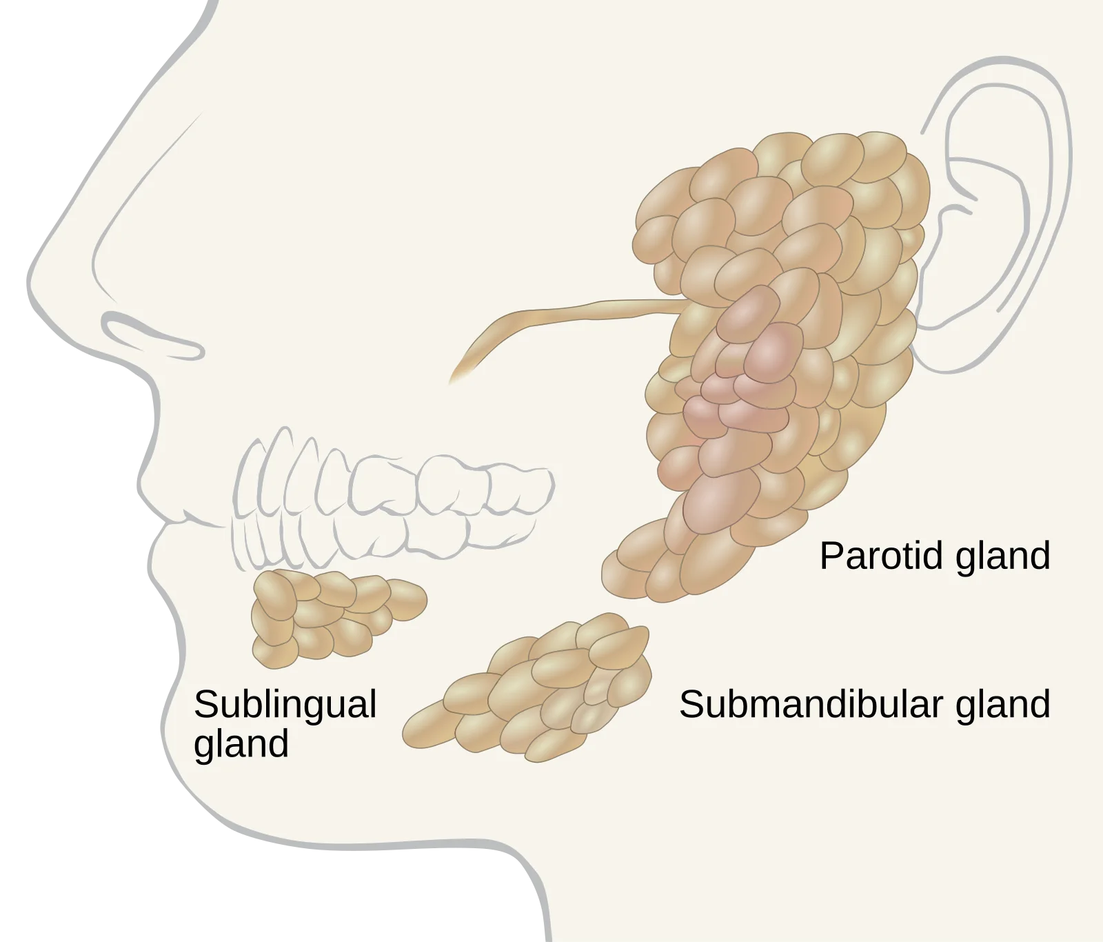
The three major salivary glands. The parotid (largest, serous/watery), submandibular (mixed), and sublingual (mostly mucous) secrete saliva containing amylase (starch digestion) and lingual lipase (fat digestion). Credit: Wikimedia Commons, CC BY-SA 4.0
Chewing (mastication) is the only voluntary step of mechanical digestion. Your teeth cut, tear, and grind food into a soft mass called a bolus. This increases the surface area available for enzymes - a whole cracker has far less exposed surface than a chewed-up cracker.
Saliva: More Than Just Water
Saliva is produced by three pairs of salivary glands:
Gland
Location
Main Contribution
Parotid
In front of each ear
Serous (watery) fluid rich in salivary amylase
Submandibular
Below the jaw
Mixed serous and mucous secretion
Sublingual
Under the tongue
Primarily mucous secretion
Saliva contains:
Water and mucus - moisten food and lubricate the bolus for swallowing
Salivary amylase - begins starch digestion
Lingual lipase - begins fat digestion (secreted by glands at the back of the tongue)
Lysozyme and IgA - antimicrobial defense
Bicarbonate - buffers acids to protect tooth enamel
Saliva production is controlled by the autonomic nervous system. Parasympathetic stimulation (rest and digest) increases saliva flow. Sympathetic stimulation (fight or flight) decreases it - that is why your mouth goes dry when you are nervous.
Salivary Amylase: Starch Digestion Starts Here
Salivary amylase hydrolyzes the alpha-1,4-glycosidic bonds in starch, breaking it into maltose and maltotriose (shorter chains). This enzyme works best at a neutral pH (~6.8) and is inactivated once it reaches the acidic stomach.
Try this: hold a plain cracker in your mouth for 30 seconds without swallowing. It starts to taste sweet. That sweetness is maltose being released as salivary amylase breaks down the starch in real time.
Lingual Lipase: Fat Digestion Also Starts Here
Lingual lipase is secreted by glands at the back of the tongue. It begins triglyceride digestion in the mouth, although its activity continues in the stomach because lingual lipase is acid-stable (it works at low pH). It is most important for digesting milk fat in infants.
What Does NOT Get Digested in the Mouth?
Protein digestion does not begin until the stomach (pepsin requires acidic pH). If you mix saliva with protein in a test tube, nothing happens to the protein - there are no proteases in saliva.
A student mixes saliva with a solution containing starch, triglycerides, and polypeptides. After 30 minutes at 37°C, which macromolecule(s) remain completely intact?
Click to reveal answer
Polypeptides remain intact. Salivary amylase partially digests starch into maltose. Lingual lipase partially digests triglycerides into fatty acids. But there are no proteases in saliva, so polypeptides are untouched. This is a common MCAT experimental question.
Why does salivary amylase stop working once food reaches the stomach?
Click to reveal answer
The stomach's low pH (~1.5-2) denatures salivary amylase. Salivary amylase has an optimal pH near 6.8. The highly acidic gastric environment inactivates it, halting starch digestion until pancreatic amylase takes over in the duodenum.
The esophagus is a simple transport tube - no digestion or absorption happens here. Its only job is to move the food bolus from the pharynx to the stomach. But that job involves an elegant reflex that keeps food out of your lungs.
The Swallowing Reflex (Deglutition)
Swallowing has three phases:
1. Voluntary (oral) phase - Your tongue pushes the bolus to the back of the mouth toward the pharynx. This is the only voluntary part of swallowing.
2. Pharyngeal phase (involuntary) - Once the bolus touches the pharynx, a reflex takes over:
The soft palate rises to close off the nasal passages (prevents food from going up your nose)
The epiglottis folds down to cover the trachea (prevents food from entering the airway)
The upper esophageal sphincter relaxes to let the bolus enter the esophagus
Breathing is briefly inhibited
3. Esophageal phase (involuntary) - Peristaltic waves push the bolus down the esophagus toward the stomach. This takes about 8-10 seconds. Gravity helps but is not required - you can swallow while upside down because peristalsis does the work.
Esophageal Anatomy
The esophagus is about 25 cm long and runs behind the trachea. Key features:
Upper third - contains skeletal muscle (voluntary-ish, used in the initial swallowing push)
Middle third - mix of skeletal and smooth muscle
Lower third - smooth muscle only (completely involuntary)
No serosa - the esophagus has adventitia instead (unlike most of the GI tract, which has serosa)
The Lower Esophageal Sphincter (LES)
At the junction between the esophagus and stomach sits the lower esophageal sphincter (also called the cardiac sphincter). It stays contracted to prevent stomach acid from splashing back into the esophagus, and relaxes only when a peristaltic wave delivers a bolus.
When the LES fails to close properly, stomach acid enters the esophagus. This causes the burning sensation of acid reflux.
An astronaut in zero gravity swallows a bite of food. Will it reach the stomach? Why or why not?
Click to reveal answer
Yes - peristalsis moves the bolus, not gravity. The esophageal smooth muscle generates peristaltic waves that actively push food toward the stomach. Gravity assists on Earth but is not necessary. Astronauts eat and swallow normally in microgravity.
The stomach is a muscular sac that serves as a holding tank and processing plant. It stores food, mixes it with acid and enzymes, and slowly releases the resulting slurry (chyme) into the small intestine. The stomach’s acid is so strong it could dissolve a nail - yet the stomach lining survives because of a constantly renewed mucus shield.
Stomach Anatomy
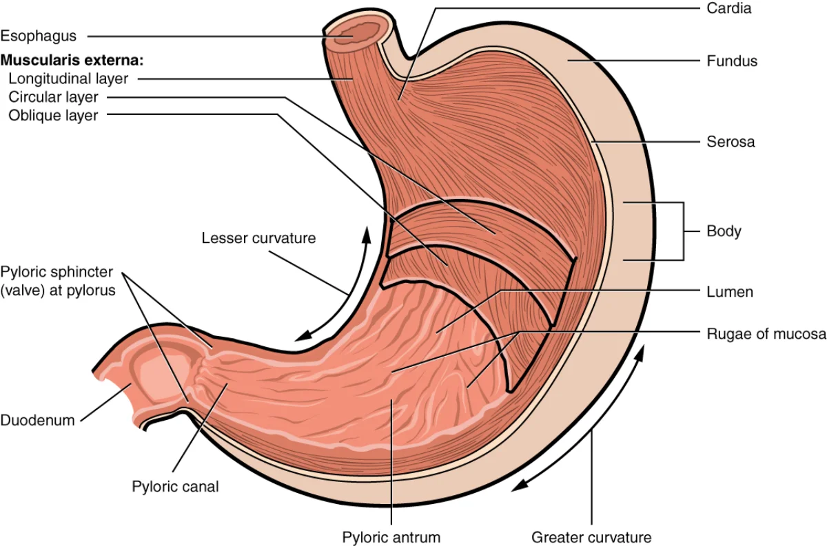
Stomach anatomy showing the four regions and sphincters. Rugae (folds) on the inner surface flatten as the stomach expands. Credit: OpenStax Anatomy and Physiology 2e, CC BY 4.0
The stomach has four main regions:
Cardia - where the esophagus connects; just below the lower esophageal sphincter
Fundus - the upper dome that often collects swallowed air
Body - the largest region; main site of mixing and digestion
Pylorus - the lower region that connects to the duodenum via the pyloric sphincter
The inner surface is thrown into folds called rugae, which flatten as the stomach expands (like an accordion). This allows the stomach to stretch from ~50 mL (empty) to ~1-1.5 L (after a large meal).
Gastric Glands: The Acid Factory
Scattered across the stomach lining are millions of gastric pits - small depressions that lead down to gastric glands. These glands contain four specialized cell types:
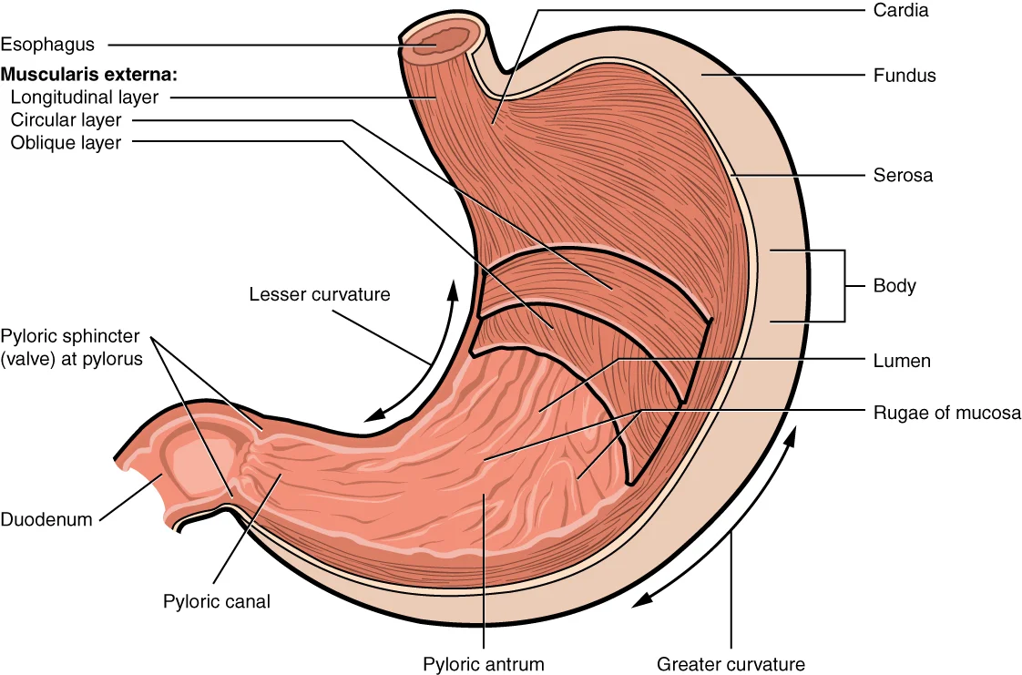
Stomach anatomy with regions and muscle layers. Focus on: the four regions (cardia, fundus, body, pylorus), the three muscle layers, and the key cell types: parietal cells (HCl + intrinsic factor), chief cells (pepsinogen), G cells (gastrin). Credit: OpenStax College, CC BY 3.0
Cell Type
Secretion
Function
Parietal cells
HCl, intrinsic factor
HCl activates pepsinogen, kills bacteria; intrinsic factor needed for B12 absorption in ileum
Chief cells
Pepsinogen, gastric lipase
Pepsinogen is the inactive zymogen of pepsin; gastric lipase digests some fat
Mucous neck cells
Alkaline mucus
Coats the stomach lining with a bicarbonate-rich barrier, protecting it from acid
G cells
Gastrin (hormone)
Stimulates parietal cells to produce more HCl; stimulates stomach motility
HCl: The Chemical Weapon
Parietal cells secrete HCl into the stomach lumen using the H+/K+ ATPase (proton pump). This pump exchanges H+ ions (into the lumen) for K+ ions (into the cell), using ATP. The result is a lumen pH of about 1.5-2 - acidic enough to kill most bacteria and denature proteins.
The HCl does three things:
Activates pepsinogen into pepsin (the active protease)
Kills pathogens - acts as a first line of defense against ingested bacteria
Pepsinogen to Pepsin: The Zymogen Cascade Begins
Chief cells secrete pepsinogen, the inactive form of the protease pepsin. This is a critical safety mechanism - if chief cells secreted active pepsin, the enzyme would digest the cells that made it.
Once pepsinogen contacts HCl in the lumen, the acid cleaves off a small peptide fragment, converting pepsinogen into active pepsin. Pepsin then:
Digests proteins into shorter peptide fragments
Works optimally at pH ~2 (and is inactive above pH ~6)
Can activate more pepsinogen (positive feedback loop)
Intrinsic Factor: The B12 Key
Parietal cells also secrete intrinsic factor, a glycoprotein that binds vitamin B12 in the stomach. The intrinsic factor-B12 complex is absorbed later in the ileum (the last segment of the small intestine). Without intrinsic factor, B12 cannot be absorbed, which leads to a form of anemia caused by B12 deficiency.
Gastric Motility and Chyme Formation
The stomach’s three smooth muscle layers (an extra oblique layer compared to the rest of the GI tract) churn and mix food with gastric juice. After 2-6 hours of mechanical and chemical digestion, food is converted into a thick, acidic paste called chyme.
Chyme is released into the duodenum in small squirts through the pyloric sphincter. The rate of gastric emptying is regulated by:
Duodenal feedback - fat and acid in the duodenum slow gastric emptying (via CCK and secretin)
Gastrin - speeds up gastric emptying
Volume and composition - liquids empty faster than solids; carbohydrates faster than proteins; fats are the slowest
Why are proteases like pepsin secreted as inactive zymogens rather than in their active form?
Click to reveal answer
To prevent self-digestion. Active proteases would digest the cells that produce them. By secreting inactive zymogens (pepsinogen, trypsinogen, etc.), the enzyme only becomes active after it reaches the lumen, where it can safely digest food without harming the secreting tissue.
A patient with autoimmune destruction of parietal cells would develop deficiencies in which two substances?
Click to reveal answer
HCl and intrinsic factor. Loss of parietal cells means no HCl (impaired pepsinogen activation and protein digestion) and no intrinsic factor (impaired B12 absorption in the ileum, leading to pernicious anemia).
The small intestine is the MVP of digestion. Despite its name (“small” refers to diameter, not length), it stretches about 6-7 meters and is where 90% of digestion and absorption occur. Its secret weapon is surface area - an internal architecture of folds, fingers, and micro-fingers that gives it the absorptive area of a tennis court packed into a tube you could coil inside your abdomen.
The Three Segments
The Duodenum: Where Chemistry Happens
The duodenum is the first and shortest segment (~25 cm). It receives:
Acidic chyme from the stomach through the pyloric sphincter
Bile from the liver/gallbladder via the common bile duct
Pancreatic juice from the pancreas via the pancreatic duct
Both the common bile duct and pancreatic duct empty into the duodenum at the ampulla of Vater (hepatopancreatic ampulla), controlled by the sphincter of Oddi.
The duodenum is the principal site for chemical digestion. It neutralizes stomach acid with pancreatic bicarbonate, emulsifies fats with bile, and unleashes a battery of pancreatic and brush-border enzymes.
The duodenum also has an endocrine role: it secretes secretin (in response to acid) and cholecystokinin (CCK) (in response to fats and proteins).
The Jejunum: Absorption Powerhouse
The jejunum is the middle segment and the primary site of nutrient absorption. Its walls are densely packed with villi, giving it a velvety appearance. Most amino acids, monosaccharides, fatty acids, and water-soluble vitamins are absorbed here.
Water-soluble nutrients (amino acids, monosaccharides) are absorbed into capillaries within each villus and travel via the hepatic portal vein to the liver for processing.
Fats take a different route - they enter lacteals (lymphatic vessels within each villus) and travel through the lymphatic system before entering the bloodstream.
The Ileum: The Final Stretch
The ileum is the longest segment and connects to the large intestine at the ileocecal valve. It has two specialized absorption jobs:
Vitamin B12 - the intrinsic factor-B12 complex formed in the stomach is absorbed exclusively in the ileum by receptor-mediated endocytosis
Bile salts - recycled back to the liver via the hepatic portal vein in a process called enterohepatic circulation (bile salts are reused 6-8 times per day)
The ileum also contains Peyer’s patches - clusters of lymphoid tissue that monitor intestinal bacteria and mount immune responses against pathogens. They are part of the gut-associated lymphoid tissue (GALT).
The Surface Area Amplification System
The small intestine achieves its enormous surface area through three levels of folding:
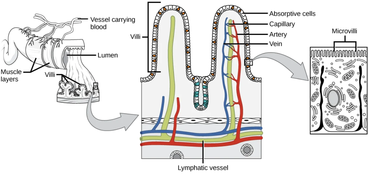
The three levels of surface area amplification in the small intestine: circular folds, villi, and microvilli. Each villus contains a capillary network and a central lacteal. Credit: Lumen Learning / OpenStax Anatomy and Physiology, CC BY 4.0
Level
Structure
Surface Area Increase
1
Plicae circulares (circular folds)
~3x the flat surface
2
Villi (finger-like projections, ~1 mm tall)
~10x more
3
Microvilli (brush border on each enterocyte)
~20x more
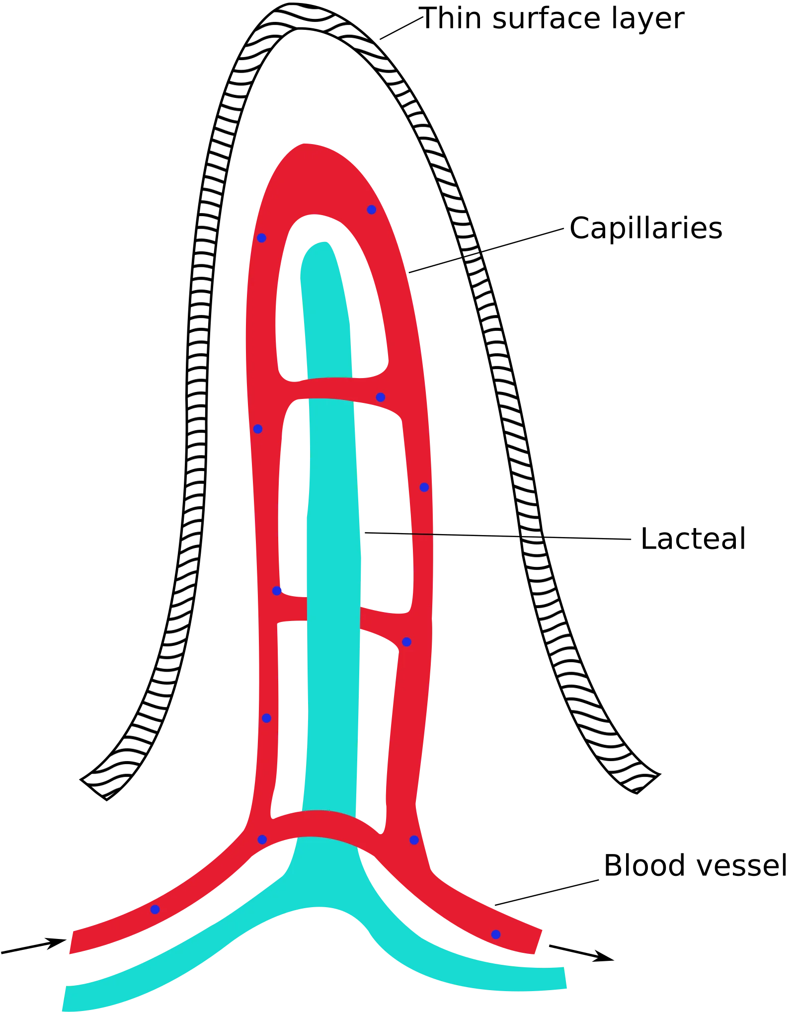
Each villus contains:
A capillary network for absorbing water-soluble nutrients
A lacteal (lymphatic vessel) for absorbing fats
Smooth muscle that contracts to “pump” absorbed nutrients into vessels
Intestinal Crypts: The Cell Factory
Structure of a single intestinal villus. Each villus contains a capillary network (absorbs amino acids, sugars) and a lacteal (absorbs fats as chylomicrons). The brush border of microvilli further increases surface area. Credit: Wikimedia Commons, Public Domain
At the base of each villus are crypts of Lieberkuhn (intestinal crypts). These contain stem cells that constantly produce new enterocytes. The intestinal epithelium renews itself every 3-5 days - one of the fastest cell turnover rates in the body.
A patient has the terminal ileum surgically removed. Which two substances will they be unable to absorb?
Click to reveal answer
Vitamin B12 and bile salts. B12-intrinsic factor complex is absorbed exclusively in the ileum. Bile salts are also reabsorbed in the ileum (enterohepatic circulation). Without the ileum, the patient will develop B12 deficiency (pernicious anemia) and fat malabsorption (bile salt depletion).
Absorbed amino acids and glucose enter capillaries in the villus. Where do they go next - directly to the heart, or somewhere else first?
Click to reveal answer
To the liver first, via the hepatic portal vein. Water-soluble nutrients (amino acids, monosaccharides) travel from intestinal capillaries to the hepatic portal vein to the liver for processing, detoxification, and storage before entering general circulation. Fats bypass this route - they enter lacteals and travel through the lymphatic system.
The small intestine is where the heavy enzymatic machinery lives. Two sources supply the enzymes: the pancreas (secreted into the lumen) and the brush border of the intestinal epithelium itself. Together, they finish the digestion of every macromolecule into its absorbable building blocks.
Pancreatic Enzymes: The Heavy Artillery
The exocrine pancreas produces a cocktail of digestive enzymes delivered to the duodenum through the pancreatic duct. These enzymes work at a slightly alkaline pH (~7-8), which is maintained by pancreatic bicarbonate that neutralizes the acidic chyme from the stomach.
The Trypsin Activation Cascade
This is one of the most important enzyme cascades for the MCAT. All pancreatic proteases are secreted as inactive zymogens to prevent the pancreas from digesting itself.
The cascade works like a row of dominoes:
Enteropeptidase (also called enterokinase) - an enzyme embedded in the duodenal brush border - cleaves trypsinogen into active trypsin
Trypsin then activates everything else:
Chymotrypsinogen → chymotrypsin
Procarboxypeptidase → carboxypeptidase
Proelastase → elastase
More trypsinogen → trypsin (autocatalysis)
Complete Pancreatic Enzyme Table
Enzyme
Zymogen Form
Substrate
Products
Trypsin
Trypsinogen
Proteins/peptides
Shorter peptides (cleaves after Arg, Lys)
Chymotrypsin
Chymotrypsinogen
Proteins/peptides
Shorter peptides (cleaves after Phe, Trp, Tyr)
Carboxypeptidase A & B
Procarboxypeptidase
Peptides
Removes C-terminal amino acids
Elastase
Proelastase
Elastin, proteins
Shorter peptides
Pancreatic amylase
(secreted active)
Starch, glycogen
Maltose, maltotriose, limit dextrins
Pancreatic lipase
(active form secreted)
Triglycerides
2 fatty acids + 1 monoglyceride
Nucleases
(secreted active)
DNA, RNA
Nucleotides
Note that amylase, lipase, and nucleases are secreted in their active forms - they are not zymogens. Only the proteases need to be secreted as zymogens because only proteases pose a self-digestion risk.
Brush-Border Enzymes: The Finishing Touches
Brush-border enzymes are embedded in the microvilli membrane of enterocytes throughout the small intestine. They perform the final digestion steps, breaking dimers and short chains into individual monomers ready for absorption.
Disaccharidases
Enzyme
Substrate
Products
Maltase
Maltose
Glucose + glucose
Isomaltase
Isomaltose (alpha-1,6 branch points)
Glucose
Sucrase
Sucrose
Glucose + fructose
Lactase
Lactose
Glucose + galactose
Peptidases
Enzyme
Substrate
Products
Aminopeptidase
Peptides
Removes N-terminal amino acid
Dipeptidase
Dipeptides
Two free amino acids
Other Brush-Border Enzymes
Enteropeptidase - converts trypsinogen to trypsin (the cascade starter)
Nucleotidases and nucleosidases - break nucleotides into bases, sugars, and phosphate groups
The Complete Digestion Summary
Protein digestion pathway: pepsin starts in the stomach, then trypsin, chymotrypsin, and brush-border peptidases finish the job in the small intestine. Credit: OpenStax Anatomy and Physiology 2e, CC BY 4.0
If enteropeptidase were deficient, which pancreatic enzymes would remain inactive?
Click to reveal answer
ALL pancreatic proteases would remain inactive. Enteropeptidase activates trypsinogen to trypsin. Trypsin activates chymotrypsinogen, procarboxypeptidase, and proelastase. Without enteropeptidase, no trypsin is formed, so the entire protease cascade fails. Pancreatic amylase and lipase are unaffected (they are secreted in active form).
A patient has severe chronic pancreatitis with almost no pancreatic enzyme output. Which macromolecule digestion would be MOST impaired?
Click to reveal answer
Fat digestion. While protein digestion can partially occur in the stomach (pepsin) and carbohydrate digestion starts in the mouth (salivary amylase), fat digestion relies almost entirely on pancreatic lipase (plus bile). Lingual and gastric lipase handle only 10-30% of fat digestion. Without pancreatic lipase, most dietary fat passes through unabsorbed, causing steatorrhea (fatty stools).
The liver is the body’s largest internal organ and the central processing hub for everything you absorb from the GI tract. The gallbladder is its sidekick - a small storage pouch that concentrates bile and squirts it into the duodenum on demand. Together, they make fat digestion possible.
Bile: Dish Soap for Fat
Bile is produced by hepatocytes (liver cells) and modified by bile duct cells. It contains:
Component
Function
Bile salts
Amphipathic molecules that emulsify fat (the active ingredient)
Cholesterol
Precursor to bile salts; also excreted in bile
Phospholipids
Help form micelles
Bilirubin (conjugated)
Waste product from hemoglobin breakdown; gives bile its yellow-green color
Water, ions
Carrier fluid
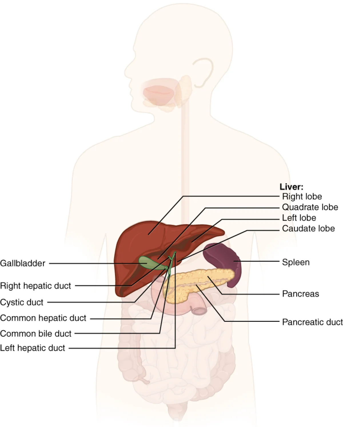
The accessory organs and bile duct system. The common bile duct and pancreatic duct merge at the ampulla of Vater before entering the duodenum. Credit: OpenStax Anatomy and Physiology 2e, CC BY 4.0
Critical distinction: Bile does NOT enzymatically digest fat. Emulsification is physical, not chemical - no covalent bonds are broken. Bile salts simply coat large fat globules with their amphipathic structure and split them into tiny droplets (and later micelles), increasing the surface area available for pancreatic lipase to act. Without bile, lipase can only attack the thin outer surface of a large fat glob, and fat digestion plummets. This is one of the clearest MCAT test points: bile is a detergent, not an enzyme.
The Bile Duct System
Bile flows through a branching duct system:
Hepatocytes produce bile and secrete it into tiny bile canaliculi
Canaliculi merge into the right and left hepatic ducts
These join to form the common hepatic duct
The cystic duct branches off to the gallbladder (for storage)
Below the cystic duct junction, the common bile duct carries bile to the duodenum
The common bile duct merges with the pancreatic duct at the ampulla of Vater
The sphincter of Oddi controls flow into the duodenum
The Gallbladder: Storage and Concentration
Between meals, the sphincter of Oddi is closed, so bile backs up into the gallbladder via the cystic duct. The gallbladder concentrates bile by absorbing water and ions - making it 5-10x more concentrated than freshly produced hepatic bile.
When fatty chyme enters the duodenum, CCK is released. CCK causes the gallbladder to contract and the sphincter of Oddi to relax, squirting concentrated bile into the duodenum right when it is needed.
Enterohepatic Circulation: Recycling Bile
About 95% of bile salts are reabsorbed in the terminal ileum and returned to the liver via the hepatic portal vein. The liver recycles them into new bile. This recycling loop is called enterohepatic circulation, and bile salts may be reused 6-8 times per day.
The Hepatic Portal System: First Pass Through the Liver
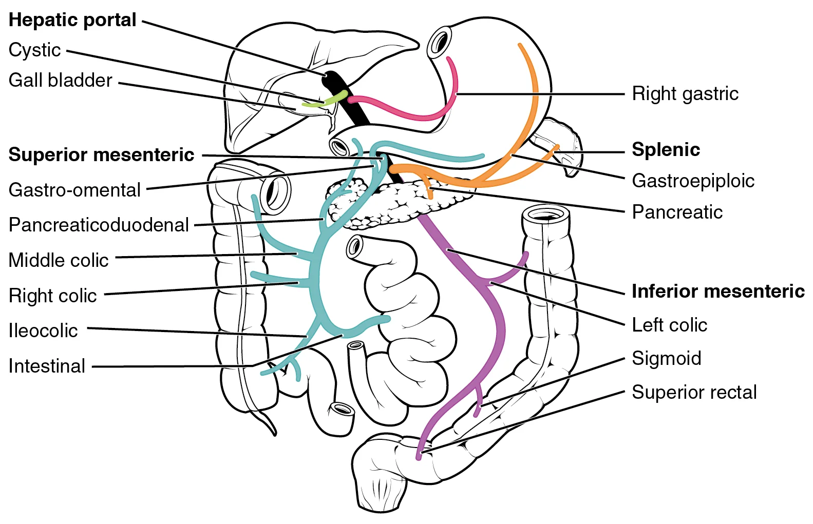
The hepatic portal system with tributaries labeled. Focus on: a portal system connects two capillary beds in series. The MCAT tests three: hepatic portal (GI → liver), hypothalamic-hypophyseal portal (hypothalamus → anterior pituitary), and renal portal (glomerulus → peritubular capillaries). Credit: OpenStax College, CC BY 3.0
All water-soluble nutrients absorbed from the GI tract (amino acids, monosaccharides, water-soluble vitamins) enter intestinal capillaries and drain into the hepatic portal vein, which carries them directly to the liver before they enter general circulation.
Process absorbed nutrients before distributing them to the body
Bilirubin and Bile Pigments
Bilirubin is a yellow breakdown product of hemoglobin from old red blood cells. The liver conjugates (makes water-soluble) bilirubin and excretes it in bile. In the intestine, bacteria convert bilirubin into pigments that give stool its brown color. Some of these pigments are reabsorbed and excreted in urine, giving it a yellow color.
Bile emulsifies fat but does not digest it. What is the difference, and why does emulsification matter for digestion?
Click to reveal answer
Emulsification is physical, not chemical. Bile salts break large fat globules into tiny micelles (physical change - no bonds broken). Digestion is chemical - pancreatic lipase hydrolyzes triglyceride ester bonds. Emulsification matters because it dramatically increases the surface area available for lipase, making fat digestion much faster and more complete.
Cholesterol is the precursor to bile salts. What happens to blood cholesterol levels if bile salt reabsorption in the ileum is blocked?
Click to reveal answer
Blood cholesterol decreases. When bile salts are not reabsorbed (enterohepatic circulation is interrupted), the liver must synthesize new bile salts from cholesterol. This pulls cholesterol out of the blood, lowering serum cholesterol levels.
The pancreas is a dual-function organ - part digestive gland, part hormone factory. Its exocrine portion (about 99% of the organ) produces digestive enzymes and bicarbonate. Its endocrine portion (tiny clusters called the Islets of Langerhans) produces hormones that regulate blood glucose. The MCAT expects you to know both sides.
Exocrine Pancreas: Enzymes and Bicarbonate
The exocrine pancreas consists of acinar cells organized into grape-like clusters. These cells produce pancreatic juice containing:
Digestive enzymes - proteases (as zymogens), lipase, amylase, nucleases (covered in detail in the enzyme section)
Bicarbonate (HCO3-) - produced by duct cells, neutralizes acidic chyme entering the duodenum, raising the pH from ~2 to ~7-8 where pancreatic enzymes work optimally
Pancreatic juice is delivered to the duodenum through the pancreatic duct, which merges with the common bile duct at the ampulla of Vater.
What Triggers Pancreatic Secretion?
Two duodenal hormones control the exocrine pancreas:
Hormone
Trigger
Pancreatic Response
Secretin
Acid in the duodenum
Duct cells release bicarbonate (neutralize acid)
CCK
Fat and protein in the duodenum
Acinar cells release digestive enzymes
Endocrine Pancreas: The Islets of Langerhans
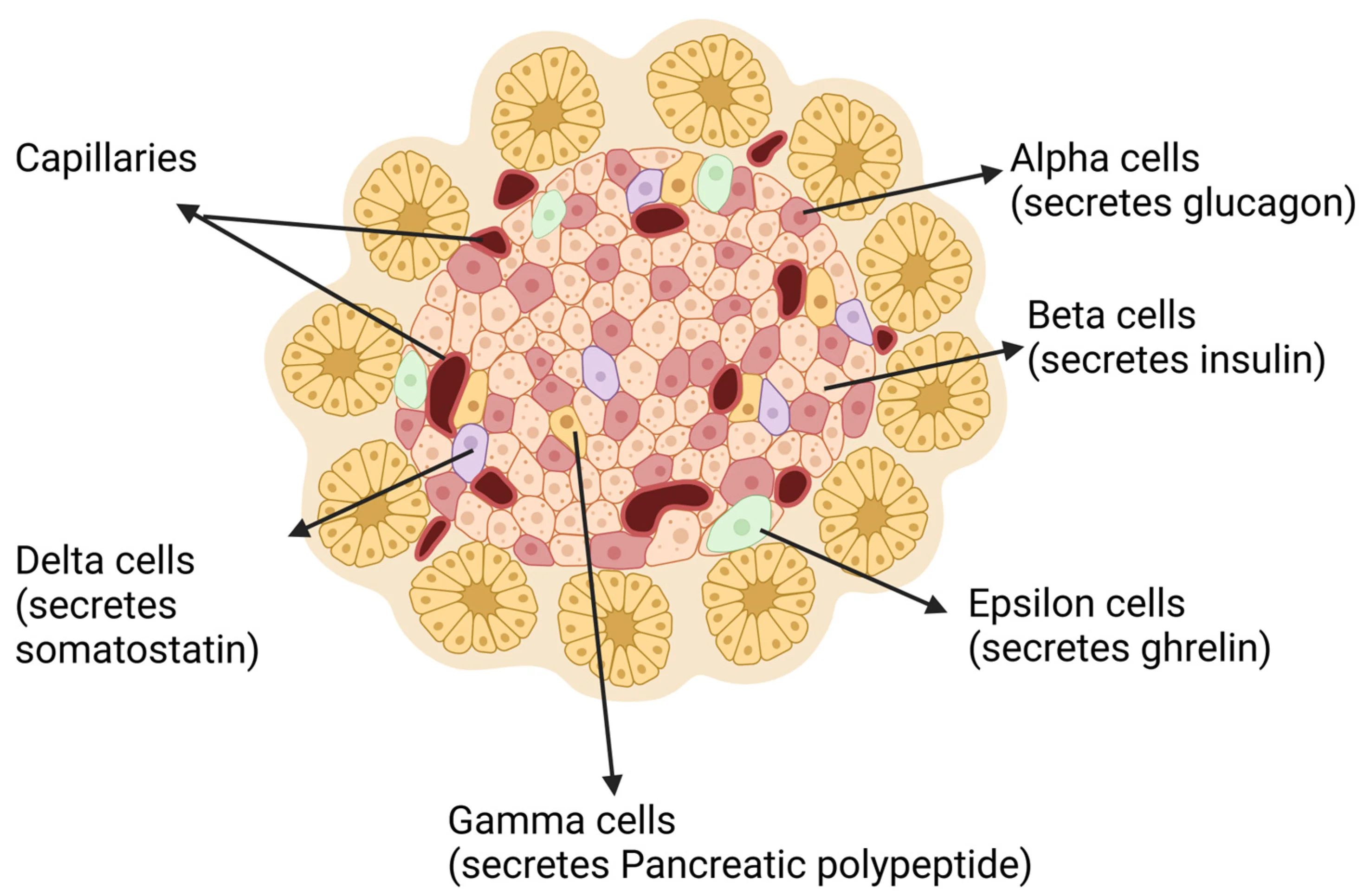
Scattered among the acinar cells are about 1-2 million tiny cell clusters called the Islets of Langerhans. They make up only ~1-2% of the pancreas by mass but are critical for metabolic regulation.
An Islet of Langerhans with its cell types labeled. Beta cells (insulin) are the most abundant, alpha cells produce glucagon, and delta cells produce somatostatin. The islets make up only 1-2% of pancreatic mass. Credit: Wikimedia Commons, CC BY 4.0
Inhibits both insulin and glucagon release; slows GI motility and secretion
Blood Glucose Regulation: Insulin vs. Glucagon
After a carbohydrate-rich meal (fed state):
Blood glucose rises
Beta cells release insulin
Insulin promotes: glucose uptake by cells (via GLUT4 in muscle/fat), glycogenesis in liver and muscle, lipogenesis, protein synthesis
Blood glucose falls back to normal (~70-100 mg/dL)
During fasting or exercise:
Blood glucose drops
Alpha cells release glucagon
Glucagon promotes: glycogenolysis (glycogen to glucose) in the liver, gluconeogenesis (making glucose from non-carbohydrate precursors like lactate, glycerol, amino acids)
Blood glucose rises back to normal
A patient has a tumor that destroys only the Islets of Langerhans. Will they have problems digesting food?
Click to reveal answer
No - digestion will be normal. The Islets of Langerhans are the endocrine pancreas (insulin, glucagon). Digestive enzyme production is handled by the exocrine pancreas (acinar cells), which remains intact. The patient will have blood glucose regulation problems (diabetes) but normal digestion.
Secretin and CCK both act on the pancreas. How do their effects differ?
Click to reveal answer
Secretin stimulates bicarbonate release (from duct cells); CCK stimulates enzyme release (from acinar cells). Secretin responds to acid in the duodenum and triggers alkaline bicarbonate to neutralize it. CCK responds to fats and proteins in the duodenum and triggers digestive enzyme secretion. Different triggers, different targets within the pancreas, complementary functions.
Digestion breaks food into tiny building blocks. Absorption is the process of moving those building blocks across the intestinal epithelium into the blood or lymph. Each type of nutrient uses a specific transport mechanism, and the MCAT expects you to know them.
Interactive 3D Intestinal Villi. See how villi project into the lumen to maximize surface area. Each villus contains capillaries and a lacteal for nutrient absorption.Credit: zames1992 via Sketchfab, CC BY
Carbohydrate Absorption
After digestion, carbohydrates exist as monosaccharides: glucose, galactose, and fructose. They are absorbed primarily in the jejunum.
Monosaccharide
Apical Membrane (Lumen → Cell)
Basolateral Membrane (Cell → Blood)
Glucose
SGLT1 (Na+-dependent secondary active transport)
GLUT2 (facilitated diffusion)
Galactose
SGLT1 (same transporter as glucose)
GLUT2 (facilitated diffusion)
Fructose
GLUT5 (facilitated diffusion - no Na+ needed)
GLUT2 (facilitated diffusion)
Note that glucose and galactose absorption is Na+-dependent (secondary active transport), while fructose absorption is Na+-independent (facilitated diffusion through GLUT5).
Protein Absorption
Proteins are digested into amino acids, dipeptides, and tripeptides. All three forms can be absorbed:
Amino acids - absorbed by Na+-dependent secondary active transport (similar to glucose) through specific amino acid transporters
Dipeptides and tripeptides - absorbed by H+-dependent cotransport, then hydrolyzed into amino acids inside the enterocyte
Once inside the cell, amino acids exit via facilitated diffusion into capillaries
All absorbed amino acids enter the hepatic portal vein and travel to the liver.
Lipid Absorption: The Special Route
Fat absorption is the most complex and the most frequently tested on the MCAT.
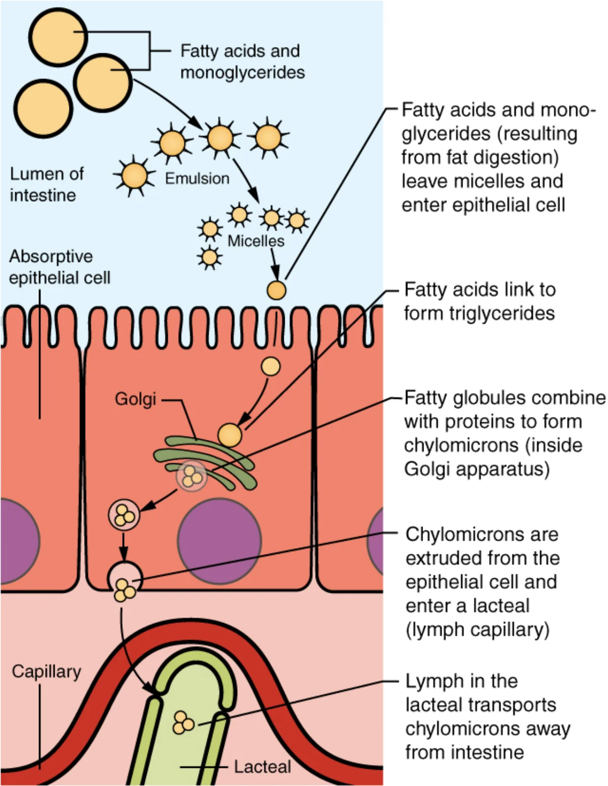
Lipid absorption pathway: bile salts emulsify fat into micelles, lipase digests triglycerides, products enter enterocytes, are repackaged into chylomicrons, and exported into lacteals. Credit: OpenStax Anatomy and Physiology 2e, CC BY 4.0
The full pathway:
Bile salts emulsify large fat globules into tiny micelles in the duodenal lumen
These products are carried to the enterocyte surface by micelles
Fatty acids and monoglycerides diffuse across the enterocyte membrane (they are hydrophobic - no transporter needed for long-chain fatty acids)
Inside the enterocyte’s smooth ER, fatty acids and monoglycerides are re-esterified back into triglycerides
Triglycerides are packaged with cholesterol and phospholipids in the Golgi to form chylomicrons
Chylomicrons are exocytosed from the basolateral membrane into lacteals (lymphatic vessels)
Lacteals drain into the lymphatic system, which empties into the thoracic duct and then the left subclavian vein
Vitamin Absorption
Vitamin Type
Examples
Absorption Mechanism
Transport
Fat-soluble (A, D, E, K)
Retinol, calciferol, tocopherol, phylloquinone
Absorbed with dietary fat in micelles
Packaged into chylomicrons; require bile
Water-soluble (B vitamins, C)
Thiamine, riboflavin, ascorbic acid
Various transporters; mostly in jejunum
Enter capillaries, travel via hepatic portal vein
Vitamin B12
Cobalamin
Binds intrinsic factor in stomach; complex absorbed in terminal ileum by receptor-mediated endocytosis
Portal circulation to liver
Water and Ion Absorption
The GI tract processes about 9 liters of fluid per day (2 L ingested + 7 L secreted). About 98% is reabsorbed:
Small intestine absorbs most water and ions (~7.5 L) by osmosis following solute absorption
Large intestine absorbs the remaining ~1.4 L
Only ~100-200 mL is lost in feces
Sodium absorption drives water absorption. Na+ enters enterocytes via:
SGLT1 (co-transported with glucose)
Na+/amino acid cotransporters
Na+/H+ exchangers
Na+ channels in the colon (regulated by aldosterone)
Water follows Na+ by osmosis through tight junctions and aquaporins.
A patient with a bile duct obstruction would have impaired absorption of which vitamins?
Click to reveal answer
Fat-soluble vitamins A, D, E, and K. Without bile, fat cannot be emulsified into micelles, and fat-soluble vitamins cannot be absorbed (they rely on micelles for transport to the enterocyte surface). Water-soluble vitamins would be unaffected. Clinically, vitamin K deficiency presents first (bleeding/bruising), since the body has smaller stores of vitamin K.
Why do absorbed fats enter the lymphatic system (lacteals) instead of the blood capillaries like amino acids and glucose?
Click to reveal answer
Chylomicrons are too large to enter blood capillaries. After re-esterification inside enterocytes, triglycerides are packaged into large chylomicron particles (~75-1200 nm). Blood capillaries have fenestrations too small for chylomicrons, but lacteals have larger openings that allow chylomicron entry. The lymphatic system then delivers them to the bloodstream via the thoracic duct.
By the time chyme reaches the large intestine, digestion is essentially complete. The large intestine’s job is not to digest, but to reclaim water and electrolytes, house trillions of bacteria, and compact the leftovers into feces. It is shorter than the small intestine (~1.5 meters) but wider in diameter - hence the name.
Anatomy: Three Main Regions
1. The Cecum
The cecum is a pouch-like structure at the junction of the small and large intestines. Material enters from the ileum through the ileocecal valve, which prevents backflow. Attached to the cecum is the appendix.
The appendix was long considered a vestigial organ, but current evidence suggests it serves as a reservoir for beneficial gut bacteria. After a bout of diarrhea that flushes the colon, the appendix may help repopulate it with normal flora. It also contains lymphoid tissue for immune surveillance.
Inflammation of the appendix (appendicitis) is a common clinical condition that may appear as passage context on the MCAT.
2. The Colon
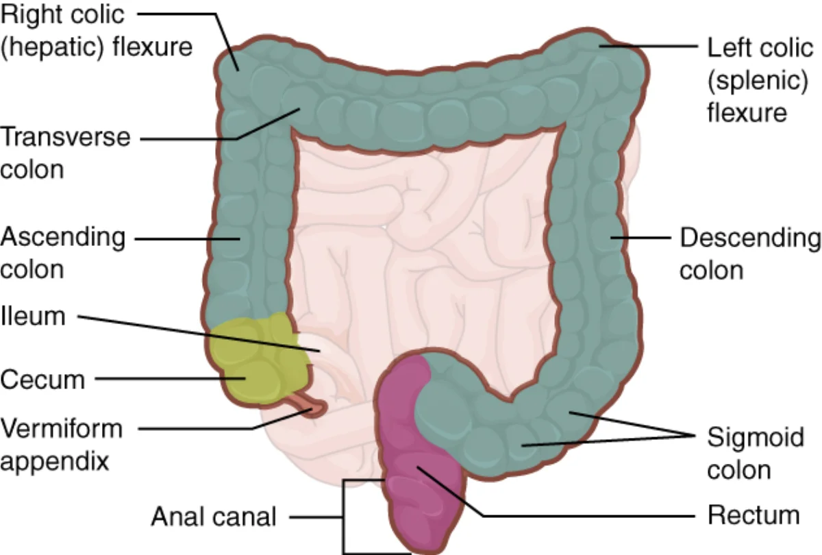
Anatomy of the large intestine. The four segments of the colon frame the small intestine in the abdominal cavity. Credit: OpenStax Anatomy and Physiology 2e, CC BY 4.0
The colon has four segments that form an inverted U-shape:
Ascending colon - runs up the right side
Transverse colon - crosses horizontally
Descending colon - runs down the left side
Sigmoid colon - S-shaped curve connecting to the rectum
3. The Rectum and Anus
The rectum stores feces until defecation. Two sphincters control the exit:
Sphincter
Muscle Type
Control
Internal anal sphincter
Smooth muscle
Involuntary (autonomic)
External anal sphincter
Skeletal muscle
Voluntary (somatic)
The defecation reflex is initiated when the rectum is stretched by feces. The internal sphincter relaxes automatically (parasympathetic), but the external sphincter remains under voluntary control - allowing you to choose when to defecate.
Water Absorption: Turning Liquid to Solid
Chyme entering the large intestine is mostly liquid. The colon absorbs about 1.4 liters of water per day along with sodium, chloride, and other ions. This converts the liquid chyme into semi-solid feces.
Sodium is absorbed via Na+ channels (regulated by aldosterone) and Na+/H+ exchangers
Water follows sodium by osmosis
Too much absorption = constipation (hard, dry stools)
Too little absorption (or excess secretion) = diarrhea
Goblet Cells: Mucus Protection
The large intestine is lined with goblet cells that secrete mucus. This mucus:
Lubricates the fecal mass for easier passage
Protects the epithelium from mechanical abrasion
Shields the lining from bacterial products and toxins
The mucus does not aid in digestion - its role is purely protective and lubricating.
Gut Microbiota: Your 100 Trillion Roommates
The large intestine hosts a vast community of bacteria (the gut microbiota). These organisms:
Ferment undigested carbohydrates (fiber), producing short-chain fatty acids (acetate, propionate, butyrate) that provide ~10% of daily caloric intake
Synthesize vitamin K and certain B vitamins (biotin, folate) - a key source since dietary vitamin K alone is often insufficient
Compete with pathogenic bacteria for nutrients and space (colonization resistance)
Train the immune system - gut flora stimulate development of GALT (Peyer’s patches, isolated lymphoid follicles) and teach the immune system to distinguish harmless microbes from pathogens
Bacteria and their byproducts make up a significant fraction of fecal mass.
What’s in Feces?
Normal feces contain:
Water (~75%)
Bacteria (living and dead)
Undigested fiber and food residue
Bile pigments (stercobilin - gives brown color)
Sloughed epithelial cells
Small amounts of fat, protein, and inorganic matter
A patient on long-term broad-spectrum antibiotics develops vitamin K deficiency and bleeding. Why?
Click to reveal answer
Antibiotics killed the gut bacteria that synthesize vitamin K. The gut microbiota in the large intestine produce vitamin K as a metabolic byproduct. Broad-spectrum antibiotics destroy these bacteria, eliminating this vitamin K source. Since vitamin K is required for clotting factor synthesis (factors II, VII, IX, X), deficiency leads to impaired coagulation and bleeding.
A patient with ulcerative colitis has chronic watery diarrhea. Which function of the large intestine is impaired?
Click to reveal answer
Water absorption. Ulcerative colitis causes chronic inflammation and damage to the mucosal lining of the colon. The damaged epithelium cannot efficiently absorb water and electrolytes, so excess water remains in the lumen, producing watery diarrhea. The inflammation is limited to the mucosa (unlike Crohn's disease, which is transmural).
The GI tract is the largest endocrine organ in the body. Specialized cells scattered throughout the stomach and small intestine release hormones that coordinate digestion like a relay team - each hormone passes the baton to trigger the next step. These hormones use the same signaling mechanisms as other endocrine hormones. The MCAT expects you to know the four major GI hormones, their triggers, and their effects.
The Big Four GI Hormones
Gastrin
Source: G cells in the stomach antrum (pyloric region)
Trigger: Stomach distension, peptides/amino acids in the stomach, vagal stimulation (parasympathetic)
Effects:
Stimulates parietal cells to secrete HCl
Stimulates chief cells to secrete pepsinogen
Stimulates gastric motility
Promotes growth of gastric mucosa (trophic effect)
Gastrin operates in a negative feedback loop: it stimulates HCl production, and when the stomach becomes sufficiently acidic (pH <2), gastrin release is suppressed.
Secretin
Source: S cells in the duodenal mucosa
Trigger: Acidic chyme entering the duodenum (pH <4.5)
Effects:
Stimulates pancreatic duct cells to release bicarbonate (neutralizes acid)
Stimulates bile secretion from the liver
Inhibits gastrin release and gastric acid secretion (slows stomach activity)
Inhibited by: Rising duodenal pH (the acid has been neutralized - job done)
Cholecystokinin (CCK)
Source: I cells in the duodenal and jejunal mucosa
Trigger: Fatty acids and amino acids in the duodenum
Stimulates pancreatic acinar cells to release digestive enzymes
Slows gastric emptying (gives the duodenum time to process)
Stimulates satiety (reduces appetite via hypothalamus)
Relaxes the sphincter of Oddi
Inhibited by: Completion of fat and protein digestion
CCK is the reason fatty meals make you feel full faster and stay in your stomach longer.
Gastric Inhibitory Peptide (GIP)
Also called glucose-dependent insulinotropic peptide (same abbreviation - GIP).
Source: K cells in the duodenum and jejunum
Trigger: Glucose, fatty acids, and amino acids in the duodenum
Effects:
Stimulates insulin release from pancreatic beta cells (the “incretin” effect)
Inhibits gastric acid secretion and motility (slows stomach)
Slows gastric emptying
Key concept: Oral glucose causes more insulin release than the same dose of IV glucose, because GIP from the gut amplifies the pancreatic response (the “incretin effect”)
Signals the hypothalamus that energy stores are sufficient
Reduces appetite, increases energy expenditure
Long-term regulator (body weight over weeks/months)
Does NOT act directly on the stomach or intestines
A patient eats a fatty meal. Which hormone causes the gallbladder to contract and release bile?
Click to reveal answer
CCK (cholecystokinin). Fat (and protein) in the duodenum triggers I cells to release CCK, which stimulates gallbladder contraction, pancreatic enzyme secretion, and slows gastric emptying. Secretin does not contract the gallbladder - it triggers bicarbonate release.
Why does oral glucose cause more insulin release than the same dose of IV glucose?
Click to reveal answer
The incretin effect. Oral glucose stimulates GIP release from intestinal cells. GIP travels to the pancreas and amplifies insulin secretion from beta cells above what blood glucose alone would trigger. IV glucose bypasses the gut, so no GIP is released, and the insulin response is lower.
Your gut has its own brain. The enteric nervous system (ENS) contains about 100 million neurons - more than the spinal cord - embedded in the walls of the GI tract from esophagus to anus. It can operate completely independently of the brain and spinal cord, earning it the nickname “the second brain.”
The Two Plexuses
The ENS is organized into two interconnected nerve networks (plexuses):
Myenteric plexus (Auerbach’s plexus)
Located between the inner circular and outer longitudinal muscle layers of the muscularis externa
Stimulation increases gut motility; inhibition relaxes sphincters and slows movement
Submucosal plexus (Meissner’s plexus)
Located in the submucosa
Controls secretion and absorption: regulates enzyme release, mucus production, and local blood flow
Also responds to luminal stretch and chemical stimuli
The ENS Contains All Three Neuron Types
The enteric nervous system is a complete neural circuit:
Sensory (afferent) neurons - detect mechanical stretch, chemical composition (pH, osmolarity, nutrients), and temperature in the lumen
Interneurons - process information and coordinate responses
Motor (efferent) neurons - control smooth muscle contraction, gland secretion, and blood vessel dilation
This means the ENS can carry out complete reflex arcs without any input from the brain or spinal cord. A chemical stimulus in the lumen can trigger enzyme secretion or a peristaltic wave entirely through local ENS circuits.
Autonomic Modulation: Parasympathetic vs. Sympathetic
While the ENS operates autonomously, the central nervous system modulates its activity through the autonomic nervous system:
The vagus nerve provides parasympathetic innervation to most of the GI tract (esophagus through the transverse colon). The distal colon, rectum, and internal anal sphincter receive parasympathetic input from pelvic splanchnic nerves (S2-S4).
”Rest and Digest” vs. “Fight or Flight”
After a meal, parasympathetic activity dominates:
Increased salivation
Increased gastric acid and enzyme secretion
Increased peristalsis and motility
Increased blood flow to the GI tract
This is why you feel drowsy after a big meal - blood is diverted to the gut (“food coma”)
During stress or exercise, sympathetic activity dominates:
Decreased salivation (dry mouth)
Decreased secretion and motility
Blood diverted away from the GI tract to skeletal muscles and brain
Sphincters contract (no time for digestion when running from danger)
The Defecation Reflex
Defecation involves both the ENS and the CNS:
Feces distend the rectum, activating stretch receptors
Sensory signals travel to the sacral spinal cord (S2-S4) via pelvic nerves
Parasympathetic motor signals return, causing:
Increased colonic and rectal motility
Relaxation of the internal anal sphincter (involuntary)
The external anal sphincter (skeletal muscle, under voluntary somatic control) can be consciously relaxed to allow defecation or kept contracted to delay it
The Three Phases of Gastric Regulation
GI secretion is controlled in three overlapping phases:
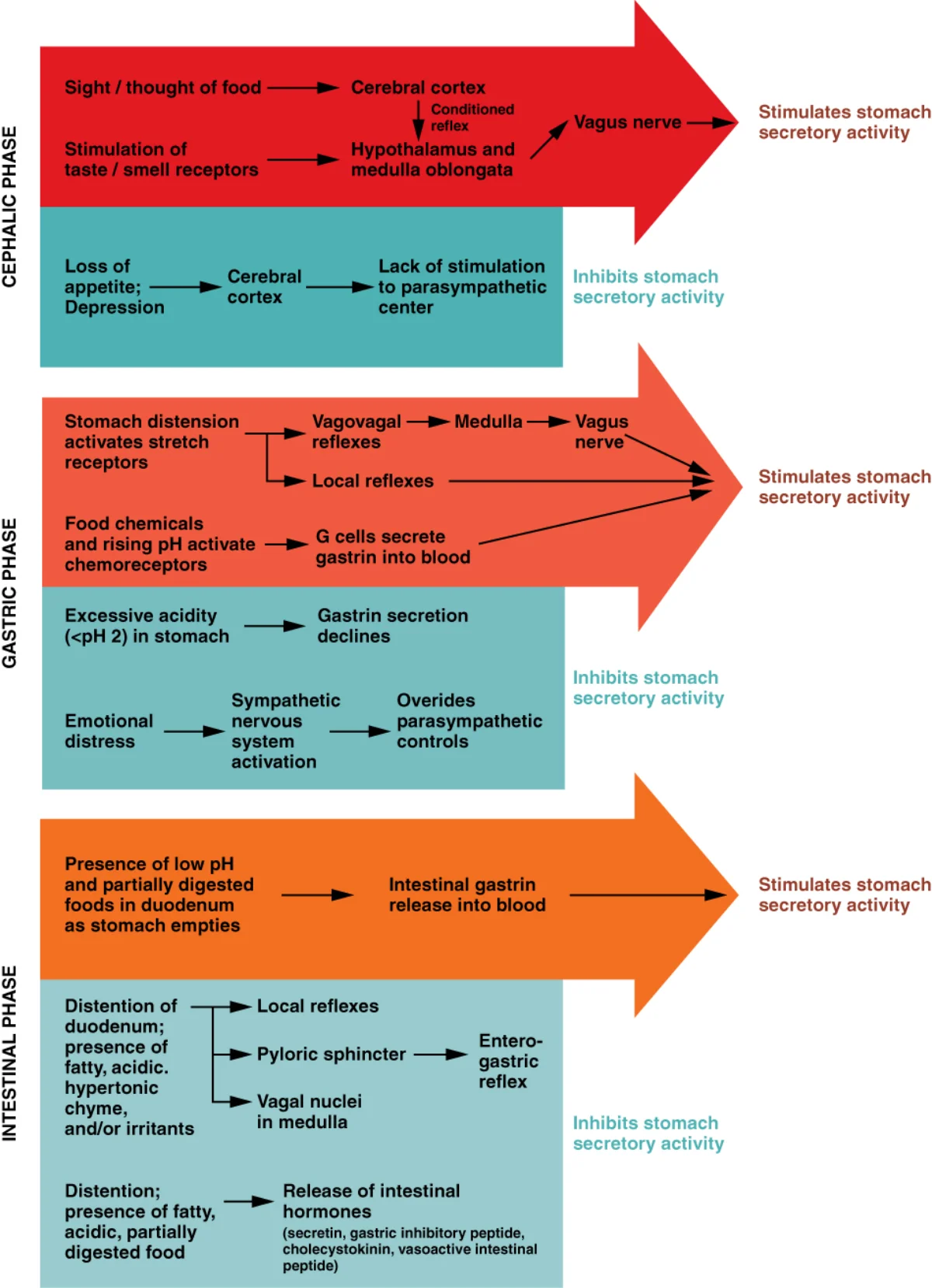
The three phases of gastric regulation. The cephalic phase begins before food arrives; the gastric phase is triggered by food in the stomach; the intestinal phase coordinates duodenal responses. Credit: OpenStax Anatomy and Physiology 2e, CC BY 4.0
Phase
Trigger
Mediator
Effect
Cephalic phase
Sight, smell, thought, taste of food
Vagus nerve (parasympathetic)
Stimulates gastric secretion before food arrives (~30% of total)
Gastric phase
Food in stomach (distension, peptides, pH changes)
Gastrin, local reflexes
Main stimulation of HCl and pepsinogen (~60% of total)
A patient has a severed vagus nerve (vagotomy). What happens to gastric acid secretion and gastric motility?
Click to reveal answer
Both decrease. The vagus nerve provides parasympathetic innervation that stimulates gastric acid secretion (directly via ACh on parietal cells, and indirectly by stimulating gastrin release from G cells) and gastric motility. Vagotomy was historically used to treat peptic ulcers by reducing acid output. The cephalic phase of gastric secretion is eliminated entirely.
Why might a person experience diarrhea during a stressful exam, even though sympathetic activation normally slows GI motility?
Click to reveal answer
The gut-brain axis and stress hormones. While acute sympathetic activation slows upper GI motility, stress hormones (CRH, cortisol) can paradoxically stimulate colonic motility and secretion via the gut-brain axis. The ENS responds to stress signals by increasing lower GI activity, causing cramping and diarrhea. This is the physiological basis of stress-induced irritable bowel syndrome.