
Immune Disorders
The immune system is a powerful weapon. When it works correctly, it protects you from infections and cancer. When it malfunctions, it can be just as destructive - attacking your own tissues, failing to fight infections, or overreacting to harmless substances.
Immune disorders fall into three categories: the system attacks self (autoimmunity), the system is too weak (immunodeficiency), or the system overreacts (hypersensitivity).
Autoimmune Diseases: Friendly Fire
In autoimmune diseases, the immune system fails to distinguish self from non-self and attacks the body’s own healthy tissues. This happens when self-tolerance breaks down - meaning self-reactive lymphocytes that should have been eliminated during development (negative selection) or suppressed by regulatory T cells escape these checkpoints.
| Disease | Target | Immune Mechanism |
|---|---|---|
| Type 1 diabetes | Insulin-producing beta cells of the pancreas | T cells destroy beta cells |
| Rheumatoid arthritis | Joint synovial membranes | Antibodies and T cells attack joints |
| Multiple sclerosis (MS) | Myelin sheath of neurons (CNS) | T cells attack myelin |
| Systemic lupus erythematosus (SLE) | DNA and multiple organs | Anti-DNA antibodies form immune complexes that deposit in tissues |
| Graves’ disease | TSH receptors on thyroid | Antibodies stimulate thyroid (hyperthyroidism) |
Immunodeficiency: A Weakened Defense
Immunodeficiency means the immune system is too weak to protect the body effectively. It can be congenital (present from birth) or acquired (developed later in life).
Primary (congenital) immunodeficiencies:
The most important example is Severe Combined Immunodeficiency (SCID) - a defect in lymphocyte development that results in no functional B or T cells. Without treatment, SCID is fatal because the patient has essentially no adaptive immunity (the “bubble boy” disease).
Other examples you may see in MCAT passages include thymus defects that prevent T cell maturation and B cell defects that prevent antibody production. The key concept: if you know which branch of immunity is missing, you can predict which types of infections the patient will be vulnerable to.
Secondary (acquired) immunodeficiency:
The most important example is HIV/AIDS:
- HIV is a retrovirus that specifically infects and destroys CD4+ helper T cells
- HIV binds to CD4 on helper T cells
- As CD4+ T cell counts drop, the immune system progressively weakens
- AIDS (Acquired Immunodeficiency Syndrome) is diagnosed when CD4+ count falls below 200 cells/microliter (normal: 500-1500)
- Without helper T cells, both humoral and cell-mediated immunity collapse
- Patients become vulnerable to opportunistic infections (e.g., Pneumocystis pneumonia, Kaposi’s sarcoma, thrush) that healthy immune systems easily control
Hypersensitivity Reactions: The Overreaction
Hypersensitivity reactions are immune responses that are disproportionate to the actual threat. There are four types, classified by mechanism and timing. This is one of the most commonly tested immune topics on the MCAT.
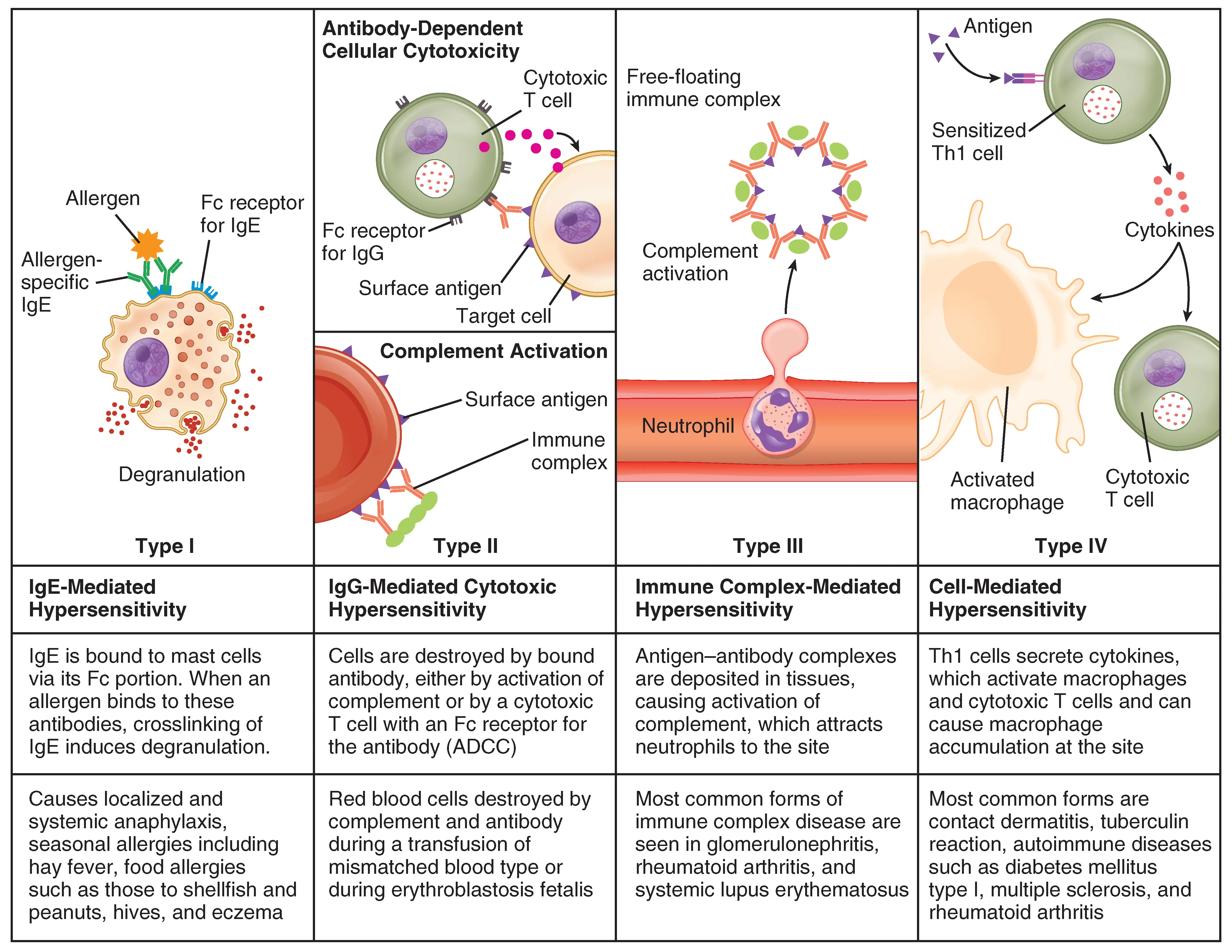
Type I: Immediate/Anaphylactic Hypersensitivity
Mechanism: IgE-mediated. On first exposure, IgE antibodies are produced and bind to mast cells/basophils. On re-exposure, the allergen cross-links IgE on mast cells, triggering degranulation and release of histamine, leukotrienes, and prostaglandins.
Timing: Minutes (immediate)
Examples: Allergic rhinitis (hay fever), asthma, food allergies, bee sting reactions, anaphylaxis
Key molecules: IgE, histamine, mast cells
Treatment: Antihistamines, epinephrine (for anaphylaxis), corticosteroids
Anaphylaxis is the most severe form - systemic vasodilation causes a dangerous drop in blood pressure, while airway constriction makes breathing difficult. Epinephrine (EpiPen) reverses these effects by causing vasoconstriction and bronchodilation.
Type II: Cytotoxic Hypersensitivity
Mechanism: IgG or IgM antibodies bind to antigens on the surface of the body’s own cells, marking them for destruction by complement or phagocytes.
Timing: Hours
Examples:
- Hemolytic disease of the newborn (Rh incompatibility) - maternal antibodies attack fetal RBCs
- Transfusion reactions (ABO incompatibility) - antibodies attack transfused RBCs
- Autoimmune hemolytic anemia
- Graves’ disease and myasthenia gravis (antibodies bind cell surface receptors)
Key molecules: IgG, IgM, complement
Type III: Immune Complex Hypersensitivity
Mechanism: Antigen-antibody complexes (immune complexes) form in the blood and deposit in tissues (joints, kidneys, blood vessel walls), triggering complement activation and inflammation at the deposition site.
Timing: Hours to days
Examples:
- Systemic lupus erythematosus (SLE) - immune complexes deposit in kidneys, joints, skin
- Serum sickness - reaction to foreign proteins (e.g., antivenom)
- Post-streptococcal glomerulonephritis
Key molecules: IgG, immune complexes, complement
Type IV: Delayed-Type Hypersensitivity (DTH)
Mechanism: T cell-mediated (no antibodies involved). Sensitized T cells encounter the antigen and release cytokines that recruit macrophages, causing inflammation and tissue damage.
Timing: 24-72 hours (delayed)
Examples:
- Tuberculin skin test (PPD test) - positive reaction = swelling at injection site after 48-72 hours
- Contact dermatitis (poison ivy, nickel allergy)
- Transplant rejection
- Type 1 diabetes (T cells destroying beta cells)
Key molecules: T cells (CD4+ helper T cells, macrophages), cytokines
Hypersensitivity Master Table
| Type | Name | Mediator | Timing | Mechanism | Classic Example |
|---|---|---|---|---|---|
| I | Anaphylactic | IgE | Minutes | Mast cell degranulation | Allergies, anaphylaxis |
| II | Cytotoxic | IgG/IgM | Hours | Antibodies target cell surfaces | Transfusion reaction, Rh disease |
| III | Immune complex | IgG | Hours-days | Immune complexes deposit in tissues | Lupus (SLE), serum sickness |
| IV | Delayed | T cells | 48-72 hrs | T cells recruit macrophages | TB skin test, poison ivy, transplant rejection |
Transplant Rejection
When a patient receives an organ from a donor, the recipient’s immune system may recognize the donor’s MHC/HLA molecules as foreign, triggering an immune attack on the transplanted organ. This is why doctors match HLA types between donor and recipient and prescribe immunosuppressive drugs that suppress T cell activation.
Transplant rejection can involve both antibody-mediated (Type II) and T cell-mediated (Type IV) hypersensitivity, depending on whether the recipient has pre-existing antibodies against the donor’s MHC or whether T cells mount a new response against the foreign tissue.