
Overview of the GI Tract
Think of the GI tract as one long conveyor belt running through a factory - except instead of building something, it tears things apart. Food enters at one end (mouth) and waste exits at the other (anus), passing through specialized stations along the way.
The technical name for this tube is the alimentary canal. It runs about 9 meters (30 feet) from mouth to anus. Here is a crucial insight: the lumen (interior) of this tube is technically outside your body. Food sitting in your stomach has not actually entered your tissues yet. It must be digested and then absorbed across the intestinal wall to truly enter the body.
The Route: Station by Station
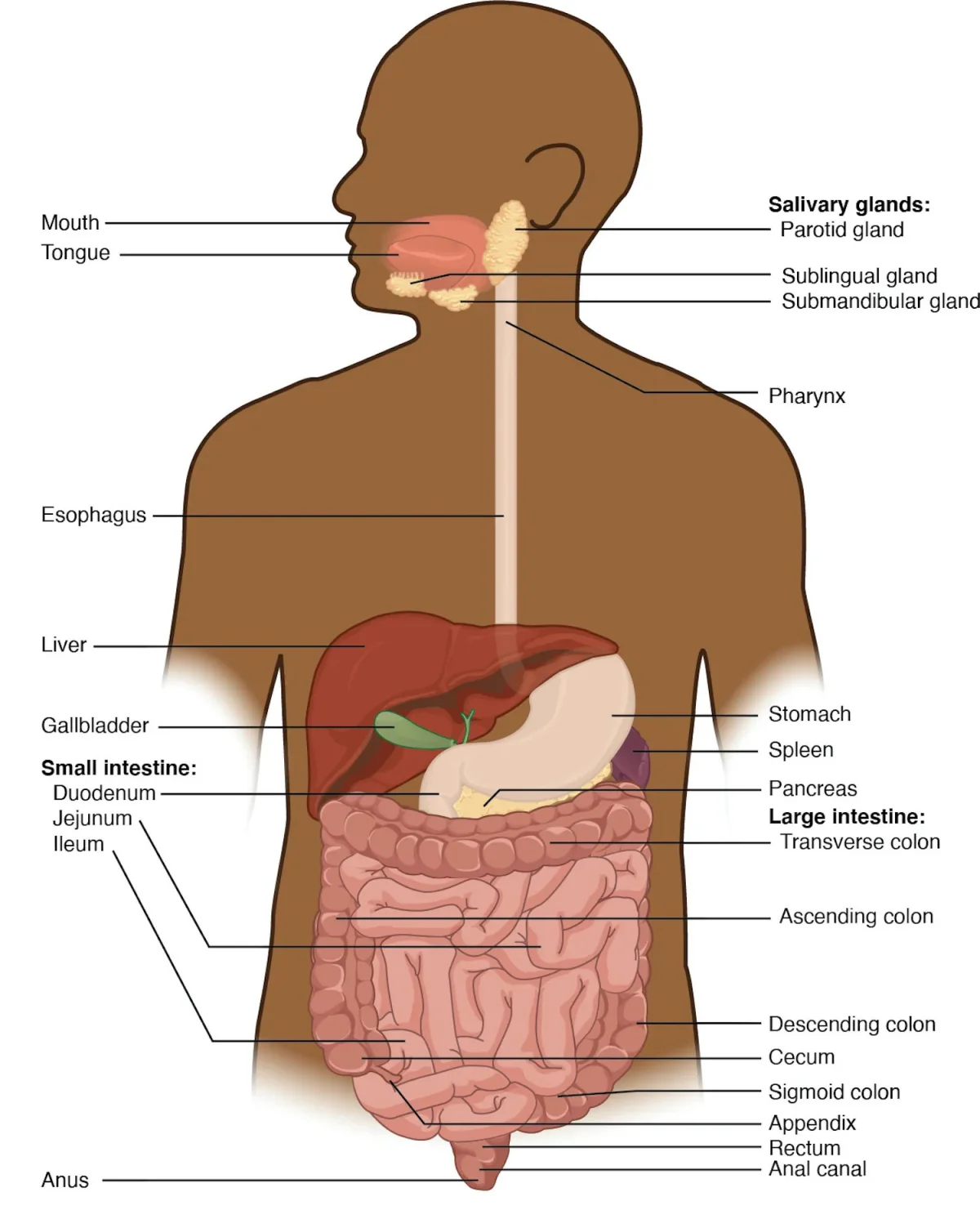
Food follows this path, in order:
- Oral cavity (mouth) - mechanical and chemical digestion begin
- Pharynx - shared passage with respiratory tract
- Esophagus - transport tube to stomach
- Stomach - acid bath and protein digestion
- Small intestine (duodenum, jejunum, ileum) - most digestion and absorption
- Large intestine (cecum, colon, rectum) - water absorption, feces formation
- Anus - elimination
Four accessory organs contribute secretions but food never passes through them: salivary glands, liver, gallbladder, and pancreas.
Two Types of Digestion
Mechanical digestion physically breaks food into smaller pieces without changing its chemical structure. Chewing is the most obvious example, but the stomach’s churning and the small intestine’s segmentation contractions also count. The purpose is to increase surface area so enzymes can work faster.
Chemical digestion uses enzymes to break covalent bonds in macromolecules:
| Macromolecule | Broken Into | Key Enzymes (Preview) |
|---|---|---|
| Carbohydrates (starch) | Monosaccharides (glucose, fructose, galactose) | Amylase, disaccharidases |
| Proteins | Amino acids | Pepsin, trypsin, chymotrypsin |
| Lipids (triglycerides) | Fatty acids + monoglycerides | Lipase (with bile assistance) |
| Nucleic acids | Nucleotides | Nucleases |
The Four Histological Layers
Every region of the alimentary canal shares the same four-layer wall structure, from innermost to outermost:
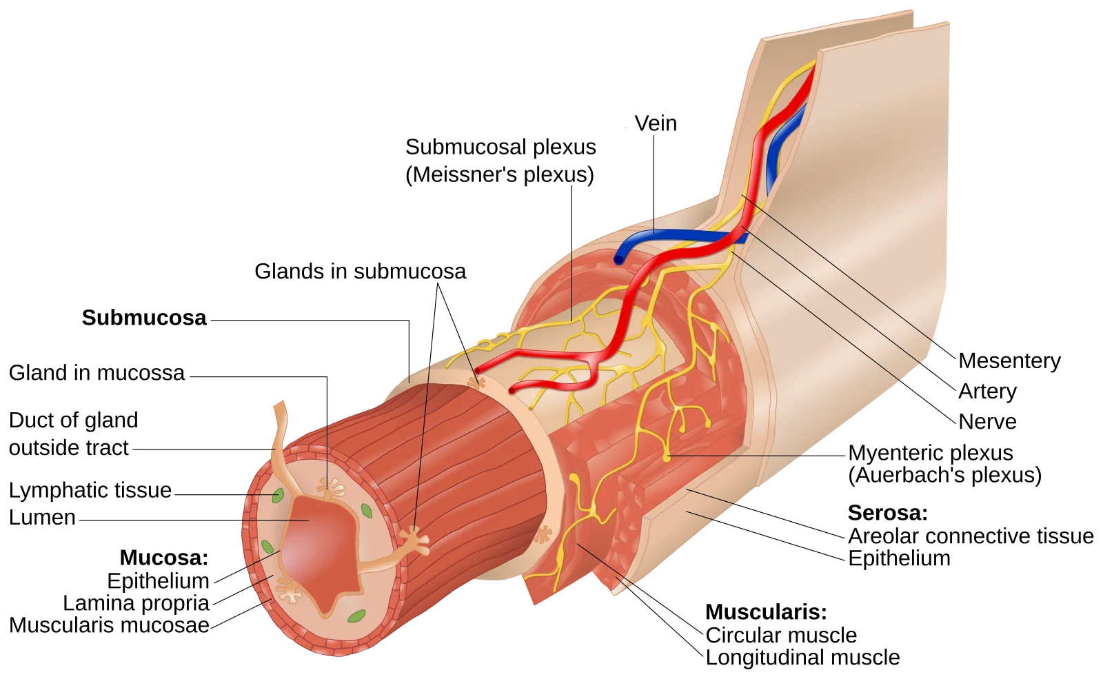
- Mucosa - innermost layer; contains the epithelium that contacts food, plus the lamina propria (connective tissue with blood vessels and lymph) and muscularis mucosae (thin smooth muscle)
- Submucosa - dense connective tissue with blood vessels, lymphatics, and the submucosal plexus (Meissner’s plexus) of the enteric nervous system
- Muscularis externa - two layers of smooth muscle (inner circular, outer longitudinal) responsible for peristalsis; contains the myenteric plexus (Auerbach’s plexus) between the muscle layers
- Serosa (or adventitia) - outermost connective tissue covering; serosa where the organ is surrounded by peritoneum, adventitia where it is not (esophagus)
Peristalsis and Segmentation
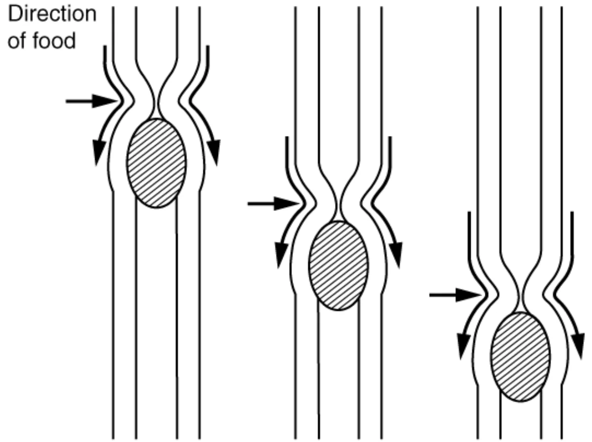
Peristalsis is the wave-like contraction that pushes food forward through the GI tract. Behind the food bolus, circular muscle contracts (squeezing); ahead of it, longitudinal muscle contracts (shortening, which widens the lumen). This coordinated wave moves food in one direction - mouth to anus.
Segmentation is a back-and-forth contraction pattern found mainly in the small intestine. Unlike peristalsis, segmentation does not push food forward. It chops and mixes the chyme with digestive juices, ensuring maximum contact between enzymes and food.
Sphincters: The Gatekeepers
Sphincters are rings of smooth muscle that act as one-way valves between GI regions:
| Sphincter | Location | Function |
|---|---|---|
| Upper esophageal | Top of esophagus | Prevents air from entering esophagus |
| Lower esophageal (cardiac) | Esophagus-stomach junction | Prevents acid reflux into esophagus |
| Pyloric | Stomach-duodenum junction | Controls gastric emptying rate |
| Ileocecal valve | Ileum-cecum junction | Prevents backflow from large intestine |
| Internal anal | Upper anus | Involuntary; maintains continence |
| External anal | Lower anus | Voluntary; controls defecation |