Before your body ever manufactures a single antibody or trains a single T cell, it has an entire defense system already in place - one that was ready the day you were born. This is innate immunity: fast, nonspecific, and always on duty.
Think of it like a building’s security system. The locked doors, security cameras, and fire alarms do not need to know who the intruder is. They just detect “something is wrong” and respond immediately. Innate immunity works the same way - it recognizes general danger signals rather than specific invaders.
First Line of Defense: Physical and Chemical Barriers
Your body’s outermost defenses prevent pathogens from ever entering in the first place.
Skin is the largest organ and the most important barrier. The epidermis is a layer of tightly packed dead cells that most pathogens simply cannot penetrate. The skin also actively fights microbes by secreting:
Lysozyme (in sweat) - an enzyme that breaks down bacterial cell walls
Sebum - an oily secretion from sebaceous glands with antimicrobial properties
Antimicrobial peptides (in sweat) - small proteins that inhibit pathogen growth
Mucous membranes line the respiratory, gastrointestinal, and urogenital tracts. Mucus is a thick, sticky fluid that physically traps pathogens and contains lysozyme to destroy bacterial cell walls.
Stomach acid (HCl, pH ~1.5-2) destroys most ingested bacteria and viruses by denaturing their proteins. This is why food poisoning requires a large enough dose of bacteria to overwhelm the acid.
Cilia in the respiratory tract beat in coordinated waves to sweep mucus (and trapped pathogens) upward toward the throat, where it is swallowed or expelled. Smoking paralyzes cilia - which is why smokers are more susceptible to respiratory infections.
Normal microbiota - the trillions of beneficial bacteria on your skin and in your gut - outcompete pathogenic microbes for nutrients and binding sites. Some even produce toxins that kill pathogens. Think of them as the “regulars” at a bar who take up all the seats so troublemakers cannot sit down.
Pathogen Recognition: PAMPs and PRRs
How does the innate immune system know what to attack? It recognizes pathogen-associated molecular patterns (PAMPs) - molecular features found on microbes but never on human cells. Examples include:
Lipopolysaccharide (LPS) on gram-negative bacterial cell walls
Peptidoglycan on gram-positive bacterial cell walls
Double-stranded RNA (dsRNA) from replicating viruses
Flagellin from bacterial flagella
Immune cells detect PAMPs using pattern recognition receptors (PRRs), the most important being Toll-like receptors (TLRs) on the surface of macrophages and dendritic cells. When a TLR binds a PAMP, it triggers an immediate immune response - no prior exposure required.
Second Line of Defense: The Inflammatory Response
When pathogens breach the physical barriers, the body launches inflammation - a rapid, localized response designed to contain the threat and recruit immune cells. Here is what happens step by step:
Tissue damage or pathogen detection - Damaged cells release chemical signals. Macrophages in the tissue detect PAMPs and release cytokines.
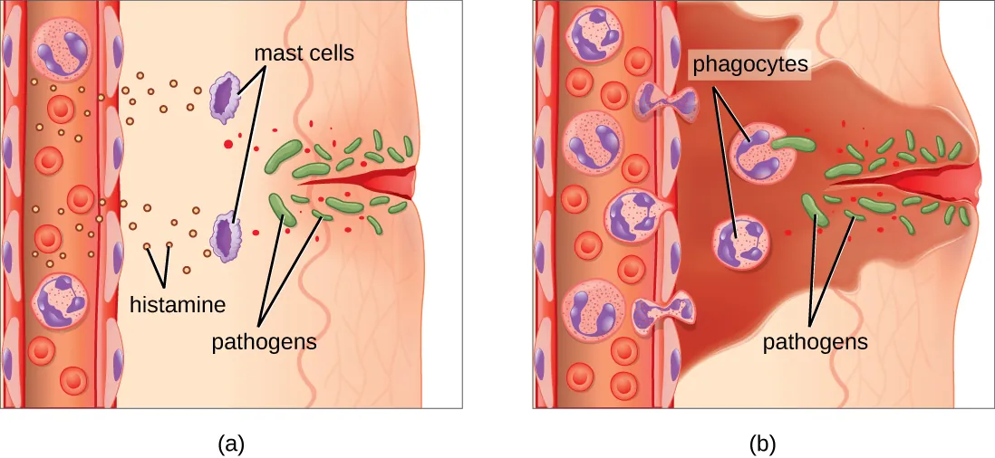
Mast cell activation - Mast cells stationed in tissues release histamine, which causes nearby blood vessels to dilate (widen).
Vasodilation and increased permeability - Dilated vessels bring more blood to the area. Vessel walls become “leaky,” allowing fluid and immune cells to exit the blood and enter the tissue.
Immune cell recruitment - White blood cells such as neutrophils are the first responders, arriving within minutes. They squeeze between endothelial cells (diapedesis) and follow chemical gradients (chemotaxis) to the infection site.
Phagocytosis and pathogen destruction - Neutrophils and macrophages engulf and destroy pathogens using enzymes and reactive oxygen species.
The four classic signs of inflammation are easy to remember:
Sign
Cause
Redness
Vasodilation brings more blood
Heat
Increased blood flow raises local temperature
Swelling
Fluid leaks from permeable vessels into tissue
Pain
Swelling puts pressure on nerve endings
The inflammatory response. Tissue damage triggers mast cell histamine release, causing vasodilation and increased vascular permeability. Neutrophils squeeze through vessel walls (diapedesis) and follow chemical gradients (chemotaxis) to the infection site. Credit: OpenStax Microbiology, CC BY 4.0
Fever is a systemic extension of inflammation. Pyrogens (fever-inducing molecules) released by immune cells or pathogens reset the hypothalamic thermostat to a higher temperature. Fever helps fight infection by inhibiting bacterial and viral reproduction and by increasing the metabolic rate of immune cells.
Interferons are antiviral cytokines. A virus-infected cell secretes interferons that bind neighbors and switch them into an antiviral state (slowing protein synthesis, degrading viral RNA). The infected cell often sacrifices itself, but the interferon signal stops the virus from spreading. Think of it as a burning building warning the whole block.
What are the four cardinal signs of inflammation, and what causes each one?
Click to reveal answer
Redness (vasodilation), Heat (increased blood flow), Swelling (fluid leaking from permeable vessels), Pain (pressure on nerve endings from swelling). All are triggered by histamine release from mast cells causing vasodilation and increased vascular permeability.
The Complement System
The complement system is a set of ~30 proteins circulating in the blood in inactive form. When activated, they work in a cascade (each protein activates the next) to destroy pathogens through three mechanisms:
Three activation pathways all converge on the same endpoint:
Pathway
Trigger
Key Detail
Classical
Antibody bound to pathogen (antigen-antibody complex)
Links adaptive immunity to complement; starts with C1
Opsonization - C3b coats the pathogen surface, making it much easier for phagocytes to recognize and engulf it. Think of opsonization as “painting a target” on the enemy.
Inflammation - Small complement fragments (C3a, C5a) attract immune cells to the site and promote inflammation by triggering mast cell degranulation and chemotaxis.
Membrane Attack Complex (MAC) - Proteins C5b, C6, C7, C8, and multiple C9s (the C5b-9 complex) assemble into a ring-shaped pore in the pathogen’s membrane, causing lysis and death. Gram-negative bacteria are especially vulnerable; gram-positive bacteria are usually protected by their thick peptidoglycan wall.
Name the three complement activation pathways and their triggers.
Click to reveal answer
Classical - triggered by antibody-antigen complexes (requires adaptive immunity). Alternative - triggered by spontaneous C3 cleavage on pathogen surfaces (no antibodies needed). Lectin - triggered by mannose-binding lectin recognizing pathogen surface sugars. All three converge to produce opsonization, inflammation, and the MAC.
A patient with a liver disease has reduced production of complement proteins. Which immune function would be MOST directly impaired?
Click to reveal answer
The complement cascade - because complement proteins are synthesized primarily in the liver. Reduced complement means impaired opsonization, reduced MAC formation, and weaker inflammatory signaling through C3a/C5a. Dendritic cell function, interferon secretion, and chemotaxis are not directly dependent on liver-synthesized complement.
All immune cells originate from hematopoietic stem cells in the bone marrow. These stem cells differentiate down two major lineages:
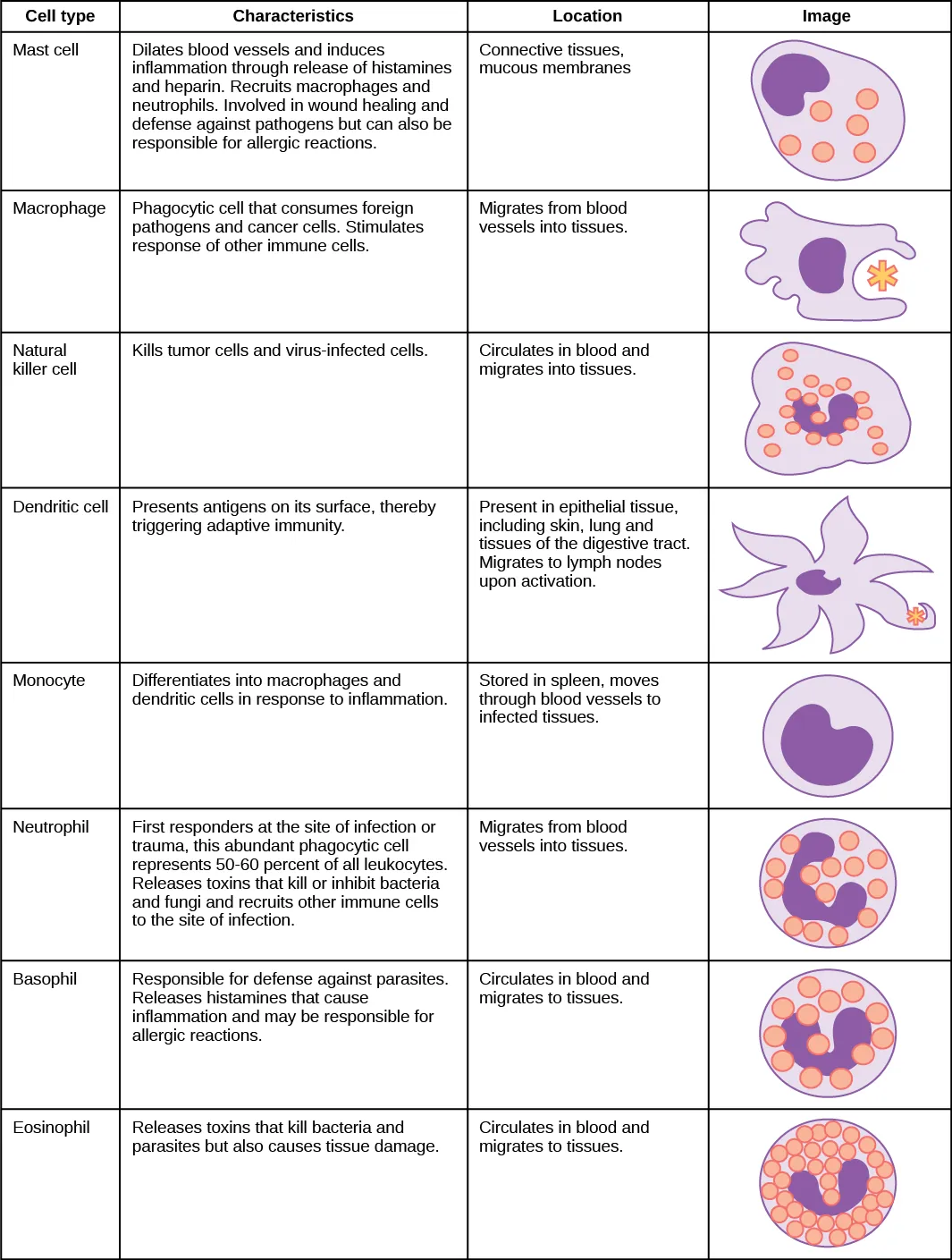
Myeloid lineage - produces most innate immune cells: neutrophils, eosinophils, basophils, mast cells, monocytes (which become macrophages and dendritic cells)
Lymphoid lineage - produces lymphocytes: T cells, B cells, and Natural Killer (NK) cells
NK cells are the exception that proves the rule - they are lymphoid cells but function in innate immunity. The MCAT loves this distinction.
Immune cells derive from hematopoietic stem cells via myeloid and lymphoid lineages. Myeloid cells include most innate immune cells; lymphoid cells include T cells, B cells, and NK cells. Credit: OpenStax Biology 2e, CC BY 4.0
Granulocytes: Cells with Granules
Granulocytes are white blood cells with visible cytoplasmic granules packed with enzymes and antimicrobial substances. There are three types, and they are easy to remember by what they fight.
Neutrophils - the most abundant white blood cell in circulation (~60-70% of all WBCs). They are the first responders to infection, arriving within minutes via chemotaxis. Their primary weapon is phagocytosis - engulfing and digesting pathogens using digestive enzymes inside the cell. Neutrophils are short-lived and die at the infection site. Pus is mostly dead neutrophils.
Eosinophils - specialized for fighting large multicellular parasites (like helminths/worms) that are too big to phagocytize. Instead, eosinophils attach to the parasite and dump cytotoxic granule contents onto its surface, destroying it from the outside. Eosinophils also play a role in allergic responses. Elevated eosinophils on a blood test suggest either a parasitic infection or allergies.
Basophils - the rarest granulocyte in blood. Functionally similar to mast cells, they release histamine and other inflammatory mediators. They amplify allergic responses and inflammation.
Agranulocytes: Cells Without Visible Granules
Monocytes are large cells that circulate in the blood as precursors. When they migrate into tissues, they differentiate into either macrophages or dendritic cells depending on the signals they receive. Think of monocytes as “rookies in transit” who become specialized once they arrive at their post.
The migration process from blood to tissue involves chemotaxis (following chemical gradients toward the infection) and diapedesis (squeezing between the endothelial cells lining blood vessel walls to exit into the tissue).
Macrophages: The Pac-Man of the Immune System
Macrophages (“big eaters”) are phagocytes stationed throughout the body, lying in wait. When they encounter a pathogen, they:
Recognize it via pattern recognition receptors (PRRs) that detect PAMPs
Engulf it into a vesicle called a phagosome
Destroy it by fusing the phagosome with a lysosome (creating a phagolysosome), where enzymes and acid digest the pathogen
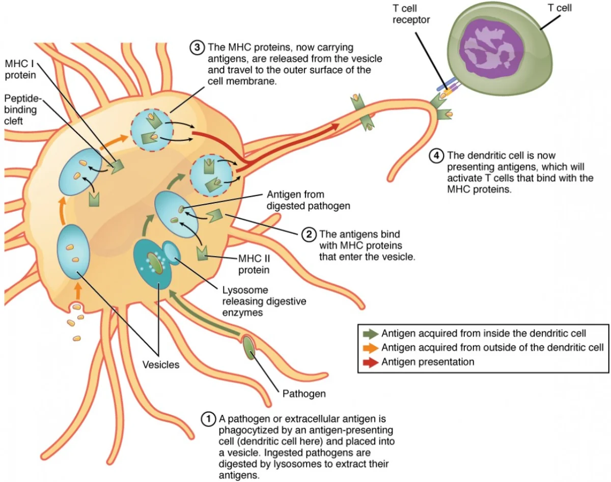
Present fragments of the pathogen on their surface using MHC II molecules - this activates the adaptive immune system
Macrophages also secrete cytokines to recruit more immune cells and promote tissue repair after the infection is cleared.
Macrophages go by different names depending on where they live. For example, they are called microglia in the brain and Kupffer cells in the liver. If an MCAT passage mentions a tissue-resident phagocyte by an unfamiliar name, it is probably a macrophage.
Dendritic Cells: The Messengers
Dendritic cells are the most important antigen-presenting cells (APCs). They are stationed in tissues that contact the external environment - skin, lungs, gut lining - where they capture pathogens via phagocytosis.
What makes dendritic cells special is what happens next: after capturing an antigen, they migrate to the nearest lymph node and present it to T cells. This is the critical bridge between innate and adaptive immunity. Macrophages stay in the tissue; dendritic cells travel to deliver the message.
Dendritic cells present antigens using both MHC I (to CD8+ T cells) and MHC II (to CD4+ T cells).
Natural Killer (NK) Cells
NK cells are the immune system’s quality control inspectors. Every nucleated cell in your body displays MHC I molecules on its surface - like wearing an employee ID badge. NK cells patrol the body, checking these badges.
When a virus infects a cell, it often downregulates MHC I expression to hide from T cells. Cancer cells do the same thing. But this “hiding” strategy backfires because NK cells detect the absence of MHC I. No badge? You get destroyed.
NK cells kill target cells by releasing:
Perforin - punches holes in the target cell’s membrane
Granzymes - enter through the holes and trigger apoptosis (programmed cell death)
Mast Cells
Mast cells are strategically positioned in tissues near blood vessels, especially in skin, lungs, and the gut. They are loaded with granules containing histamine and other inflammatory mediators.
When mast cells detect tissue damage or bind IgE antibodies (in allergic reactions), they degranulate - releasing histamine that triggers vasodilation, increased vascular permeability, and recruitment of other immune cells. Mast cells are the “alarm pullers” of the immune system.
Complete Immune Cell Summary
Cell
Lineage
Key Function
Special Feature
Neutrophil
Myeloid
Phagocytosis of bacteria
Most abundant WBC; first responder; pus
Eosinophil
Myeloid
Kill parasites; allergy role
Dump granules on large targets
Basophil
Myeloid
Release histamine
Rarest granulocyte; like mast cells in blood
Mast cell
Myeloid
Release histamine; trigger inflammation
Stationed in tissues; key in allergy
Monocyte
Myeloid
Precursor cell
Becomes macrophage or dendritic cell
Macrophage
Myeloid
Phagocytosis + antigen presentation (MHC II)
“Big eater”; tissue-resident
Dendritic cell
Myeloid
Antigen presentation to T cells
Migrates to lymph nodes; bridges innate/adaptive
NK cell
Lymphoid
Kill cells missing MHC I
Uses perforin + granzymes; no MHC needed
A virus infects a host cell and downregulates MHC I expression. Which immune cell would MOST likely destroy this infected cell?
Click to reveal answer
Natural Killer (NK) cells. NK cells detect cells with reduced or absent MHC I expression. Cytotoxic T cells (CD8+) require MHC I to present foreign peptides, so downregulation of MHC I helps infected cells evade T cells but makes them targets for NK cells. This is a common MCAT distractor - do not confuse NK cell and CD8+ T cell recognition.
What is the key difference between how macrophages and dendritic cells contribute to adaptive immunity?
Click to reveal answer
Both are antigen-presenting cells (APCs) that display pathogen fragments on MHC II. The key difference: dendritic cells migrate to lymph nodes to present antigens to naive T cells, initiating the adaptive response. Macrophages remain in the tissue and primarily present to already-activated T cells at the infection site. Dendritic cells are the most potent activators of naive T cells.
Innate immunity buys time. Adaptive immunity finishes the job.
The adaptive immune system is slower to activate (taking days to weeks on first exposure), but it has two superpowers that innate immunity lacks: specificity (it targets one particular pathogen) and memory (it remembers that pathogen forever).
Two Arms, Two Targets
Adaptive immunity splits into two branches based on the type of threat:
Humoral immunity (“humor” = body fluid) uses antibodies produced by B cells to fight pathogens circulating freely in the blood, lymph, and tissue fluids. If the enemy is floating around outside of cells - bacteria in the bloodstream, toxins, viruses before they enter cells - humoral immunity handles it.
Cell-mediated immunity uses T cells to fight pathogens hiding inside cells. If a virus has already infected a cell and is replicating inside it, antibodies cannot reach it. Instead, cytotoxic T cells recognize the infected cell and destroy it, sacrificing the cell to kill the pathogen within.
Extracellular vs. Intracellular Pathogens
This distinction is critical for MCAT reasoning:
Pathogen Location
Examples
Primary Defense
Extracellular (in body fluids)
Most bacteria, fungi, parasites, viral particles before cell entry
Humoral immunity (antibodies from B cells)
Intracellular (inside host cells)
Viruses (replicating), some bacteria (e.g., Mycobacterium tuberculosis), some parasites (e.g., Plasmodium)
Cell-mediated immunity (cytotoxic T cells)
Important nuance: viruses exist in both states. Before entering a cell, a virus is an extracellular particle (virion) targetable by antibodies. Once inside a cell, it can only be reached by cell-mediated immunity. This is why both arms are needed for a complete antiviral response.
Antigen processing and presentation bridges innate and adaptive immunity. APCs capture pathogens, process them into peptide fragments, and present them on MHC molecules to activate T cells. Credit: Lumen Learning / OpenStax Anatomy and Physiology, CC BY 4.0
B Cells and T Cells: Origin and Maturation
Both B cells and T cells originate from hematopoietic stem cells in the bone marrow, but they mature in different locations:
B cells mature in the Bone marrow
T cells migrate to and mature in the Thymus
After maturation, both cell types are mature but naive - they are fully functional but have not yet encountered their specific antigen. Think of them as trained soldiers who have graduated boot camp but have never seen combat. They circulate through the blood and lymph, waiting in lymph nodes and the spleen until they encounter their matching antigen.
The Adaptive Response Timeline
On first exposure to a new pathogen (primary response):
Innate immunity responds immediately (hours)
Dendritic cells capture antigens and migrate to lymph nodes (hours to days)
Naive T and B cells that match the antigen are activated (days)
Clonal expansion produces an army of identical effector cells (days to weeks)
Antibodies appear in the blood and effector T cells deploy (~7-10 days)
The pathogen is cleared
Most effector cells die, but memory cells persist
On second exposure (secondary response):
Memory cells recognize the pathogen immediately
Response is faster (1-2 days), stronger (more antibodies, higher affinity), and longer-lasting
The person may never develop symptoms
This is exactly why vaccines work - they trigger the primary response without causing disease, so the secondary response is ready when the real pathogen arrives.
How the Two Arms Cooperate
Humoral and cell-mediated immunity are not independent - they work together, with helper T cells (CD4+) acting as the coordinator:
Dendritic cells present antigen to helper T cells via MHC II
Activated helper T cells release cytokines that:
Stimulate B cells to produce antibodies (activating humoral immunity)
Stimulate cytotoxic T cells to kill infected cells (activating cell-mediated immunity)
Enhance macrophage killing ability
Without helper T cells, both branches of adaptive immunity collapse. This is precisely why HIV - which destroys CD4+ helper T cells - is so devastating. The entire adaptive immune system depends on them.
Feature
Humoral Immunity
Cell-Mediated Immunity
Effector cells
B cells (plasma cells)
T cells (CD8+ cytotoxic)
Coordinator
Helper T cells (CD4+)
Helper T cells (CD4+)
Weapon
Antibodies
Perforin + granzymes
Target
Extracellular pathogens
Intracellular pathogens
Works against
Bacteria, toxins, free viruses
Virus-infected cells, cancer cells
Memory?
Yes (memory B cells)
Yes (memory T cells)
A patient with HIV has a severely depleted CD4+ T cell count. Which aspects of adaptive immunity are impaired?
Click to reveal answer
Both humoral AND cell-mediated immunity are impaired. CD4+ helper T cells are required to activate B cells (humoral) and to fully activate CD8+ cytotoxic T cells (cell-mediated). Without helper T cells, B cells cannot undergo class switching or produce high-affinity antibodies, and cytotoxic T cells are not effectively stimulated. This is why AIDS patients are susceptible to both extracellular and intracellular infections.
Why are antibodies ineffective against a virus that has already infected a host cell?
Click to reveal answer
Antibodies circulate in body fluids and bind to targets on extracellular surfaces. Once a virus is inside a host cell, it is shielded from antibodies by the cell membrane. Only cell-mediated immunity (cytotoxic CD8+ T cells) can recognize viral peptides displayed on MHC I on the infected cell's surface and destroy the cell to eliminate the virus.
B cells are the weapons factory of the adaptive immune system. When activated, they transform into plasma cells that mass-produce antibodies - Y-shaped proteins that tag, neutralize, and help destroy specific pathogens. Understanding antibody structure is essential for the MCAT because the structure directly explains the function.
Antibody Structure: The Y-Shape
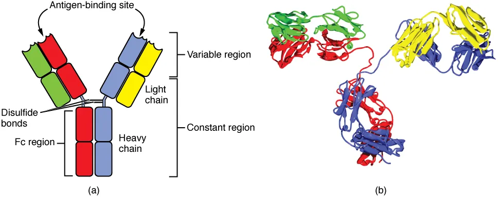
Every antibody (also called an immunoglobulin, Ig) is a Y-shaped protein made of four polypeptide chains:
2 identical heavy chains (the long chains forming the stem and inner arms of the Y)
2 identical light chains (the shorter chains forming the outer portion of the arms)
The chains are held together by disulfide bonds.
The Y-shape creates two functionally distinct regions:
Fab region (Fragment, antigen-binding) - the two arms of the Y. Each arm has a binding site at its tip that recognizes and binds to a specific antigen. Because there are two arms, each antibody has two identical antigen-binding sites.
Fc region (Fragment, crystallizable) - the stem of the Y. This is the part that interacts with the rest of the immune system: it binds to Fc receptors on phagocytes (triggering engulfment), activates complement (classical pathway), and determines the antibody class (IgG, IgA, IgM, etc.).
Antibody (immunoglobulin) structure. The Y-shape consists of two heavy chains and two light chains linked by disulfide bonds. The Fab regions (tips) bind antigens; the Fc region (stem) interacts with immune cells and complement. Credit: OpenStax Anatomy and Physiology 2e, CC BY 4.0
Interactive 3D Antibody. Rotate to see the Y-shape: two Fab arms (antigen-binding) and the Fc stem (immune cell signaling).Credit: truekit via Sketchfab, CC BY
Variable vs. Constant Regions
Each chain (heavy and light) has two types of regions:
Variable (V) region - located at the tips of the Y arms. The amino acid sequence here differs between antibodies, giving each antibody its unique antigen specificity. This is the “lock” that fits a specific antigen “key.” The variable regions of one heavy chain and one light chain together form one antigen-binding site.
Constant (C) region - the rest of the chain. This sequence is the same for all antibodies of the same class. The constant region of the heavy chain determines the antibody’s class (isotype) and its effector functions.
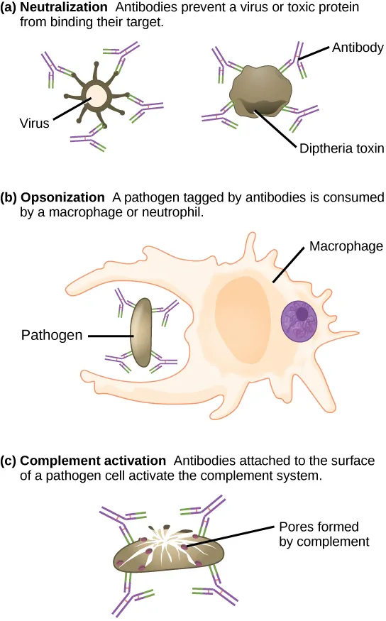
The four main mechanisms by which antibodies eliminate pathogens: neutralization, opsonization, complement activation, and agglutination. Credit: OpenStax Biology 2e, CC BY 4.0
How Antibodies Eliminate Pathogens
Antibodies do not directly kill pathogens (with the exception of toxin neutralization). Instead, they employ four main mechanisms:
1. Neutralization - Antibodies bind to the surface of a pathogen or toxin, physically blocking it from attaching to host cells. A virus coated in antibodies cannot bind its receptor on a target cell. A toxin bound by antibodies cannot interact with its target. This is the simplest mechanism - just cover the enemy so it cannot function.
2. Opsonization - Antibodies coat the pathogen surface. The Fc regions of these antibodies are recognized by Fc receptors on macrophages and neutrophils, dramatically enhancing phagocytosis. Think of it as putting a “please eat me” sign on the pathogen.
3. Complement activation - When IgG or IgM binds a pathogen, the Fc region activates the classical complement pathway, leading to MAC formation and pathogen lysis. The Fab region finds the target; the Fc region triggers destruction.
4. Agglutination - Because each antibody has two binding sites, antibodies can cross-link multiple pathogens into clumps. These large clumps are much easier for phagocytes to find and engulf than individual bacteria. IgM, with its 10 binding sites (pentamer), is especially effective at agglutination.
Mechanism
What Happens
Key Detail
Neutralization
Antibody blocks pathogen from binding host cells
Prevents infection; works on toxins too
Opsonization
Antibody coats pathogen; Fc signals phagocytes
”Eat me” tag; Fc receptors on macrophages
Complement activation
Fc triggers classical pathway
Leads to MAC and lysis
Agglutination
Antibodies cross-link pathogens into clumps
IgM (pentamer) is best at this
B Cell Development and Activation
Each B cell carries ~10,000 copies of a single unique antibody on its surface as a B cell receptor (BCR). All copies are identical - each B cell recognizes only one antigen.
How does the immune system produce millions of different B cells, each with a unique antibody, from only ~20,000 genes? Through somatic recombination (also called V(D)J recombination). During B cell development in the bone marrow, gene segments encoding the variable region are randomly shuffled and combined, generating an enormous diversity of antibody specificities. This is like shuffling a deck of cards - the number of possible combinations far exceeds the number of cards.
Activation process:
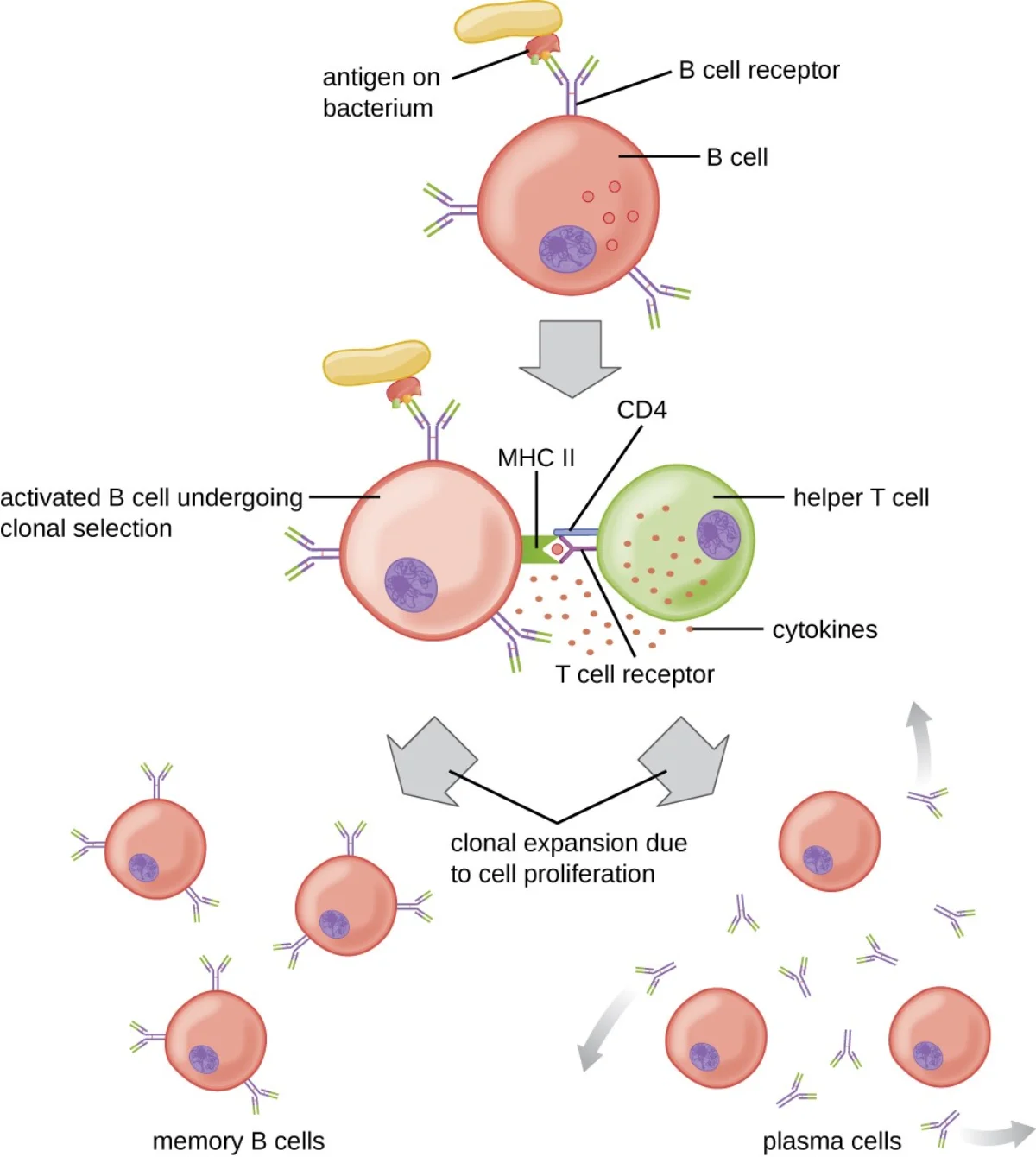
A naive B cell encounters its matching antigen (the BCR binds to it)
The B cell internalizes the antigen, processes it, and presents it on MHC II
A helper T cell (CD4+) that recognizes the same antigen binds to the B cell and releases cytokines
The B cell is now fully activated and undergoes clonal expansion
After activation, B cells differentiate into:
Plasma cells (effector B cells) - antibody factories that secrete thousands of antibodies per second. They live for days to weeks.
Memory B cells - long-lived cells that persist for years or decades. On re-exposure to the same antigen, they rapidly differentiate into plasma cells, producing a faster and stronger response.
Which part of the antibody determines its class (IgG, IgM, etc.), and which part determines its antigen specificity?
Click to reveal answer
The constant region of the heavy chain determines the antibody class (isotype). The variable regions of the heavy and light chains (together forming the Fab tips) determine antigen specificity. Class switching changes the constant region (and thus the class) while keeping the variable region the same, so the antibody targets the same antigen but has different effector functions.
How can the human genome, with only ~20,000 genes, produce billions of unique antibody specificities?
Click to reveal answer
Through somatic recombination (V(D)J recombination). During B cell development, gene segments encoding the variable region are randomly shuffled and combined, creating an enormous number of unique combinations from a limited set of gene segments. Additional diversity comes from somatic hypermutation after activation. This combinatorial approach generates far more antibody types than there are genes.
All antibodies share the same basic Y-shaped structure, but the constant region of the heavy chain comes in five varieties, creating five antibody classes (isotypes). Each class has a different shape, location, and job. The MCAT expects you to know all five.
The Five Antibody Classes
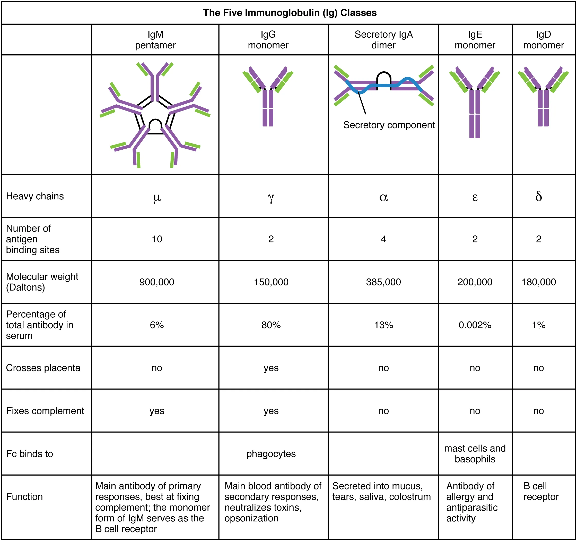
The five antibody classes (isotypes) differ in structure and function. IgG is a monomer, IgA forms dimers in secretions, IgM forms pentamers in blood, IgD sits on B cell surfaces, and IgE binds mast cells. Credit: OpenStax Anatomy and Physiology 2e, CC BY 4.0
IgG - The All-Purpose Workhorse
IgG is the most abundant antibody in the blood (~75% of serum immunoglobulins). It is the primary antibody of the secondary immune response - when your body encounters a pathogen it has seen before, IgG levels surge.
Key features:
Monomer (single Y-shape)
Only antibody that crosses the placenta - provides passive immunity to the fetus
Longest half-life of any antibody (~21 days)
Effective at neutralization, opsonization, and complement activation
Dominant antibody after class switching from IgM
IgA - The Secretory Guardian
IgA is found primarily in mucosal secretions - saliva, tears, mucus, breast milk, and the lining of the respiratory and GI tracts. It exists as a dimer (two Y-shapes joined by a J chain) in secretions.
Key features:
Guards the mucosal surfaces that are the body’s main entry points for pathogens
Found in breast milk - provides passive immunity to nursing infants through the GI tract
Prevents pathogen attachment to epithelial surfaces (neutralization)
Does not activate complement efficiently
IgM - The First Responder
IgM is the first antibody produced during a primary immune response. It exists as a pentamer (five Y-shapes joined by a J chain) in the blood, giving it 10 antigen-binding sites - making it excellent at agglutination.
Key features:
First antibody made by naive B cells before class switching occurs
Very effective at complement activation (classical pathway)
Also found as a monomer on the surface of naive B cells (part of the BCR)
Short half-life (~5 days) - levels decline as IgG takes over
Elevated IgM = sign of recent/current infection
IgE - The Allergy Antibody
IgE is present in very low concentrations in blood but plays an outsized role in allergic reactions and defense against parasites.
Key features:
Monomer that binds to Fc receptors on mast cells and basophils
When an allergen cross-links two IgE molecules on a mast cell, the mast cell degranulates, releasing histamine
This is the mechanism behind Type I hypersensitivity (allergies, anaphylaxis)
Also important in defense against parasitic worms (helminths) - eosinophils have Fc receptors for IgE
Elevated IgE = allergies or parasitic infection
IgD - The Mystery Antibody
IgD is the least understood antibody class. It exists in tiny amounts in the blood and is found primarily on the surface of naive, mature B cells as part of the B cell receptor complex (along with IgM).
Key features:
Monomer on B cell surface
Signals B cell activation when antigen binds
Not secreted in significant amounts
No well-defined role in pathogen destruction
Master Comparison Table
Class
Structure
Location
Key Function
High-Yield Fact
IgG
Monomer
Blood, tissue fluids
Opsonization, neutralization, complement
Crosses placenta; most abundant; secondary response
IgA
Dimer (secreted)
Mucous membranes, secretions, breast milk
Mucosal defense, neutralization
In All secretions; protects newborns via breast milk
IgM
Pentamer (secreted), Monomer (BCR)
Blood, B cell surface
Agglutination, complement activation
First responder; largest; indicates new infection
IgE
Monomer
Bound to mast cells/basophils
Allergic response, anti-parasite defense
Triggers histamine release; Type I hypersensitivity
IgD
Monomer
B cell surface
B cell activation signaling
Least understood; on naive B cells
Isotype (Class) Switching
When a B cell is first activated, it produces IgM. But with help from cytokines released by helper T cells, the B cell can undergo class switching (isotype switching) - changing the constant region of its heavy chain to produce a different antibody class (IgG, IgA, or IgE).
Critical detail: class switching changes the Fc region (and therefore the effector function) but keeps the variable region identical. The antibody still targets the exact same antigen - it just communicates differently with the immune system.
Class switching requires helper T cell signals (cytokines) that direct the B cell to rearrange its heavy chain DNA, swapping out one constant region for another.
A newborn has not been exposed to any pathogens but has detectable IgG in their blood. Where did these antibodies come from?
Click to reveal answer
Maternal IgG crossed the placenta. IgG is the only antibody class that can cross the placenta, providing passive immunity to the fetus. The newborn also receives IgA through breast milk, but IgA does not enter the blood - it protects the GI tract mucosa. This maternal IgG gradually declines over the first ~6 months as the infant's own immune system begins producing antibodies.
During class switching, a B cell changes from producing IgM to producing IgG. What changes and what stays the same?
Click to reveal answer
Changes: The constant region of the heavy chain (determines antibody class and effector function). Stays the same: The variable region (determines antigen specificity). The antibody still recognizes and binds the same antigen, but now it has different effector properties - IgG is better at opsonization and has a longer half-life than IgM. Class switching is triggered by helper T cell cytokines.
If B cells are the weapons factory, T cells are the field commanders and assassins of adaptive immunity. There are three main types, each with a distinct role.
Helper T Cells (CD4+ / Th)
Helper T cells are the coordinators of the adaptive immune system. They do not kill anything directly. Instead, they release cytokines (signaling molecules) that activate and direct other immune cells.
What helper T cells activate:
B cells - stimulate clonal expansion, class switching, and antibody production
Cytotoxic T cells - promote their activation and proliferation
Macrophages - enhance their killing ability
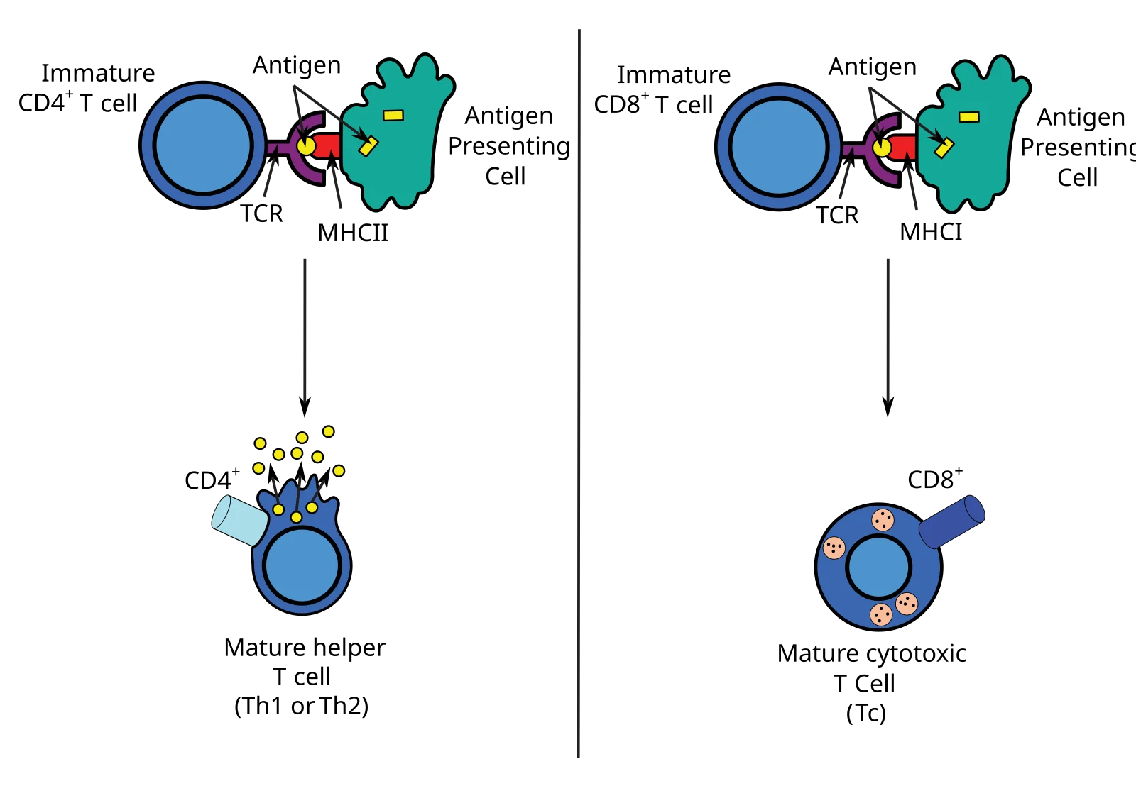
Surface marker: CD4 glycoprotein, which binds to MHC II on antigen-presenting cells.
Helper T cells only activate when they encounter their specific antigen presented on MHC II by an APC (dendritic cell, macrophage, or B cell). They also require a co-stimulatory signal from the APC to prevent accidental activation.
Cytotoxic T Cells (CD8+ / Tc / CTLs)
Cytotoxic T cells are the executioners. They recognize and kill host cells that are infected with viruses, harbor intracellular bacteria, or have become cancerous.
Surface marker: CD8 glycoprotein, which binds to MHC I on target cells.
How they kill:
The CD8+ T cell recognizes a foreign peptide displayed on MHC I of an infected cell
The T cell binds tightly to the target cell
The T cell releases:
Perforin - forms pores (holes) in the target cell’s membrane
Granzymes - enzymes that enter through the pores and trigger apoptosis (programmed cell death)
The infected cell dies in a controlled way, preventing the release of more pathogens
This is the same killing mechanism used by NK cells. The difference is the recognition system: CD8+ T cells check MHC I for foreign peptides (adaptive), while NK cells check for missing MHC I (innate).
Regulatory T Cells (Treg / Suppressor T Cells)
Regulatory T cells are the brakes of the immune system. Their job is to suppress immune responses that are no longer needed or that are mistakenly targeting the body’s own tissues.
Surface marker: CD4 (same as helper T cells, but Tregs are functionally distinct)
Functions:
Suppress overactive immune responses after an infection is cleared
Prevent autoimmune reactions by maintaining self-tolerance
Secrete inhibitory cytokines that calm down other immune cells
When regulatory T cells fail, the immune system attacks healthy tissues - this is the basis of autoimmune diseases.
Antigen presentation to T cells. Focus on: MHC I presents to CD8+ cytotoxic T cells (kill infected cells), MHC II presents to CD4+ helper T cells (activate B cells and other immune cells). Credit: Wikimedia Commons, CC BY-SA 3.0
T Cell Receptor (TCR) and Activation
Every T cell has a unique T cell receptor (TCR) on its surface that recognizes one specific antigen-MHC combination. Unlike antibodies (which can bind free-floating antigens), TCRs can only recognize antigens that are presented on an MHC molecule. This means T cells are useless against free pathogens in the blood - they need an APC to “show” them the antigen.
T cell activation requires two signals:
Signal 1: TCR binds to the antigen-MHC complex (specificity check)
Signal 2: A co-stimulatory signal from the APC (safety check)
Without both signals, the T cell becomes unresponsive rather than activated. This two-signal requirement prevents accidental activation against self-antigens.
T Cell Maturation: Positive and Negative Selection
T cells mature in the thymus, where the body builds central tolerance by killing any T cell that cannot work or that might attack self. Only ~2% of developing T cells survive.
Positive selection (thymic cortex):
Tests whether the TCR can bind self-MHC at all
Cells that bind MHC I become CD8+; cells that bind MHC II become CD4+
Cells that bind no MHC die by apoptosis (useless)
Negative selection (thymic medulla):
Tests whether the TCR binds self-antigens too strongly
Strongly self-reactive cells die or are diverted into the regulatory T cell lineage
This is the core mechanism of self vs non-self tolerance; failure here is a direct route to autoimmunity
The MHC-CD Math Trick
Which T cell talks to which MHC? There is a multiplication trick:
MHC I presents to CD8+ T cells: 1 x 8 = 8
MHC II presents to CD4+ T cells: 2 x 4 = 8
Both products equal 8. If the product is 8, the pairing is correct.
T Cell Type
Surface Marker
Recognizes
Function
Analogy
Helper (Th)
CD4
MHC II + antigen
Coordinates response via cytokines
General
Cytotoxic (Tc)
CD8
MHC I + antigen
Kills infected/cancerous cells
Sniper
Regulatory (Treg)
CD4
Self-antigens
Suppresses immune response
Referee
A developing T cell in the thymus binds strongly to a self-antigen presented on MHC. What happens to this T cell?
Click to reveal answer
The T cell is eliminated by negative selection (undergoes apoptosis) or may be converted into a regulatory T cell. Negative selection occurs in the thymic medulla and removes T cells whose TCRs react strongly to self-antigens. This prevents these cells from entering circulation and attacking the body's own tissues (autoimmunity). If negative selection fails, autoimmune disease can result.
Why does T cell activation require two signals (TCR-MHC binding AND co-stimulation)?
Click to reveal answer
The two-signal requirement is a safety mechanism to prevent autoimmunity. Signal 1 (TCR binding antigen-MHC) ensures specificity. Signal 2 (co-stimulation from the APC) confirms that the antigen was presented by a legitimate APC that has detected a real threat. Without co-stimulation, the T cell becomes unresponsive, preventing activation against harmless self-antigens that might be presented on MHC.
The adaptive immune system cannot detect pathogens directly. T cells can only see antigens that are chopped into small peptide fragments and displayed on the cell surface by MHC molecules (Major Histocompatibility Complex). Understanding MHC I and MHC II is essential - this is one of the most heavily tested topics in MCAT immunology.
The ID Badge System
MHC I (endogenous pathway) presents intracellular peptides to CD8+ T cells. MHC II (exogenous pathway) presents extracellular peptides to CD4+ T cells. Note the different processing compartments and cell types involved. Credit: OpenStax Microbiology, CC BY 4.0
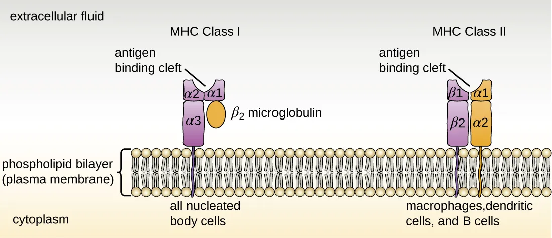
MHC I: Present on All Nucleated Cells
Who has it: Every nucleated cell in the body (NOT red blood cells, which lack a nucleus)
What it presents: Peptides from proteins made inside the cell (endogenous antigens)
Who reads it: CD8+ cytotoxic T cells
The endogenous pathway:
Proteins inside the cell (both normal and foreign, if the cell is infected) are chopped into small peptide fragments
Peptide fragments are loaded onto MHC I molecules in the endoplasmic reticulum (ER)
The MHC I-peptide complex is transported to the cell surface
CD8+ T cells scan the displayed peptides
If the peptide is a normal self-protein - the CD8+ T cell ignores the cell (it passed the badge check).
If the peptide is foreign (viral, bacterial, or mutant cancer protein) - the CD8+ T cell kills the cell.
MHC II: Present on Professional APCs Only
Who has it: Only professional antigen-presenting cells - dendritic cells, macrophages, and B cells (remembered as “the Big Three APCs”)
What it presents: Peptides from proteins ingested from outside the cell (exogenous antigens)
Who reads it: CD4+ helper T cells
The exogenous pathway:
The APC engulfs a pathogen via phagocytosis or receptor-mediated endocytosis
The pathogen is degraded in a phagolysosome into peptide fragments
MHC II molecules (assembled in the ER) are transported to the phagolysosome
Peptide fragments are loaded onto MHC II
The MHC II-peptide complex is transported to the cell surface
CD4+ helper T cells scan the displayed peptides and activate if they recognize the antigen
This activation of helper T cells is the starting point for the entire adaptive immune response - it triggers both B cell activation (humoral) and cytotoxic T cell activation (cell-mediated).
Side-by-Side Comparison
Feature
MHC I
MHC II
Found on
All nucleated cells
APCs only (dendritic cells, macrophages, B cells)
Presents
Endogenous antigens (from inside the cell)
Exogenous antigens (from outside the cell)
Recognized by
CD8+ cytotoxic T cells
CD4+ helper T cells
Pathway name
Endogenous pathway
Exogenous pathway
Processing
Proteins degraded in cytoplasm, loaded in ER
Phagolysosome
Purpose
”Is this cell infected or cancerous?"
"What pathogen did this APC encounter?”
Math trick
MHC I x CD8 = 8
MHC II x CD4 = 8
HLA: The Human Version
In humans, MHC molecules are specifically called HLA (Human Leukocyte Antigen) molecules. HLA genes are the most polymorphic genes in the human genome - they have thousands of variants in the population.
This extreme diversity is why organ transplant matching is so difficult. HLA molecules differ between individuals, and the recipient’s immune system may recognize a donor organ’s HLA molecules as foreign, leading to transplant rejection. Immunosuppressive drugs suppress T cell activity to prevent this.
A cell is infected by a virus. Through which pathway and on which MHC class will viral peptides be displayed?
Click to reveal answer
The endogenous pathway via MHC I. Viral proteins are synthesized inside the infected cell, degraded into peptide fragments, loaded onto MHC I in the ER, and displayed on the cell surface. CD8+ cytotoxic T cells recognize the foreign peptide and kill the infected cell. All nucleated cells can present via MHC I because any nucleated cell can be infected.
Why don't red blood cells express MHC I molecules?
Click to reveal answer
Mature red blood cells lack a nucleus (and most organelles). MHC I expression requires ongoing gene transcription and protein synthesis, which RBCs cannot perform. This is actually advantageous - RBCs are not susceptible to most intracellular infections because they lack the machinery viruses need to replicate. Without MHC I, they also avoid destruction by CD8+ T cells. However, this also means pathogens like Plasmodium (malaria) can hide inside RBCs without being detected by T cells.
Your body contains millions of unique B and T cells, each recognizing a different antigen. When a pathogen arrives, only the tiny fraction of lymphocytes that match that specific antigen are activated. This process - selecting and amplifying the right cells - is called clonal selection, and it is the engine that drives the entire adaptive immune response.
Clonal Selection Theory
The core claims of the theory:
Pre-existing diversity - Before any infection, the body already has millions of unique lymphocytes, each with a different antigen receptor (generated by somatic recombination during development).
Antigen selects, not instructs - The pathogen does not teach the lymphocyte what shape to make. The matching receptor already exists. The antigen simply finds and activates the cell that already fits.
Clonal expansion - The selected lymphocyte divides rapidly, producing a large population of identical clones, all specific to that one antigen.
Differentiation - Clones differentiate into effector cells (which fight the current infection) and memory cells (which prepare for future encounters).
Self-reactive clones are deleted - Lymphocytes that recognize self-antigens are eliminated during development (negative selection in the thymus for T cells, clonal deletion in the bone marrow for B cells).
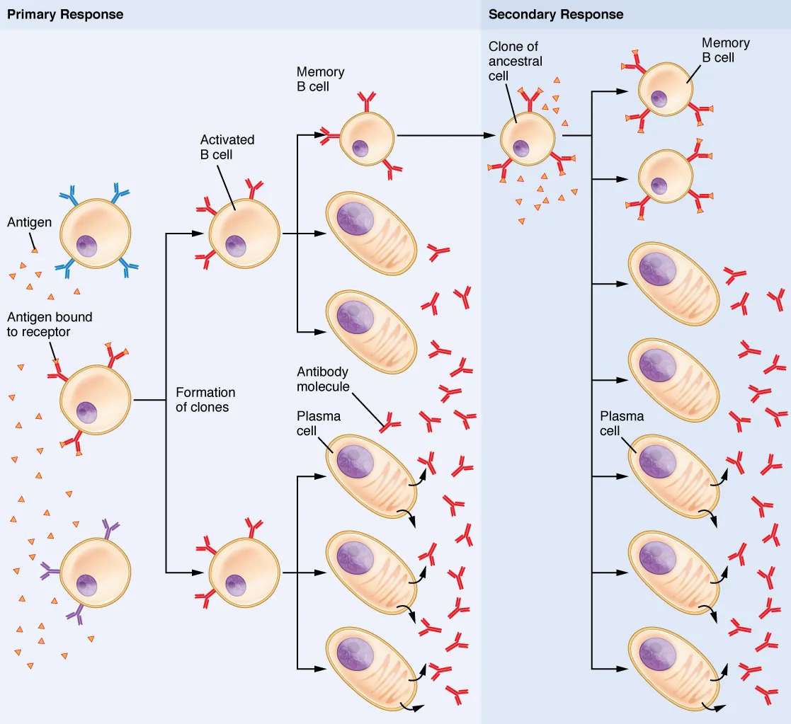
Clonal selection of B cells. An antigen selects the B cell with the matching receptor. That cell undergoes clonal expansion, producing plasma cells (which secrete antibodies) and memory cells (which provide long-term immunity). Credit: OpenStax Anatomy and Physiology 2e, CC BY 4.0
The Steps of Clonal Selection
Step 1: Antigen encounter - A naive B or T cell encounters its matching antigen. For B cells, the antigen binds directly to the BCR. For T cells, the antigen must be presented on MHC by an APC.
Step 2: Activation - The lymphocyte receives activation signals (antigen binding + co-stimulation + helper T cell cytokines for B cells).
Step 3: Clonal expansion - The activated cell divides rapidly by mitosis, producing thousands of identical daughter cells (clones). All clones have the same antigen specificity.
Step 4: Differentiation - Clones differentiate into two populations:
Effector cells - fight the current infection (plasma cells for B cells, armed cytotoxic T cells for CD8+)
Memory cells - long-lived cells that persist after the infection is cleared
Step 5: Contraction - After the pathogen is eliminated, most effector cells undergo apoptosis (programmed death). The immune response winds down. Memory cells remain.
Affinity Maturation
During clonal expansion of B cells, something remarkable happens. Activated B cells in the germinal centers of lymph nodes undergo somatic hypermutation - random point mutations in the genes encoding the variable region of their antibodies.
Most of these mutations are useless or harmful. But some improve the antibody’s binding affinity for the antigen. B cells with higher-affinity antibodies receive stronger survival signals, while those with lower affinity die. This is essentially natural selection happening inside your lymph nodes.
The result: each round of mutation and selection produces antibodies that bind the antigen more tightly. This process is called affinity maturation, and it is why the secondary immune response produces higher-quality antibodies than the primary response.
Primary vs. Secondary Immune Response
The existence of memory cells creates a dramatically different response on re-exposure:
Feature
Primary Response
Secondary Response
Timing
Slow (~7-10 days to peak)
Fast (~1-3 days to peak)
Antibody produced first
IgM
IgG (already class-switched)
Antibody levels
Lower
Much higher (10-100x)
Antibody affinity
Lower
Higher (affinity maturation)
Duration
Shorter
Longer
Cells involved
Naive lymphocytes
Memory lymphocytes
The secondary response is faster because memory cells are already present in greater numbers than naive cells, they are pre-selected for the antigen, and they require less co-stimulation to activate. This is the entire basis of vaccination.
Memory Cells: The Long Game
Memory B cells and memory T cells can survive for decades - some persist for your entire life. They circulate through blood and lymph, residing in lymph nodes and the spleen, waiting.
When the same antigen appears again, memory cells:
Activate much faster than naive cells
Require less co-stimulation
Are already present in larger numbers
(For B cells) Already produce high-affinity, class-switched antibodies
This is why you only get chickenpox once. Your memory cells mount such a rapid and overwhelming response on re-exposure that the virus is destroyed before it can cause symptoms.
During the primary immune response, IgM levels rise first, followed by IgG. During the secondary response, IgG rises rapidly with minimal IgM. Explain why.
Click to reveal answer
In the primary response, naive B cells first produce IgM (the default antibody). With helper T cell signals, they undergo class switching to IgG. In the secondary response, memory B cells have already undergone class switching and affinity maturation. They are primed to produce IgG immediately upon re-exposure, and they activate much faster than naive cells. IgM barely rises because the memory cells skip the IgM stage.
What is the difference between somatic recombination and somatic hypermutation?
Click to reveal answer
Somatic recombination (V(D)J recombination) occurs during B cell development in the bone marrow. It randomly rearranges gene segments to create an initial, diverse repertoire of antibody specificities. Somatic hypermutation occurs after B cell activation, in the germinal centers of lymph nodes. It introduces random point mutations in the variable region to fine-tune antibody affinity. Recombination creates diversity; hypermutation optimizes it.
Not all immunity is created the same way. Some types you build yourself; others are borrowed. Some last a lifetime; others fade in weeks. The MCAT expects you to classify immunity along two dimensions: active vs. passive and natural vs. artificial.
The Two-by-Two Grid
The fastest way to organize immunity types:
Natural
Artificial
Active (you make your own antibodies)
Getting infected and recovering
Vaccination
Passive (you receive someone else’s antibodies)
Maternal antibodies (placenta and breast milk)
Injection of pre-formed antibodies (antiserum)
Active Immunity: Building Your Own Defenses
Active immunity occurs when your immune system encounters an antigen and mounts its own response, producing antibodies and memory cells.
Natural active immunity - You get infected, your immune system fights off the pathogen, and you develop lasting immunity. This is what happens when you catch chickenpox and become immune for life. The downside: you have to get sick first.
Artificial active immunity (vaccination) - A vaccine introduces a harmless version of the antigen to your immune system. Your body mounts a primary immune response and creates memory cells - without the risk of full-blown disease. When the real pathogen arrives later, the secondary response destroys it before symptoms develop.
Key features of active immunity:
Slow to develop (days to weeks for primary response)
Long-lasting (years to lifelong due to memory cells)
Specific (targets one particular antigen)
Produces memory (faster, stronger response on re-exposure)
Vaccine Types
Vaccine Type
What It Contains
Examples
Key Detail
Live attenuated
Weakened (but living) pathogen
MMR, varicella, oral polio
Strongest immune response; cannot give to immunocompromised patients
Inactivated (killed)
Dead pathogen
Flu shot, rabies, hepatitis A
Safer but weaker response; may need boosters
Subunit/conjugate
Purified antigen (protein or polysaccharide)
Hepatitis B, HPV, pertussis (acellular)
Very safe; targets specific antigen
Toxoid
Inactivated toxin
Tetanus, diphtheria
Targets the toxin, not the organism itself
mRNA
mRNA encoding a pathogen protein
COVID-19 (Pfizer, Moderna)
Body’s cells produce the antigen temporarily
Passive Immunity: Borrowed Protection
Passive immunity occurs when pre-formed antibodies are transferred from one individual to another. The recipient never activates their own immune system, so no memory cells are produced.
Natural passive immunity - A mother transfers antibodies to her child:
IgG crosses the placenta during pregnancy, providing the fetus with protection
IgA is in breast milk, protecting the infant’s GI tract mucosa
This protection fades over ~3-6 months as maternal antibodies are degraded and not replaced
Artificial passive immunity - Pre-formed antibodies are injected into a patient:
Antiserum/antitoxin - used for emergencies like snakebite (antivenin), rabies post-exposure, or tetanus when there is no time to wait for active immunity
Monoclonal antibodies - engineered antibodies used to treat cancer, autoimmune diseases, and some infections
Key features of passive immunity:
Immediate protection (works right away - antibodies are already made)
Short-lived (weeks to months - antibodies degrade and are not replaced)
No memory (the recipient’s immune system was never activated)
Not specific to the recipient (antibodies were made by another organism)
Comparison Table
Feature
Active Immunity
Passive Immunity
How acquired
Immune system produces own antibodies
Receives pre-formed antibodies
Speed of onset
Slow (days to weeks)
Immediate
Duration
Long (years to lifelong)
Short (weeks to months)
Memory cells?
Yes
No
Booster needed?
Sometimes (for vaccines)
Not applicable (just re-inject)
Natural example
Recovering from infection
Maternal IgG across placenta
Artificial example
Vaccination
Antiserum injection
B cell activation leads to clonal expansion, producing plasma cells (which secrete antibodies for the current infection) and memory B cells (which provide rapid, enhanced protection on re-exposure). This is the mechanism underlying both natural and vaccine-induced active immunity. Credit: Lumen Learning / OpenStax Anatomy and Physiology, CC BY 4.0
Herd Immunity
When a large enough percentage of a population is immune to a disease (through vaccination or prior infection), the pathogen cannot spread efficiently because it keeps encountering immune individuals. This provides indirect protection to people who are not immune (infants, immunocompromised patients, unvaccinated individuals).
The threshold percentage needed for herd immunity depends on how contagious the disease is:
Measles (very contagious): ~95% immunity needed
Polio (less contagious): ~80-85% immunity needed
A patient bitten by a venomous snake receives an injection of antivenin (antibodies harvested from a horse). What type of immunity is this, and why doesn't it provide long-term protection?
Click to reveal answer
This is artificial passive immunity. The patient receives pre-formed antibodies that immediately neutralize the venom. However, the patient's own immune system was never activated, so no memory cells are produced. The injected antibodies are gradually degraded over weeks, and the patient has no lasting protection against future envenomation.
Why can live attenuated vaccines NOT be given to immunocompromised patients?
Click to reveal answer
Live attenuated vaccines contain a weakened but living pathogen. In a healthy person, the immune system easily controls the weakened pathogen and develops immunity. In an immunocompromised patient (e.g., AIDS, chemotherapy), the weakened immune system may not be able to control even the attenuated pathogen, potentially causing full-blown disease. Inactivated or subunit vaccines are safer alternatives because they contain no living organisms.
The immune system is a powerful weapon. When it works correctly, it protects you from infections and cancer. When it malfunctions, it can be just as destructive - attacking your own tissues, failing to fight infections, or overreacting to harmless substances.
Immune disorders fall into three categories: the system attacks self (autoimmunity), the system is too weak (immunodeficiency), or the system overreacts (hypersensitivity).
Autoimmune Diseases: Friendly Fire
In autoimmune diseases, the immune system fails to distinguish self from non-self and attacks the body’s own healthy tissues. This happens when self-tolerance breaks down - meaning self-reactive lymphocytes that should have been eliminated during development (negative selection) or suppressed by regulatory T cells escape these checkpoints.
Disease
Target
Immune Mechanism
Type 1 diabetes
Insulin-producing beta cells of the pancreas
T cells destroy beta cells
Rheumatoid arthritis
Joint synovial membranes
Antibodies and T cells attack joints
Multiple sclerosis (MS)
Myelin sheath of neurons (CNS)
T cells attack myelin
Systemic lupus erythematosus (SLE)
DNA and multiple organs
Anti-DNA antibodies form immune complexes that deposit in tissues
Graves’ disease
TSH receptors on thyroid
Antibodies stimulate thyroid (hyperthyroidism)
Immunodeficiency: A Weakened Defense
Immunodeficiency means the immune system is too weak to protect the body effectively. It can be congenital (present from birth) or acquired (developed later in life).
Primary (congenital) immunodeficiencies:
The most important example is Severe Combined Immunodeficiency (SCID) - a defect in lymphocyte development that results in no functional B or T cells. Without treatment, SCID is fatal because the patient has essentially no adaptive immunity (the “bubble boy” disease).
Other examples you may see in MCAT passages include thymus defects that prevent T cell maturation and B cell defects that prevent antibody production. The key concept: if you know which branch of immunity is missing, you can predict which types of infections the patient will be vulnerable to.
Secondary (acquired) immunodeficiency:
The most important example is HIV/AIDS:
HIV is a retrovirus that specifically infects and destroys CD4+ helper T cells
HIV binds to CD4 on helper T cells
As CD4+ T cell counts drop, the immune system progressively weakens
AIDS (Acquired Immunodeficiency Syndrome) is diagnosed when CD4+ count falls below 200 cells/microliter (normal: 500-1500)
Without helper T cells, both humoral and cell-mediated immunity collapse
Patients become vulnerable to opportunistic infections (e.g., Pneumocystis pneumonia, Kaposi’s sarcoma, thrush) that healthy immune systems easily control
Hypersensitivity Reactions: The Overreaction
Hypersensitivity reactions are immune responses that are disproportionate to the actual threat. There are four types, classified by mechanism and timing. This is one of the most commonly tested immune topics on the MCAT.
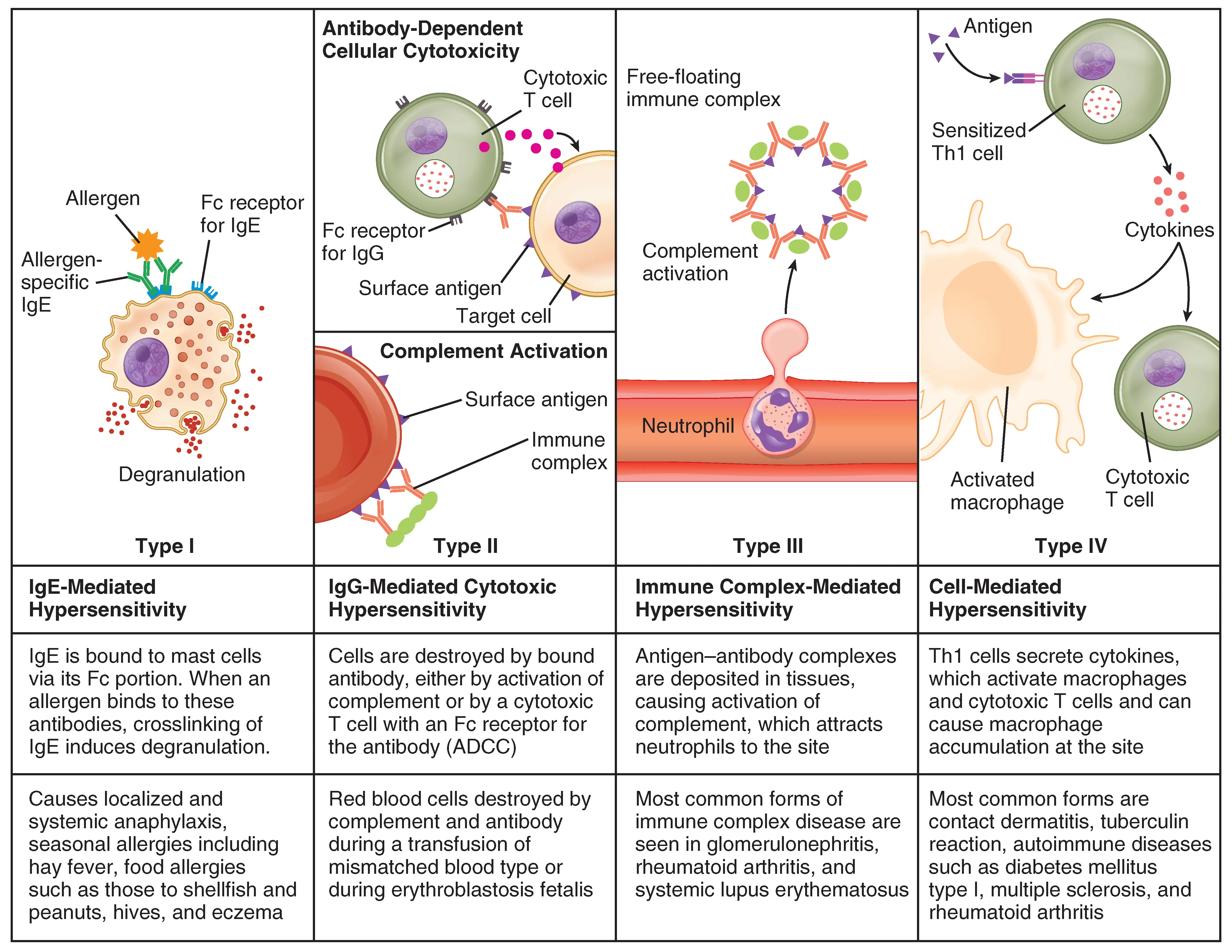
The four types of hypersensitivity reactions. Types I-III are antibody-mediated; Type IV is T cell-mediated and delayed. Knowing the mediator and timing for each type is essential for MCAT reasoning. Credit: OpenStax Anatomy and Physiology 2e, CC BY 4.0
Type I: Immediate/Anaphylactic Hypersensitivity
Mechanism: IgE-mediated. On first exposure, IgE antibodies are produced and bind to mast cells/basophils. On re-exposure, the allergen cross-links IgE on mast cells, triggering degranulation and release of histamine, leukotrienes, and prostaglandins.
Anaphylaxis is the most severe form - systemic vasodilation causes a dangerous drop in blood pressure, while airway constriction makes breathing difficult. Epinephrine (EpiPen) reverses these effects by causing vasoconstriction and bronchodilation.
Type II: Cytotoxic Hypersensitivity
Mechanism: IgG or IgM antibodies bind to antigens on the surface of the body’s own cells, marking them for destruction by complement or phagocytes.
Timing: Hours
Examples:
Hemolytic disease of the newborn (Rh incompatibility) - maternal antibodies attack fetal RBCs
Graves’ disease and myasthenia gravis (antibodies bind cell surface receptors)
Key molecules: IgG, IgM, complement
Type III: Immune Complex Hypersensitivity
Mechanism: Antigen-antibody complexes (immune complexes) form in the blood and deposit in tissues (joints, kidneys, blood vessel walls), triggering complement activation and inflammation at the deposition site.
Serum sickness - reaction to foreign proteins (e.g., antivenom)
Post-streptococcal glomerulonephritis
Key molecules: IgG, immune complexes, complement
Type IV: Delayed-Type Hypersensitivity (DTH)
Mechanism: T cell-mediated (no antibodies involved). Sensitized T cells encounter the antigen and release cytokines that recruit macrophages, causing inflammation and tissue damage.
Timing: 24-72 hours (delayed)
Examples:
Tuberculin skin test (PPD test) - positive reaction = swelling at injection site after 48-72 hours
Contact dermatitis (poison ivy, nickel allergy)
Transplant rejection
Type 1 diabetes (T cells destroying beta cells)
Key molecules: T cells (CD4+ helper T cells, macrophages), cytokines
Hypersensitivity Master Table
Type
Name
Mediator
Timing
Mechanism
Classic Example
I
Anaphylactic
IgE
Minutes
Mast cell degranulation
Allergies, anaphylaxis
II
Cytotoxic
IgG/IgM
Hours
Antibodies target cell surfaces
Transfusion reaction, Rh disease
III
Immune complex
IgG
Hours-days
Immune complexes deposit in tissues
Lupus (SLE), serum sickness
IV
Delayed
T cells
48-72 hrs
T cells recruit macrophages
TB skin test, poison ivy, transplant rejection
Transplant Rejection
When a patient receives an organ from a donor, the recipient’s immune system may recognize the donor’s MHC/HLA molecules as foreign, triggering an immune attack on the transplanted organ. This is why doctors match HLA types between donor and recipient and prescribe immunosuppressive drugs that suppress T cell activation.
Transplant rejection can involve both antibody-mediated (Type II) and T cell-mediated (Type IV) hypersensitivity, depending on whether the recipient has pre-existing antibodies against the donor’s MHC or whether T cells mount a new response against the foreign tissue.
A patient develops a rash 48 hours after touching poison ivy. What type of hypersensitivity reaction is this, and what cells are primarily responsible?
Click to reveal answer
Type IV (delayed-type) hypersensitivity. This is a T cell-mediated reaction - no antibodies are involved. Sensitized T cells (from a prior exposure) recognize the allergen (urushiol in poison ivy), release cytokines, and recruit macrophages to the area, causing inflammation and tissue damage. The 48-hour delay is the hallmark of Type IV - the time needed for T cells to migrate and activate.
Why does HIV cause such a catastrophic immunodeficiency when it only infects one type of immune cell?
Click to reveal answer
HIV targets CD4+ helper T cells, which are the central coordinators of the adaptive immune response. Without helper T cells: B cells cannot undergo class switching or produce high-affinity antibodies (humoral immunity collapses), CD8+ cytotoxic T cells are not effectively activated (cell-mediated immunity collapses), and macrophages are not enhanced. Destroying one cell type brings down the entire adaptive immune system because everything depends on CD4+ coordination.