
Innate Immune Cells
All immune cells originate from hematopoietic stem cells in the bone marrow. These stem cells differentiate down two major lineages:
- Myeloid lineage - produces most innate immune cells: neutrophils, eosinophils, basophils, mast cells, monocytes (which become macrophages and dendritic cells)
- Lymphoid lineage - produces lymphocytes: T cells, B cells, and Natural Killer (NK) cells
NK cells are the exception that proves the rule - they are lymphoid cells but function in innate immunity. The MCAT loves this distinction.
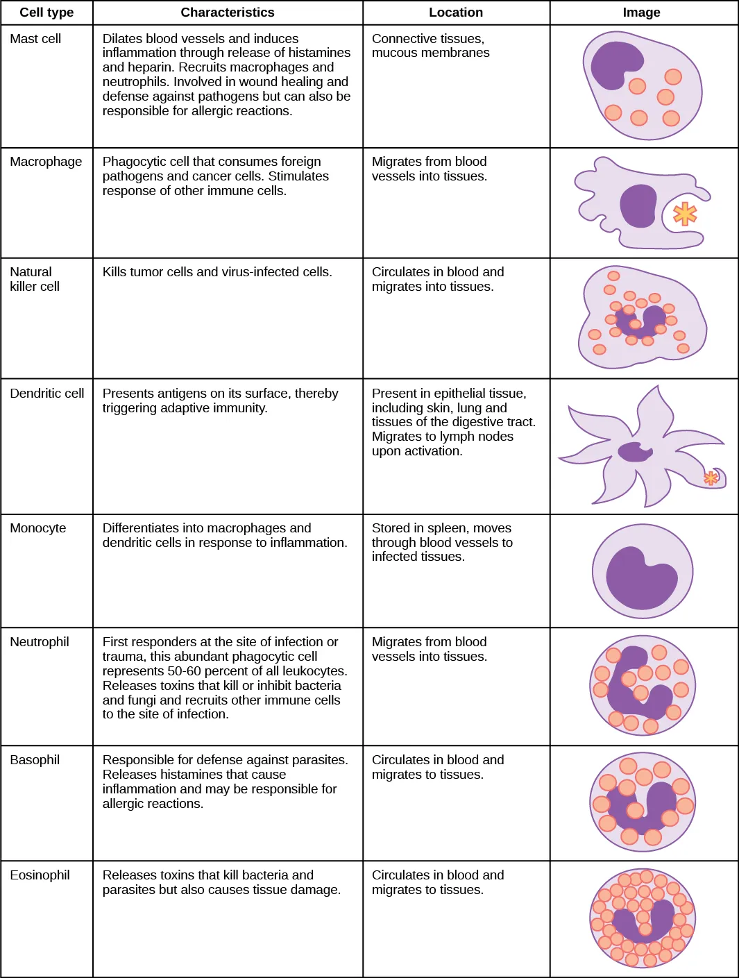
Granulocytes: Cells with Granules
Granulocytes are white blood cells with visible cytoplasmic granules packed with enzymes and antimicrobial substances. There are three types, and they are easy to remember by what they fight.
Neutrophils - the most abundant white blood cell in circulation (~60-70% of all WBCs). They are the first responders to infection, arriving within minutes via chemotaxis. Their primary weapon is phagocytosis - engulfing and digesting pathogens using digestive enzymes inside the cell. Neutrophils are short-lived and die at the infection site. Pus is mostly dead neutrophils.
Eosinophils - specialized for fighting large multicellular parasites (like helminths/worms) that are too big to phagocytize. Instead, eosinophils attach to the parasite and dump cytotoxic granule contents onto its surface, destroying it from the outside. Eosinophils also play a role in allergic responses. Elevated eosinophils on a blood test suggest either a parasitic infection or allergies.
Basophils - the rarest granulocyte in blood. Functionally similar to mast cells, they release histamine and other inflammatory mediators. They amplify allergic responses and inflammation.
Agranulocytes: Cells Without Visible Granules
Monocytes are large cells that circulate in the blood as precursors. When they migrate into tissues, they differentiate into either macrophages or dendritic cells depending on the signals they receive. Think of monocytes as “rookies in transit” who become specialized once they arrive at their post.
The migration process from blood to tissue involves chemotaxis (following chemical gradients toward the infection) and diapedesis (squeezing between the endothelial cells lining blood vessel walls to exit into the tissue).
Macrophages: The Pac-Man of the Immune System
Macrophages (“big eaters”) are phagocytes stationed throughout the body, lying in wait. When they encounter a pathogen, they:
- Recognize it via pattern recognition receptors (PRRs) that detect PAMPs
- Engulf it into a vesicle called a phagosome
- Destroy it by fusing the phagosome with a lysosome (creating a phagolysosome), where enzymes and acid digest the pathogen
- Present fragments of the pathogen on their surface using MHC II molecules - this activates the adaptive immune system
Macrophages also secrete cytokines to recruit more immune cells and promote tissue repair after the infection is cleared.
Macrophages go by different names depending on where they live. For example, they are called microglia in the brain and Kupffer cells in the liver. If an MCAT passage mentions a tissue-resident phagocyte by an unfamiliar name, it is probably a macrophage.
Dendritic Cells: The Messengers
Dendritic cells are the most important antigen-presenting cells (APCs). They are stationed in tissues that contact the external environment - skin, lungs, gut lining - where they capture pathogens via phagocytosis.
What makes dendritic cells special is what happens next: after capturing an antigen, they migrate to the nearest lymph node and present it to T cells. This is the critical bridge between innate and adaptive immunity. Macrophages stay in the tissue; dendritic cells travel to deliver the message.
Dendritic cells present antigens using both MHC I (to CD8+ T cells) and MHC II (to CD4+ T cells).
Natural Killer (NK) Cells
NK cells are the immune system’s quality control inspectors. Every nucleated cell in your body displays MHC I molecules on its surface - like wearing an employee ID badge. NK cells patrol the body, checking these badges.
When a virus infects a cell, it often downregulates MHC I expression to hide from T cells. Cancer cells do the same thing. But this “hiding” strategy backfires because NK cells detect the absence of MHC I. No badge? You get destroyed.
NK cells kill target cells by releasing:
- Perforin - punches holes in the target cell’s membrane
- Granzymes - enter through the holes and trigger apoptosis (programmed cell death)
Mast Cells
Mast cells are strategically positioned in tissues near blood vessels, especially in skin, lungs, and the gut. They are loaded with granules containing histamine and other inflammatory mediators.
When mast cells detect tissue damage or bind IgE antibodies (in allergic reactions), they degranulate - releasing histamine that triggers vasodilation, increased vascular permeability, and recruitment of other immune cells. Mast cells are the “alarm pullers” of the immune system.
Complete Immune Cell Summary
| Cell | Lineage | Key Function | Special Feature |
|---|---|---|---|
| Neutrophil | Myeloid | Phagocytosis of bacteria | Most abundant WBC; first responder; pus |
| Eosinophil | Myeloid | Kill parasites; allergy role | Dump granules on large targets |
| Basophil | Myeloid | Release histamine | Rarest granulocyte; like mast cells in blood |
| Mast cell | Myeloid | Release histamine; trigger inflammation | Stationed in tissues; key in allergy |
| Monocyte | Myeloid | Precursor cell | Becomes macrophage or dendritic cell |
| Macrophage | Myeloid | Phagocytosis + antigen presentation (MHC II) | “Big eater”; tissue-resident |
| Dendritic cell | Myeloid | Antigen presentation to T cells | Migrates to lymph nodes; bridges innate/adaptive |
| NK cell | Lymphoid | Kill cells missing MHC I | Uses perforin + granzymes; no MHC needed |