
T Cells
If B cells are the weapons factory, T cells are the field commanders and assassins of adaptive immunity. There are three main types, each with a distinct role.
Helper T Cells (CD4+ / Th)
Helper T cells are the coordinators of the adaptive immune system. They do not kill anything directly. Instead, they release cytokines (signaling molecules) that activate and direct other immune cells.
What helper T cells activate:
- B cells - stimulate clonal expansion, class switching, and antibody production
- Cytotoxic T cells - promote their activation and proliferation
- Macrophages - enhance their killing ability
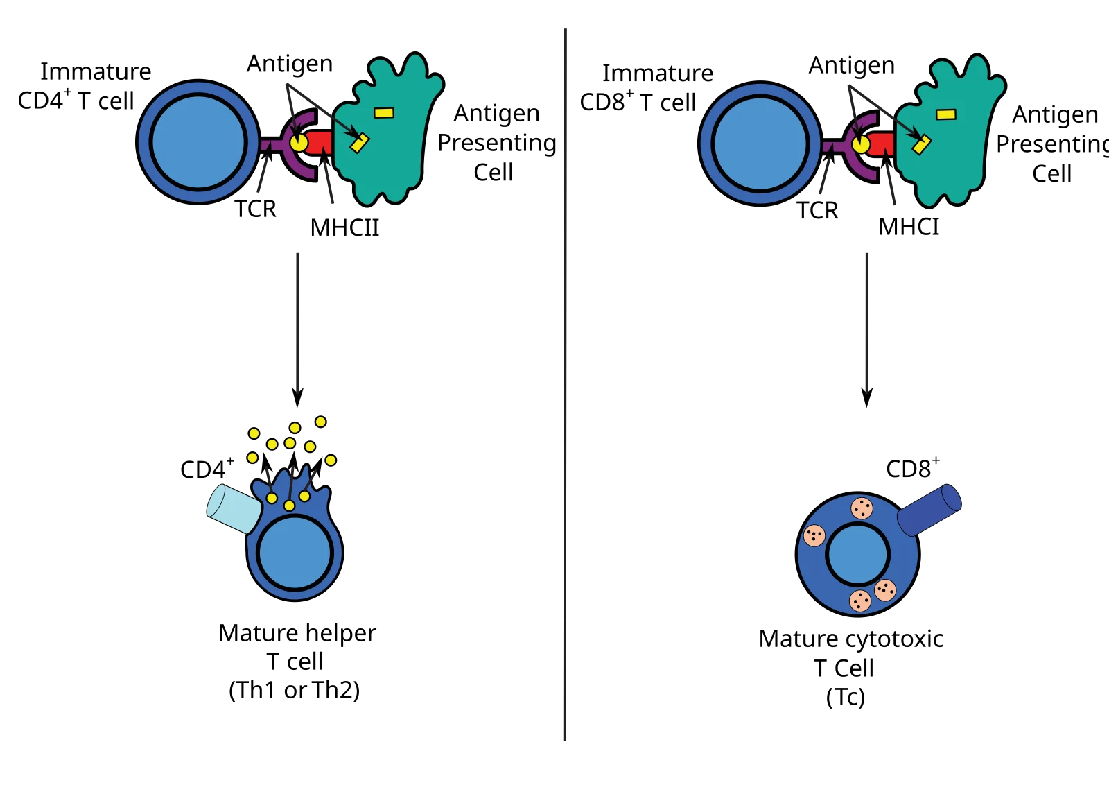
Surface marker: CD4 glycoprotein, which binds to MHC II on antigen-presenting cells.
Helper T cells only activate when they encounter their specific antigen presented on MHC II by an APC (dendritic cell, macrophage, or B cell). They also require a co-stimulatory signal from the APC to prevent accidental activation.
Cytotoxic T Cells (CD8+ / Tc / CTLs)
Cytotoxic T cells are the executioners. They recognize and kill host cells that are infected with viruses, harbor intracellular bacteria, or have become cancerous.
Surface marker: CD8 glycoprotein, which binds to MHC I on target cells.
How they kill:
- The CD8+ T cell recognizes a foreign peptide displayed on MHC I of an infected cell
- The T cell binds tightly to the target cell
- The T cell releases:
- Perforin - forms pores (holes) in the target cell’s membrane
- Granzymes - enzymes that enter through the pores and trigger apoptosis (programmed cell death)
- The infected cell dies in a controlled way, preventing the release of more pathogens
This is the same killing mechanism used by NK cells. The difference is the recognition system: CD8+ T cells check MHC I for foreign peptides (adaptive), while NK cells check for missing MHC I (innate).
Regulatory T Cells (Treg / Suppressor T Cells)
Regulatory T cells are the brakes of the immune system. Their job is to suppress immune responses that are no longer needed or that are mistakenly targeting the body’s own tissues.
Surface marker: CD4 (same as helper T cells, but Tregs are functionally distinct)
Functions:
- Suppress overactive immune responses after an infection is cleared
- Prevent autoimmune reactions by maintaining self-tolerance
- Secrete inhibitory cytokines that calm down other immune cells
When regulatory T cells fail, the immune system attacks healthy tissues - this is the basis of autoimmune diseases.
T Cell Receptor (TCR) and Activation
Every T cell has a unique T cell receptor (TCR) on its surface that recognizes one specific antigen-MHC combination. Unlike antibodies (which can bind free-floating antigens), TCRs can only recognize antigens that are presented on an MHC molecule. This means T cells are useless against free pathogens in the blood - they need an APC to “show” them the antigen.
T cell activation requires two signals:
- Signal 1: TCR binds to the antigen-MHC complex (specificity check)
- Signal 2: A co-stimulatory signal from the APC (safety check)
Without both signals, the T cell becomes unresponsive rather than activated. This two-signal requirement prevents accidental activation against self-antigens.
T Cell Maturation: Positive and Negative Selection
T cells mature in the thymus, where the body builds central tolerance by killing any T cell that cannot work or that might attack self. Only ~2% of developing T cells survive.
Positive selection (thymic cortex):
- Tests whether the TCR can bind self-MHC at all
- Cells that bind MHC I become CD8+; cells that bind MHC II become CD4+
- Cells that bind no MHC die by apoptosis (useless)
Negative selection (thymic medulla):
- Tests whether the TCR binds self-antigens too strongly
- Strongly self-reactive cells die or are diverted into the regulatory T cell lineage
- This is the core mechanism of self vs non-self tolerance; failure here is a direct route to autoimmunity
The MHC-CD Math Trick
Which T cell talks to which MHC? There is a multiplication trick:
- MHC I presents to CD8+ T cells: 1 x 8 = 8
- MHC II presents to CD4+ T cells: 2 x 4 = 8
Both products equal 8. If the product is 8, the pairing is correct.
| T Cell Type | Surface Marker | Recognizes | Function | Analogy |
|---|---|---|---|---|
| Helper (Th) | CD4 | MHC II + antigen | Coordinates response via cytokines | General |
| Cytotoxic (Tc) | CD8 | MHC I + antigen | Kills infected/cancerous cells | Sniper |
| Regulatory (Treg) | CD4 | Self-antigens | Suppresses immune response | Referee |