
MHC and Antigen Presentation
The adaptive immune system cannot detect pathogens directly. T cells can only see antigens that are chopped into small peptide fragments and displayed on the cell surface by MHC molecules (Major Histocompatibility Complex). Understanding MHC I and MHC II is essential - this is one of the most heavily tested topics in MCAT immunology.
The ID Badge System
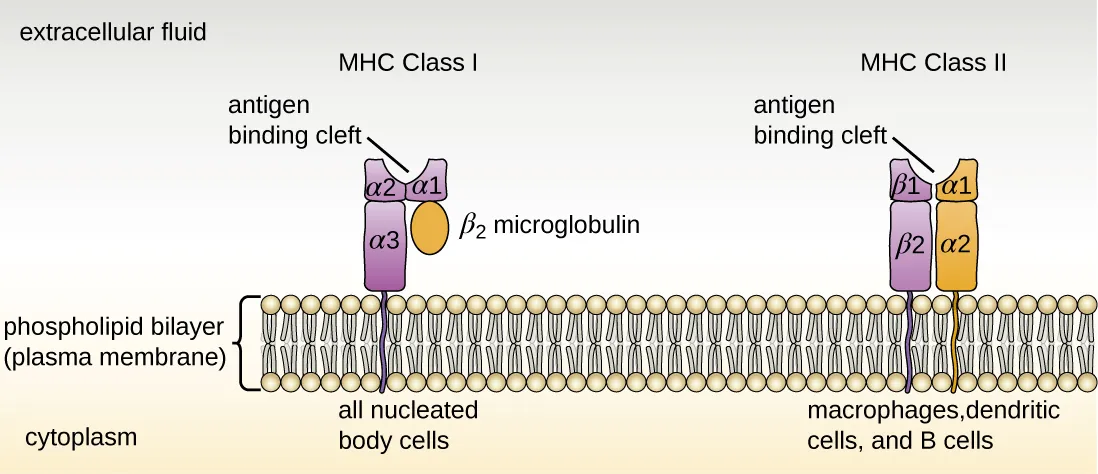
MHC I: Present on All Nucleated Cells
Who has it: Every nucleated cell in the body (NOT red blood cells, which lack a nucleus)
What it presents: Peptides from proteins made inside the cell (endogenous antigens)
Who reads it: CD8+ cytotoxic T cells
The endogenous pathway:
- Proteins inside the cell (both normal and foreign, if the cell is infected) are chopped into small peptide fragments
- Peptide fragments are loaded onto MHC I molecules in the endoplasmic reticulum (ER)
- The MHC I-peptide complex is transported to the cell surface
- CD8+ T cells scan the displayed peptides
If the peptide is a normal self-protein - the CD8+ T cell ignores the cell (it passed the badge check).
If the peptide is foreign (viral, bacterial, or mutant cancer protein) - the CD8+ T cell kills the cell.
MHC II: Present on Professional APCs Only
Who has it: Only professional antigen-presenting cells - dendritic cells, macrophages, and B cells (remembered as “the Big Three APCs”)
What it presents: Peptides from proteins ingested from outside the cell (exogenous antigens)
Who reads it: CD4+ helper T cells
The exogenous pathway:
- The APC engulfs a pathogen via phagocytosis or receptor-mediated endocytosis
- The pathogen is degraded in a phagolysosome into peptide fragments
- MHC II molecules (assembled in the ER) are transported to the phagolysosome
- Peptide fragments are loaded onto MHC II
- The MHC II-peptide complex is transported to the cell surface
- CD4+ helper T cells scan the displayed peptides and activate if they recognize the antigen
This activation of helper T cells is the starting point for the entire adaptive immune response - it triggers both B cell activation (humoral) and cytotoxic T cell activation (cell-mediated).
Side-by-Side Comparison
| Feature | MHC I | MHC II |
|---|---|---|
| Found on | All nucleated cells | APCs only (dendritic cells, macrophages, B cells) |
| Presents | Endogenous antigens (from inside the cell) | Exogenous antigens (from outside the cell) |
| Recognized by | CD8+ cytotoxic T cells | CD4+ helper T cells |
| Pathway name | Endogenous pathway | Exogenous pathway |
| Processing | Proteins degraded in cytoplasm, loaded in ER | Phagolysosome |
| Purpose | ”Is this cell infected or cancerous?" | "What pathogen did this APC encounter?” |
| Math trick | MHC I x CD8 = 8 | MHC II x CD4 = 8 |
HLA: The Human Version
In humans, MHC molecules are specifically called HLA (Human Leukocyte Antigen) molecules. HLA genes are the most polymorphic genes in the human genome - they have thousands of variants in the population.
This extreme diversity is why organ transplant matching is so difficult. HLA molecules differ between individuals, and the recipient’s immune system may recognize a donor organ’s HLA molecules as foreign, leading to transplant rejection. Immunosuppressive drugs suppress T cell activity to prevent this.