
B Cells and Antibodies
B cells are the weapons factory of the adaptive immune system. When activated, they transform into plasma cells that mass-produce antibodies - Y-shaped proteins that tag, neutralize, and help destroy specific pathogens. Understanding antibody structure is essential for the MCAT because the structure directly explains the function.
Antibody Structure: The Y-Shape
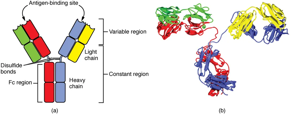
Every antibody (also called an immunoglobulin, Ig) is a Y-shaped protein made of four polypeptide chains:
- 2 identical heavy chains (the long chains forming the stem and inner arms of the Y)
- 2 identical light chains (the shorter chains forming the outer portion of the arms)
The chains are held together by disulfide bonds.
The Y-shape creates two functionally distinct regions:
Fab region (Fragment, antigen-binding) - the two arms of the Y. Each arm has a binding site at its tip that recognizes and binds to a specific antigen. Because there are two arms, each antibody has two identical antigen-binding sites.
Fc region (Fragment, crystallizable) - the stem of the Y. This is the part that interacts with the rest of the immune system: it binds to Fc receptors on phagocytes (triggering engulfment), activates complement (classical pathway), and determines the antibody class (IgG, IgA, IgM, etc.).
Variable vs. Constant Regions
Each chain (heavy and light) has two types of regions:
Variable (V) region - located at the tips of the Y arms. The amino acid sequence here differs between antibodies, giving each antibody its unique antigen specificity. This is the “lock” that fits a specific antigen “key.” The variable regions of one heavy chain and one light chain together form one antigen-binding site.
Constant (C) region - the rest of the chain. This sequence is the same for all antibodies of the same class. The constant region of the heavy chain determines the antibody’s class (isotype) and its effector functions.
How Antibodies Eliminate Pathogens
Antibodies do not directly kill pathogens (with the exception of toxin neutralization). Instead, they employ four main mechanisms:
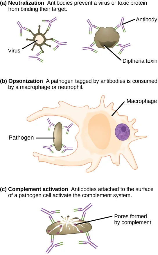
1. Neutralization - Antibodies bind to the surface of a pathogen or toxin, physically blocking it from attaching to host cells. A virus coated in antibodies cannot bind its receptor on a target cell. A toxin bound by antibodies cannot interact with its target. This is the simplest mechanism - just cover the enemy so it cannot function.
2. Opsonization - Antibodies coat the pathogen surface. The Fc regions of these antibodies are recognized by Fc receptors on macrophages and neutrophils, dramatically enhancing phagocytosis. Think of it as putting a “please eat me” sign on the pathogen.
3. Complement activation - When IgG or IgM binds a pathogen, the Fc region activates the classical complement pathway, leading to MAC formation and pathogen lysis. The Fab region finds the target; the Fc region triggers destruction.
4. Agglutination - Because each antibody has two binding sites, antibodies can cross-link multiple pathogens into clumps. These large clumps are much easier for phagocytes to find and engulf than individual bacteria. IgM, with its 10 binding sites (pentamer), is especially effective at agglutination.
| Mechanism | What Happens | Key Detail |
|---|---|---|
| Neutralization | Antibody blocks pathogen from binding host cells | Prevents infection; works on toxins too |
| Opsonization | Antibody coats pathogen; Fc signals phagocytes | ”Eat me” tag; Fc receptors on macrophages |
| Complement activation | Fc triggers classical pathway | Leads to MAC and lysis |
| Agglutination | Antibodies cross-link pathogens into clumps | IgM (pentamer) is best at this |
B Cell Development and Activation
Each B cell carries ~10,000 copies of a single unique antibody on its surface as a B cell receptor (BCR). All copies are identical - each B cell recognizes only one antigen.
How does the immune system produce millions of different B cells, each with a unique antibody, from only ~20,000 genes? Through somatic recombination (also called V(D)J recombination). During B cell development in the bone marrow, gene segments encoding the variable region are randomly shuffled and combined, generating an enormous diversity of antibody specificities. This is like shuffling a deck of cards - the number of possible combinations far exceeds the number of cards.
Activation process:
- A naive B cell encounters its matching antigen (the BCR binds to it)
- The B cell internalizes the antigen, processes it, and presents it on MHC II
- A helper T cell (CD4+) that recognizes the same antigen binds to the B cell and releases cytokines
- The B cell is now fully activated and undergoes clonal expansion
After activation, B cells differentiate into:
- Plasma cells (effector B cells) - antibody factories that secrete thousands of antibodies per second. They live for days to weeks.
- Memory B cells - long-lived cells that persist for years or decades. On re-exposure to the same antigen, they rapidly differentiate into plasma cells, producing a faster and stronger response.