Your body temperature right now is approximately 37 degrees C (98.6 degrees F). It was the same temperature yesterday. It will be the same tomorrow. Whether you are running a marathon in the desert or skiing in a blizzard, your core temperature barely budges. The same is true for your blood pH (7.35-7.45), your blood glucose (~70-100 mg/dL fasting), your blood osmolarity (~285-295 mOsm/L), and dozens of other variables.
This is not an accident. Your body is constantly measuring, adjusting, and correcting. That process - maintaining a stable internal environment despite changing external conditions - is homeostasis.
Why Homeostasis Matters for the MCAT
Homeostasis is not just one topic. It is the organizing principle behind every organ system. The endocrine system uses hormones to maintain blood glucose. The respiratory system adjusts breathing rate to maintain blood pH. The cardiovascular system changes heart rate to maintain blood pressure. The renal system adjusts urine output to maintain fluid balance.
Every time the MCAT asks “what happens when X changes,” the answer almost always involves a homeostatic mechanism restoring the variable to its set point.
The Three Components of Every Homeostatic System
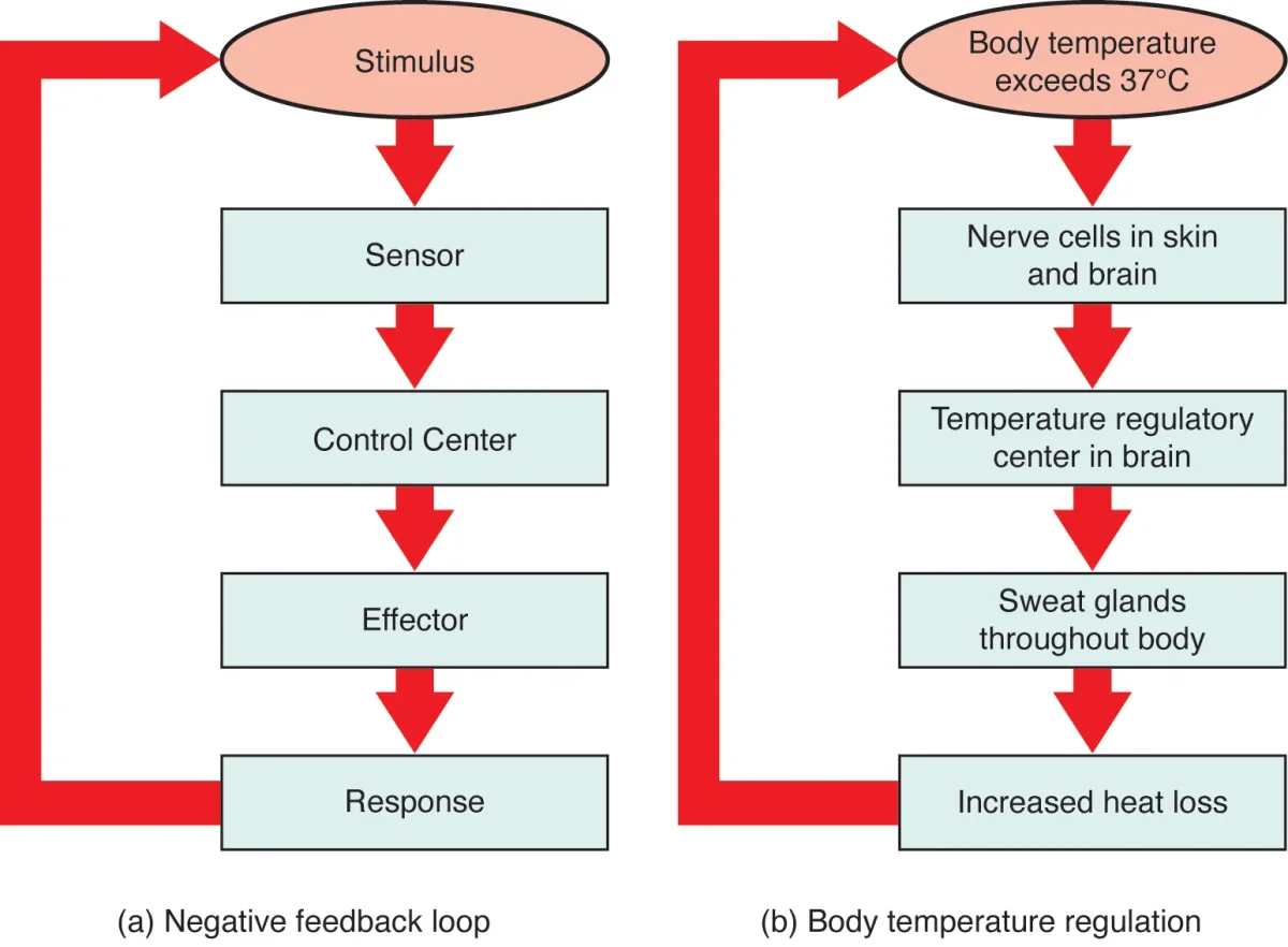
Every feedback loop has three parts:
1. Sensor (receptor) - detects the current value of the variable. Example: osmoreceptors in the hypothalamus detect blood osmolarity.
2. Control center (integrator) - compares the current value to the set point and decides what to do. Example: the hypothalamus compares detected osmolarity to the normal range.
3. Effector - carries out the correction. Example: the posterior pituitary releases ADH, which tells the kidneys to reabsorb more water.
Negative Feedback: The Default Mode
Negative feedback is the dominant control mechanism in the body. The principle: the output of a process opposes the original stimulus, pushing the variable back toward the set point.
“Negative” does not mean “bad.” It means the response negates (reverses) the change that triggered it.
How it works:
A variable deviates from its set point
The sensor detects the deviation
The control center activates the effector
The effector produces a response that opposes the original change
The variable returns toward the set point
As the variable normalizes, the stimulus weakens, and the response diminishes
The general negative feedback loop. The effector's response opposes the original stimulus, returning the variable to its set point. Credit: OpenStax Anatomy and Physiology 2e, CC BY 4.0
Classic examples:
Variable
Stimulus
Sensor
Effector
Response
Body temp
Too high
Hypothalamus
Sweat glands, blood vessels
Sweating, vasodilation (cool down)
Body temp
Too low
Hypothalamus
Skeletal muscles, blood vessels
Shivering, vasoconstriction (warm up)
Blood glucose
Too high
Beta cells (pancreas)
Liver, muscle, adipose
Insulin drives glucose into cells
Blood glucose
Too low
Alpha cells (pancreas)
Liver
Glucagon releases stored glucose
Blood osmolarity
Too high
Osmoreceptors (hypothalamus)
Kidneys (collecting duct)
ADH increases water reabsorption
Blood pressure
Too low
JGA (kidney)
Blood vessels, adrenal cortex
RAAS causes vasoconstriction + Na+ retention
Positive Feedback: The Rare Amplifier
Positive feedback is the opposite: the output amplifies the original stimulus rather than opposing it. This creates an escalating cycle that continues until an external event breaks the loop.
Positive feedback is rare because runaway amplification is dangerous. The body uses it only when a process must be driven rapidly to completion.
Key positive feedback examples for the MCAT:
Oxytocin during labor - cervical stretch triggers oxytocin release, which causes stronger contractions, which increases cervical stretch. Loop breaks when the baby is delivered.
LH surge during ovulation - rising estrogen from the dominant follicle triggers a massive LH surge (positive feedback on the anterior pituitary). Loop breaks when ovulation occurs and progesterone restores negative feedback.
Blood clotting cascade - activated platelets release signals that activate more platelets. Loop breaks when the clot seals the vessel.
Set points are not permanently fixed. During a fever, the hypothalamus raises the body temperature set point (e.g., from 37 degrees C to 39 degrees C). Now the body “thinks” 37 degrees C is too cold, so it shivers and vasoconstricts to warm up. When the fever breaks, the set point drops back to normal, and you suddenly feel hot and start sweating.
This concept explains why fever is not a failure of homeostasis - it is homeostasis working perfectly around a new, temporarily elevated set point. Pyrogens (from pathogens or immune cells) cause prostaglandin release, which acts on the hypothalamus to raise the set point.
A patient has a fever of 39 degrees C and is shivering. Is this a failure of homeostasis or an example of homeostasis in action? Explain.
Click to reveal answer
This is homeostasis in action. Pyrogens raised the hypothalamic set point to 39 degrees C. At the current body temperature (below 39 degrees C), the body "thinks" it is too cold, so it activates warming mechanisms (shivering, vasoconstriction). The feedback loop is working correctly - just around an elevated set point.
A researcher blocks all ADH receptors in the kidneys. What happens to blood osmolarity and urine volume? Which type of feedback is disrupted?
Click to reveal answer
Blood osmolarity increases and urine volume increases dramatically. Without ADH action, the collecting ducts cannot reabsorb water, so dilute urine is produced in large volumes (diabetes insipidus). The negative feedback loop for osmolarity is broken - the sensor and control center still detect high osmolarity and release ADH, but the effector cannot respond.
Before you can understand how the kidney works, you need a clear mental map of what it looks like. The kidney’s structure is not random - every anatomical feature exists because of the function it serves. The cortex is where filtration begins. The medulla is where urine gets concentrated. The pelvis is where urine collects before leaving. Once you connect structure to function, the physiology becomes much easier.
Gross Anatomy: The Kidney
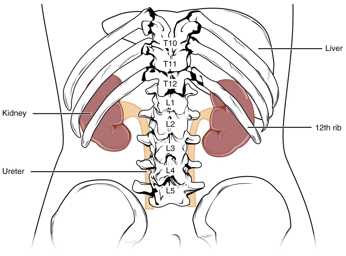
Each person has two kidneys, bean-shaped organs roughly the size of a fist, located retroperitoneally (behind the peritoneum) against the posterior abdominal wall. The right kidney sits slightly lower than the left because the liver pushes it down.
Cross-section of the kidney showing the cortex, medulla (with renal pyramids), renal pelvis, and major blood vessels entering at the hilum. Credit: OpenStax Anatomy and Physiology 2e, CC BY 4.0
Three regions of the kidney:
Renal cortex - the outer layer. Contains the glomeruli, Bowman’s capsules, proximal convoluted tubules (PCT), and distal convoluted tubules (DCT). This is where blood filtration begins and where most reabsorption occurs.
Renal medulla - the inner region. Contains the loops of Henle and collecting ducts, organized into cone-shaped structures called renal pyramids. The medulla creates the osmotic gradient that allows urine concentration. The tips of the pyramids (renal papillae) point inward toward the renal pelvis.
Renal pelvis - the funnel-shaped collecting area at the center. Urine drains from the collecting ducts through the papillae into minor calyces, which merge into major calyces, which empty into the renal pelvis. From there, urine flows into the ureter.
The Urinary Tract: From Kidney to Exit
Once urine leaves the renal pelvis, it follows a simple path:
Renal pelvis -> Ureter -> Urinary bladder -> Urethra -> outside the body
Ureters - muscular tubes (~25-30 cm long) that use peristalsis to move urine to the bladder. One from each kidney.
Urinary bladder - hollow muscular organ lined with transitional epithelium (stretches as it fills). The detrusor muscle (smooth muscle) contracts during urination.
Urethra - final passage to the exterior. In females, it is short (~4 cm) and carries only urine. In males, it is longer (~20 cm) and carries both urine and semen (at different times).
Each kidney contains over 1 million nephrons. The nephron is where all the action happens - filtration, reabsorption, secretion, and excretion. Understanding the nephron’s anatomy is essential for every renal question on the MCAT.
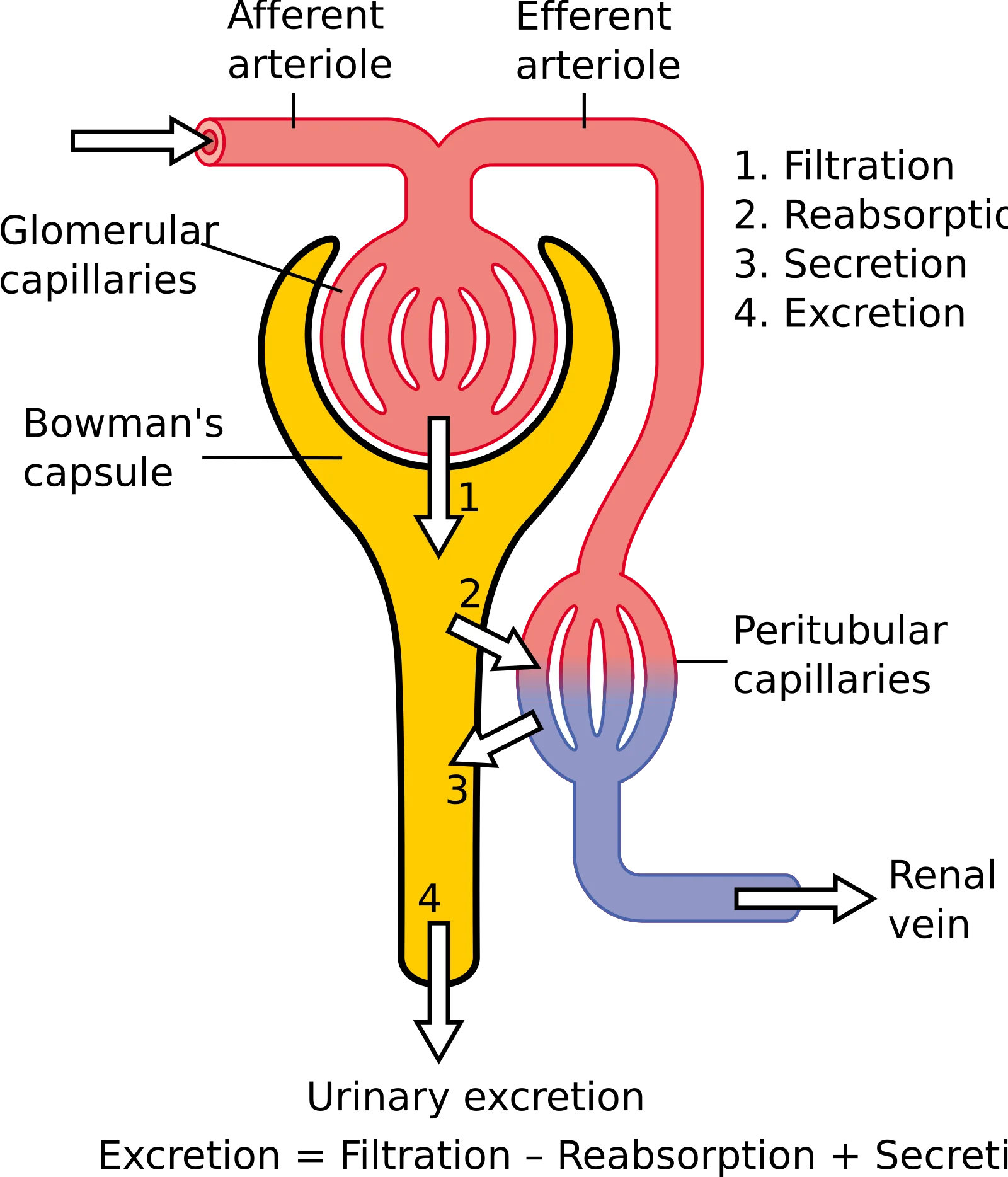
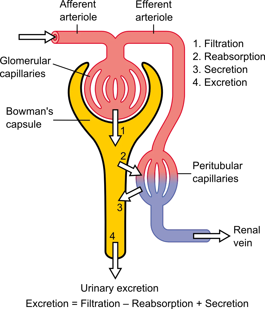
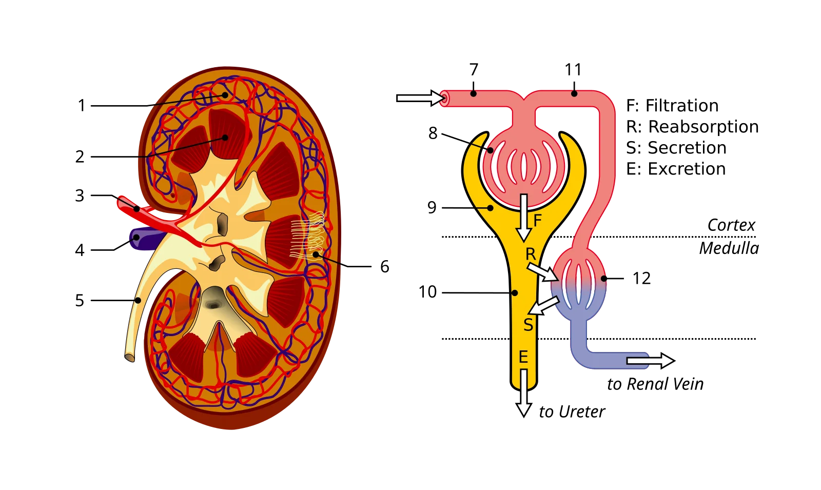
The nephron with its four key processes. Blood is filtered at the glomerulus, useful substances are reabsorbed in the tubules, waste is secreted, and the final urine is excreted. Remember: Excretion = Filtration - Reabsorption + Secretion. Credit: Wikimedia Commons, CC BY 3.0
Interactive 3D Nephron. Rotate to trace the filtrate path: glomerulus to Bowman's capsule to PCT to loop of Henle to DCT to collecting duct.Credit: iqcenter via Sketchfab, CC BY
Walk through the nephron, segment by segment:
1. Renal corpuscle (in the cortex)
Glomerulus - a ball of fenestrated capillaries where blood is filtered. High pressure forces fluid and small solutes out of the blood.
Bowman’s capsule - a cup that surrounds the glomerulus and collects the filtrate. Lined with podocytes whose foot processes form filtration slits.
2. Proximal convoluted tubule (PCT) (in the cortex)
The workhorse of reabsorption. Reabsorbs ~65% of filtered Na+, water, all glucose, all amino acids, and most bicarbonate.
Cells have a brush border (microvilli) to maximize surface area.
3. Loop of Henle (dips into the medulla)
Descending limb - permeable to water, impermeable to solutes. Water leaves by osmosis as the filtrate descends into the increasingly salty medulla.
Ascending limb - impermeable to water, actively pumps out NaCl (especially the thick ascending limb). This is what creates the medullary osmotic gradient.
4. Distal convoluted tubule (DCT) (in the cortex)
Fine-tuning station. Responds to aldosterone (reabsorbs Na+, secretes K+) and PTH (reabsorbs Ca2+). Site of additional H+ secretion for acid-base balance.
5. Collecting duct (runs from cortex through medulla)
Receives filtrate from multiple nephrons. Permeability to water is controlled by ADH - more ADH means more aquaporin channels, more water reabsorbed, more concentrated urine.
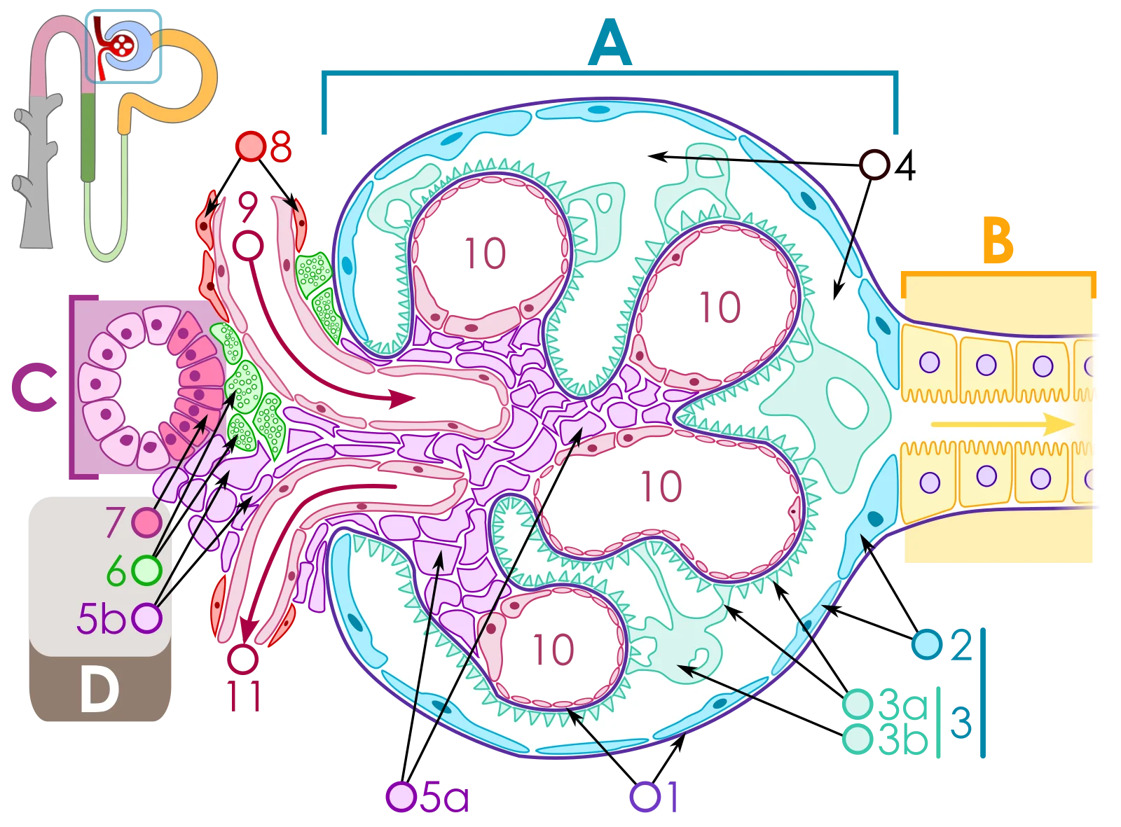
The renal corpuscle: Bowman's capsule surrounds the glomerular capillary tuft. Blood enters via the afferent arteriole and exits via the efferent arteriole. Focus on: the efferent arteriole is narrower than the afferent, maintaining high filtration pressure. Credit: Wikimedia Commons, CC BY-SA 3.0
Nephron Vasculature
The kidney’s blood supply is unusual and highly testable. Pay close attention to the arrangement:
Afferent arteriole - brings blood TO the glomerulus. “Afferent” = arriving.
Efferent arteriole - carries blood AWAY from the glomerulus. “Efferent” = exiting.
The glomerulus is a capillary bed sandwiched between TWO arterioles (not an arteriole and a venule). This is unique in the body and allows precise pressure control.
Peritubular capillaries - surround the PCT and DCT in cortical nephrons. Low-pressure, high-surface-area vessels that reabsorb solutes and water from the tubular cells back into the blood.
Vasa recta - long, straight capillaries that run alongside the loops of Henle in juxtamedullary nephrons. They maintain the medullary osmotic gradient by acting as countercurrent exchangers.
Two Types of Nephrons
Feature
Cortical Nephrons (~85%)
Juxtamedullary Nephrons (~15%)
Location of glomerulus
Outer cortex
Near cortex-medulla border
Loop of Henle
Short, barely enters medulla
Long, extends deep into medulla
Capillary network
Peritubular capillaries
Vasa recta
Primary role
Routine filtration and reabsorption
Urine concentration (creates medullary gradient)
A drug constricts the efferent arteriole. What happens to glomerular filtration rate (GFR) and why?
Click to reveal answer
GFR increases. Constricting the efferent arteriole (the "exit") traps blood in the glomerulus, raising hydrostatic pressure inside the glomerular capillaries. Higher pressure means more fluid is forced through the filtration membrane. This is similar to pinching the end of a garden hose - the water backs up and pressure increases upstream.
Which nephron segments are found in the cortex, and which extend into the medulla?
Click to reveal answer
Cortex: glomerulus, Bowman's capsule, PCT, DCT, and the beginning of the collecting duct. Medulla: loop of Henle (both limbs) and the collecting duct (which runs from cortex through medulla to the papilla). Juxtamedullary nephrons have loops that extend deep into the medulla; cortical nephrons have shorter loops.
Filtration is the first step of urine formation. It is a bulk, nonselective process - the glomerulus does not “choose” what to filter. Instead, it acts like a sieve: anything small enough passes through, and anything too large stays behind. The selectivity comes later, in the tubules, where the kidney carefully reclaims what the body needs.
Understanding what drives filtration, what determines how much gets filtered, and what the filtration barrier looks like is critical for MCAT success.
The Filtration Membrane
The filtration barrier between blood and Bowman’s capsule has three layers:
1. Fenestrated endothelium of glomerular capillaries - has pores (fenestrations) that allow most plasma components to pass but block blood cells.
2. Basement membrane - a gel-like layer of glycoproteins with a net negative charge. This charge repels most plasma proteins (which are also negatively charged at physiological pH, especially albumin).
3. Podocytes - specialized cells that wrap around the capillaries with foot processes (pedicels). The gaps between the foot processes are called filtration slits. These provide the final size barrier.
The glomerular filtration membrane. Focus on: the three layers (fenestrated endothelium, basement membrane, podocyte foot processes) and the size/charge selectivity - water, glucose, and ions pass through; proteins and blood cells do not. You do not need the molecular details of each layer. Credit: OpenStax Anatomy and Physiology 2e, CC BY 4.0
What Gets Filtered vs. What Stays Behind
Freely Filtered (passes into Bowman’s space)
NOT Filtered (stays in blood)
Water
Red blood cells
Glucose
White blood cells
Amino acids
Platelets
Urea, creatinine, uric acid
Plasma proteins (albumin, globulins)
Electrolytes (Na+, K+, Cl-, Ca2+, HCO3-)
Protein-bound substances
Small peptide hormones
Large molecules (>70 kDa)
Clinical connection: If protein or blood appears in the urine, something is wrong with the filtration membrane. Proteinuria (protein in urine) indicates damage to the basement membrane or podocytes. Hematuria (blood in urine) indicates damage to the capillary endothelium.
Starling Forces: What Drives Filtration
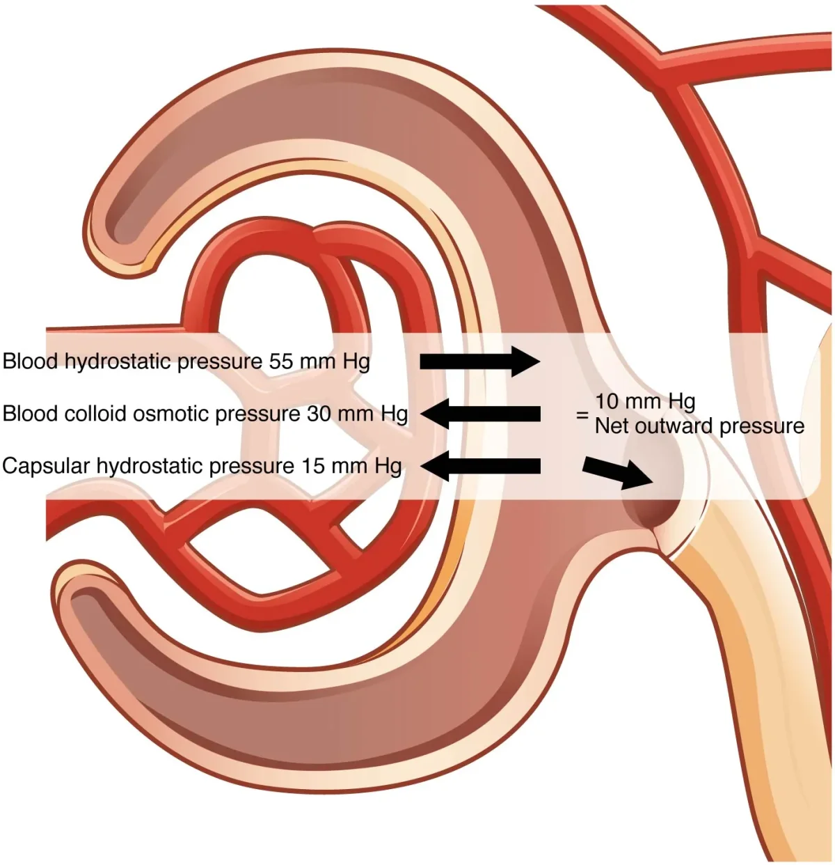
Filtration is driven by pressure. Specifically, the net filtration pressure (NFP) is determined by the balance of four forces - collectively called Starling forces:
Forces FAVORING filtration (pushing fluid out of capillaries):
Glomerular hydrostatic pressure - the blood pressure inside the glomerular capillaries. This is the main driving force and is much higher than in most capillary beds.
Forces OPPOSING filtration (pushing fluid back or keeping it in):
Bowman’s capsule hydrostatic pressure - the pressure of fluid already in Bowman’s space pushing back.
Glomerular oncotic pressure - the osmotic pull of plasma proteins in the blood (proteins cannot cross, so they pull water back).
The net filtration pressure is the balance of these forces. Under normal conditions, hydrostatic pressure greatly exceeds the opposing forces, so the net flow is always from blood into Bowman’s space.
Glomerular Filtration Rate (GFR)
GFR is the volume of filtrate produced per minute by all glomeruli in both kidneys.
Normal GFR: ~125 mL/min = ~180 L/day
That is an enormous volume - far more than your total blood volume. You only excrete ~1-2 L as urine because the tubules reabsorb 99% of the filtrate.
You cannot watch filtrate form, so GFR is estimated by measuring how fast the kidneys clear a substance from plasma. A substance that is freely filtered, not reabsorbed, and not secreted has a clearance that equals GFR.
Afferent vs Efferent: Why Direction Matters
Because the glomerulus sits between two arterioles, the kidney can control GFR with precision. Constriction at either end has opposite effects:
Afferent constriction -> less blood arrives -> glomerular pressure falls -> GFR drops.
Efferent constriction -> blood backs up in the glomerulus -> glomerular pressure rises -> GFR rises (up to a point).
Angiotensin II preferentially constricts the efferent arteriole, which protects GFR when renal perfusion is low. This is why ACE inhibitors and ARBs can drop GFR in patients with already-compromised renal blood flow: remove angiotensin II, and the efferent arteriole relaxes, dropping glomerular pressure.
Autoregulation of GFR
The kidneys maintain a stable GFR even when systemic blood pressure fluctuates (within the range of ~80-180 mmHg MAP). Two mechanisms accomplish this:
1. Myogenic mechanism - when blood pressure rises, the increased stretch on afferent arteriole walls triggers them to constrict (smooth muscle response). This prevents the pressure increase from reaching the glomerulus. When pressure drops, the arteriole relaxes.
2. Tubuloglomerular feedback (TGF) - the macula densa cells in the DCT (part of the juxtaglomerular apparatus) monitor NaCl concentration in the filtrate. If GFR increases, more NaCl reaches the macula densa. The macula densa signals the afferent arteriole to constrict, reducing blood flow and lowering GFR back to normal.
The Juxtaglomerular Apparatus (JGA)
The JGA is where the DCT brushes against its own afferent arteriole. Two cell types do the work:
JG cells in the afferent arteriole wall secrete renin when blood pressure drops or sympathetic nerves fire.
Macula densa cells in the DCT wall sense tubular NaCl and signal the JG cells.
That physical adjacency is the point - a local sensor (macula densa) sitting next to a local effector (JG cells), no hormones required for the feedback.
A patient with severe liver disease has low plasma albumin levels. Predict the effect on GFR and explain using Starling forces.
Click to reveal answer
GFR increases. Albumin is the major contributor to plasma oncotic pressure (piGC). With less albumin, oncotic pressure drops, meaning less opposition to filtration. Since NFP = PGC - PBS - piGC, a decrease in piGC increases NFP, increasing GFR. Clinically, this is why liver disease can cause edema - more fluid is filtered out of capillaries throughout the body.
If GFR is 120 mL/min and RPF is 600 mL/min, what is the filtration fraction? Is this normal?
Click to reveal answer
FF = GFR/RPF = 600120 = 0.20 = 20%. This is normal. A filtration fraction of ~20% means that about one-fifth of the plasma entering the kidney is filtered at the glomerulus. The remaining 80% continues through the efferent arteriole to the peritubular capillaries.
After filtration dumps 180 liters of fluid per day into Bowman’s space, the tubules must reclaim almost all of it. If they did not, you would lose your entire plasma volume in about 30 minutes. Reabsorption is the process of moving useful substances from the tubular filtrate back into the blood. Secretion is the reverse - moving additional waste from the blood into the filtrate for excretion.
Each nephron segment has a specific job. The MCAT expects you to know what happens where.
The four nephron processes: filtration at the glomerulus, reabsorption and secretion along the tubules, and excretion of final urine. Focus on: Excretion = Filtration - Reabsorption + Secretion. Credit: Wikimedia Commons, CC BY 3.0
The Big Picture: Three Processes
1. Filtration (glomerulus) - bulk movement of fluid and small solutes from blood into Bowman’s space. Nonselective.
2. Reabsorption (tubules) - movement of useful substances from filtrate back into peritubular capillary blood. Selective and highly regulated.
3. Secretion (tubules) - movement of additional waste, drugs, and ions from peritubular blood into the filtrate. Provides a second chance to remove substances that were not filtered or need faster elimination.
Proximal Convoluted Tubule (PCT): The Workhorse
The PCT reabsorbs the majority of everything. It is the most metabolically active segment of the nephron, packed with mitochondria and covered in microvilli (brush border) to maximize surface area.
What the PCT reabsorbs:
~65% of filtered Na+ and water
~100% of filtered glucose (via sodium-glucose co-transporters)
~100% of filtered amino acids
~80-90% of filtered bicarbonate (HCO3-)
Most phosphate, lactate, and citrate
Urea (partially - ~50% is passively reabsorbed)
What the PCT secretes:
H+ (important for bicarbonate reabsorption)
Organic acids (uric acid, drug metabolites)
Organic bases (creatinine, certain antibiotics)
NH3/NH4+ (for acid-base buffering)
How sodium reabsorption works in the PCT:
The Na+/K+ ATPase on the basolateral membrane (blood side) pumps Na+ out of the cell and into the blood, keeping intracellular Na+ low. This creates a gradient that drives Na+ into the cell from the tubular lumen through various co-transporters on the apical membrane (lumen side):
Na+-glucose co-transporter - drags glucose into the cell along with Na+
Na+-amino acid co-transporters - drags amino acids with Na+
Na+/H+ exchanger - trades Na+ in for H+ out (helps reclaim bicarbonate)
Water follows Na+ passively through aquaporins and paracellular pathways. The PCT reabsorbs water and solutes in roughly equal proportions, so the filtrate leaving the PCT is still isotonic (~300 mOsm/L) - just much reduced in volume.
Transport Maximum (Tm) and Glucose
Glucose reabsorption uses sodium-glucose co-transport carriers in the PCT. These carriers have a maximum transport rate - the transport maximum (Tm).
At normal blood glucose (~100 mg/dL), all filtered glucose is reabsorbed. But if blood glucose exceeds ~180-200 mg/dL (the renal threshold), the carriers become saturated. Glucose that exceeds the Tm cannot be reabsorbed and appears in the urine (glucosuria).
This is exactly what happens in uncontrolled diabetes mellitus. The excess glucose in the urine also drags water with it by osmosis, causing the classic symptoms of polyuria (excessive urination) and polydipsia (excessive thirst).
Loop of Henle: Creating the Medullary Gradient
The loop of Henle has two limbs with opposite permeability properties. This is the key to understanding urine concentration.
Descending limb:
Permeable to water
Impermeable to solutes (NaCl)
As filtrate descends into the increasingly salty medulla, water leaves by osmosis
Filtrate becomes progressively more concentrated (up to ~1200 mOsm/L at the hairpin turn)
Thin ascending limb:
Impermeable to water
Passively permeable to NaCl (salt diffuses out)
Filtrate becomes less concentrated as salt leaves
Thick ascending limb:
Impermeable to water
Actively pumps NaCl out via the Na+/K+/2Cl- co-transporter
This is the segment that actively creates the medullary osmotic gradient
Filtrate becomes dilute (~100 mOsm/L) by the time it reaches the DCT
Distal Convoluted Tubule (DCT): Fine-Tuning
The DCT handles the last bit of adjustable Na+ and Ca2+ reabsorption:
Thiazide-sensitive Na+/Cl- cotransporter - reabsorbs a small fraction of filtered Na+ (the site of thiazide diuretics).
PTH - increases Ca2+ reabsorption here.
Aldosterone begins to act on the late DCT but exerts its main effect on the collecting duct.
The DCT also continues H+ secretion for acid-base regulation.
A patient has a blood glucose level of 350 mg/dL. Explain why glucose appears in their urine using the concept of transport maximum.
Click to reveal answer
The glucose carriers in the PCT are saturated. At 350 mg/dL, the amount of glucose filtered exceeds the transport maximum (~375 mg/min). The excess glucose that cannot bind to carriers remains in the tubular fluid and is excreted in the urine (glucosuria). The renal threshold for glucose is ~180-200 mg/dL - above this, glucose begins to "spill over" into the urine.
Why is the filtrate leaving the thick ascending limb of the loop of Henle more dilute than plasma?
Click to reveal answer
The thick ascending limb actively pumps out NaCl but is impermeable to water. Salt leaves the filtrate, but water cannot follow. This removes solute without removing solvent, making the filtrate progressively more dilute. By the time filtrate reaches the DCT, its osmolarity is ~100 mOsm/L (compared to plasma at ~300 mOsm/L). The thick ascending limb is sometimes called the "diluting segment."
The countercurrent multiplier is one of the most elegant mechanisms in all of physiology - and one of the most commonly tested on the MCAT. It explains how the kidney creates a concentrated environment in the medulla that allows the collecting duct to produce concentrated urine. Without this gradient, you could never conserve water during dehydration.
The concept is straightforward once you see it clearly: two parallel tubes running in opposite directions, doing different things, amplifying a small difference into a large gradient.
How It Works: Step by Step
The countercurrent multiplier depends on two facts:
The descending limb is permeable to water but NOT to salt
The thick ascending limb actively pumps salt out (via Na+/K+/2Cl- co-transport) but is impermeable to water
These two limbs run side by side in opposite directions (countercurrent flow).
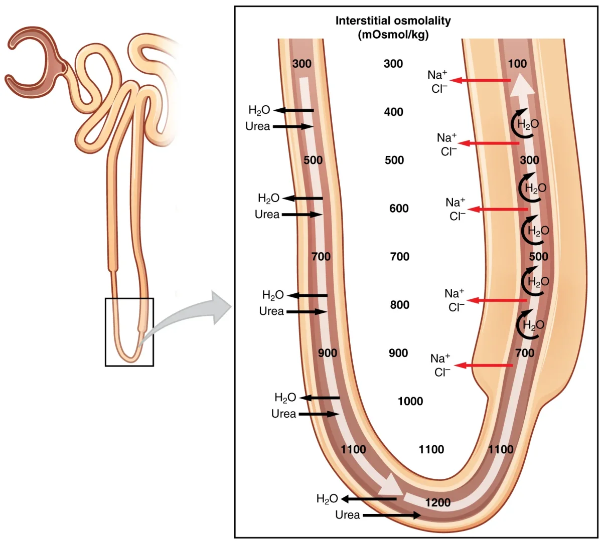
The single effect: At any horizontal level, the thick ascending limb pumps NaCl into the medullary interstitium. This raises the interstitial osmolarity by a small amount (~200 mOsm/L above the filtrate).
The multiplication: Because the descending limb is permeable to water, water leaves the descending limb (drawn out by the salty interstitium), concentrating the filtrate inside. This concentrated filtrate then flows around the hairpin turn into the ascending limb, where more salt is pumped out. Each “cycle” adds to the gradient.
The result:
Cortex: ~300 mOsm/L (isotonic with plasma)
Outer medulla: ~600 mOsm/L
Inner medulla: ~900 mOsm/L
Deepest medulla (papilla): ~1200 mOsm/L
The countercurrent multiplier system. The descending limb loses water; the ascending limb pumps out NaCl. The result is a progressively saltier medullary interstitium from cortex to papilla. Credit: OpenStax Anatomy and Physiology 2e, CC BY 4.0
Why “Countercurrent” and Why “Multiplier”?
Countercurrent - the fluid flows in opposite directions in the two limbs. Descending filtrate flows down; ascending filtrate flows up. This is critical because it means the ascending limb is always adjacent to a slightly less concentrated section of the descending limb, allowing continuous salt extraction to have a cumulative effect.
Multiplier - the single effect (a ~200 mOsm/L difference at any one level) gets multiplied along the length of the loop into a much larger total gradient (300 to 1200 mOsm/L).
If flow were concurrent (same direction in both tubes), the gradient would equilibrate and you would get a uniform, modest difference. The opposite flow directions are what allow the small single effect to be amplified.
The Vasa Recta: Countercurrent Exchanger
The vasa recta are the capillaries that run alongside the loops of Henle in juxtamedullary nephrons. They have a critical job: supply blood to the medulla WITHOUT washing away the osmotic gradient.
The vasa recta accomplish this through countercurrent exchange:
As blood descends into the salty medulla, water leaves and solutes enter (blood equilibrates with the surrounding interstitium)
As blood ascends back toward the cortex, water re-enters and solutes leave (blood re-equilibrates)
Net effect: blood delivers oxygen and nutrients to the medulla while maintaining the gradient
Urea Recycling: The Gradient Booster
Urea contributes about 50% of the medullary osmotic gradient in the inner medulla. Here is how:
Urea is filtered at the glomerulus and partially reabsorbed in the PCT
In the inner medullary collecting duct (under ADH stimulation), urea transporters allow urea to diffuse out into the medullary interstitium
Some of this urea enters the thin ascending limb of the loop of Henle (urea recycling)
The urea travels through the nephron again and re-enters the medulla through the collecting duct
This recycling keeps urea concentrated in the inner medulla, adding to the osmotic gradient that drives water reabsorption from the collecting duct.
What Happens If the Gradient Is Destroyed?
Loop diuretics block the Na+/K+/2Cl- co-transporter in the thick ascending limb. Without active NaCl pumping, the medullary gradient collapses. The collecting duct has nothing to drive water reabsorption against, so large volumes of dilute urine are produced. This is why loop diuretics are the most powerful class of diuretics - they attack the gradient at its source.
Why does the countercurrent multiplier require flow in opposite directions? What would happen if both limbs of the loop of Henle flowed in the same direction?
Click to reveal answer
Countercurrent flow allows each small single-effect difference to be multiplied along the length of the tube. The ascending limb continuously pumps NaCl out at every level, and because it is next to progressively more dilute descending-limb fluid, the gradient builds cumulatively. If flow were concurrent (same direction), the two limbs would quickly equilibrate - you would get a uniform, small difference throughout, not the 300 to 1200 mOsm/L gradient needed for urine concentration.
What is the difference between the countercurrent multiplier and the countercurrent exchanger? Which structures perform each?
Click to reveal answer
Countercurrent multiplier (Loop of Henle): actively creates the medullary osmotic gradient by pumping NaCl out of the ascending limb while water leaves the descending limb. Uses energy (ATP for active salt transport). Countercurrent exchanger (Vasa recta): passively preserves the gradient by allowing blood to equilibrate with the surrounding interstitium as it descends and re-equilibrate as it ascends. No energy input - purely passive exchange.
The collecting duct is where the final decision is made: will you produce dilute urine or concentrated urine? Everything upstream - the glomerulus, PCT, loop of Henle, DCT - has been preparing the filtrate. The collecting duct uses the medullary gradient (built by the countercurrent multiplier) to determine how much water to reclaim based on hormonal signals.
The key hormone is ADH (antidiuretic hormone, also called vasopressin). ADH is the single most important regulator of urine concentration.
ADH: The “Don’t Pee” Hormone
Where ADH comes from:
Osmoreceptors in the hypothalamus detect increased blood osmolarity (dehydration)
The hypothalamus synthesizes ADH
ADH is transported down axons to the posterior pituitary
The posterior pituitary releases ADH into the bloodstream
ADH travels to the kidneys and binds receptors on collecting duct principal cells
What ADH does at the collecting duct:
ADH binds receptors on the basolateral membrane of principal cells
This activates a cAMP signaling cascade
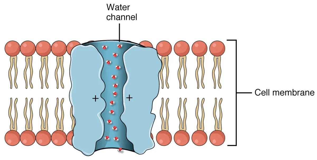
Aquaporin water channels are inserted into the apical (lumen-facing) membrane
Water flows from the tubular lumen through aquaporins, through the cell, and out the basolateral side
Water enters the hypertonic medullary interstitium and is picked up by the vasa recta
ADH triggers the insertion of aquaporin water channels into collecting duct cells, allowing water reabsorption driven by the medullary osmotic gradient. Credit: OpenStax Anatomy and Physiology 2e, CC BY 4.0
Two Scenarios: Hydrated vs. Dehydrated
When you are well-hydrated (low ADH):
Blood osmolarity is low, so little ADH is released
Collecting duct remains impermeable to water (few aquaporins)
The dilute filtrate (~100 mOsm/L from the ascending limb) passes through the collecting duct without losing water
Result: large volume of dilute urine (~50-100 mOsm/L)
When you are dehydrated (high ADH):
Blood osmolarity is high, so ADH is released
Collecting duct becomes permeable to water (aquaporins inserted)
Water is reabsorbed as the collecting duct passes through the increasingly concentrated medulla
Result: small volume of concentrated urine (up to ~1200 mOsm/L)
Condition
ADH Level
Aquaporins
Collecting Duct Permeability
Urine Volume
Urine Osmolarity
Overhydrated
Very low
Few/none
Impermeable
High (~18 L/day max)
Low (~50 mOsm/L)
Normal
Moderate
Some
Moderately permeable
Normal (~1-2 L/day)
~300-600 mOsm/L
Dehydrated
High
Many
Highly permeable
Low (~0.5 L/day)
High (~1200 mOsm/L)
Cell Types in the Collecting Duct
The collecting duct has two main cell types:
Principal cells - respond to ADH (insert aquaporins) and aldosterone (Na+ reabsorption, K+ secretion). These handle water and electrolyte balance.
Intercalated cells - handle acid-base balance.
Type A intercalated cells - secrete H+ into the lumen and reabsorb HCO3- into the blood. Active during acidosis.
Type B intercalated cells - secrete HCO3- into the lumen and reabsorb H+. Active during alkalosis.
Diabetes Insipidus: When ADH Fails
Diabetes insipidus (DI) is a condition where ADH signaling fails, resulting in massive volumes of dilute urine (up to 18-20 L/day) and severe dehydration.
Central DI - the hypothalamus/posterior pituitary does not produce enough ADH. Caused by head trauma, tumors, or surgery.
Nephrogenic DI - the kidneys do not respond to ADH (defective receptors or aquaporin channels). ADH levels are actually HIGH, but the collecting duct cannot respond.
The key distinction: in central DI, the problem is upstream (no hormone produced). In nephrogenic DI, the problem is downstream (hormone is present but the effector is broken). This is the same “primary vs. secondary” reasoning pattern you learned for endocrine disorders.
SIADH: When There Is Too Much ADH
Syndrome of Inappropriate ADH secretion (SIADH) is the opposite of DI. Too much ADH is released (often from lung tumors, brain injury, or certain drugs), causing excessive water reabsorption. The result:
Very concentrated, low-volume urine
Dilutional hyponatremia (blood Na+ drops because excess water dilutes it)
Patients may develop confusion, seizures, or cerebral edema from the low sodium
A patient drinks several alcoholic beverages over the course of an evening. Predict the effect on ADH levels, urine volume, and urine osmolarity.
Click to reveal answer
ADH levels decrease (alcohol inhibits ADH release from the posterior pituitary). Without ADH, the collecting duct becomes impermeable to water. Urine volume increases (diuresis) and urine osmolarity decreases (dilute urine). This is why alcohol consumption leads to dehydration and frequent urination.
A patient with SIADH has a blood Na+ of 120 mEq/L (normal: 135-145). Explain the pathophysiology using the ADH-collecting duct relationship.
Click to reveal answer
Excess ADH causes the collecting duct to maximally reabsorb water. The retained water dilutes all plasma solutes, including sodium, causing dilutional hyponatremia. The urine is inappropriately concentrated (high osmolarity) because the collecting duct is reabsorbing water despite already-low blood osmolarity. The problem is not sodium loss - it is water excess.
Three hormones dominate renal regulation: ADH, aldosterone, and ANP. Each responds to a different stimulus, acts on a different part of the nephron, and produces a different effect. But they all serve the same ultimate goal - maintaining blood volume, blood pressure, and electrolyte balance.
The MCAT tests these hormones constantly. If you know the trigger, target, and effect of each hormone, you can reason through almost any renal physiology question.
Kidney cross-section with nephron placement. Glomeruli and convoluted tubules sit in the cortex, while the loops of Henle dip into the medulla. Hormones (ADH, aldosterone, ANP) act on specific nephron segments to regulate water and ion balance. Credit: Wikimedia Commons, CC BY-SA 3.0
ADH (Antidiuretic Hormone / Vasopressin)
Trigger: Increased blood osmolarity (detected by hypothalamic osmoreceptors) or decreased blood volume/pressure (detected by baroreceptors in the carotid sinus and aortic arch)
Source: Synthesized in the hypothalamus, released from the posterior pituitary
Target: Receptors on collecting duct principal cells
Effect: Inserts aquaporin-2 channels into the apical membrane, increasing water reabsorption. Also promotes urea recycling in the inner medullary collecting duct.
Trigger: Angiotensin II (from RAAS), high blood K+, low blood Na+, and ACTH (minor role)
Source: Zona glomerulosa of the adrenal cortex
Target: Principal cells of the DCT and collecting duct
Effect: Increases expression of sodium channels on the apical membrane and Na+/K+ ATPase on the basolateral membrane. This increases Na+ reabsorption and K+ secretion.
Net result: Na+ and water retention (water follows sodium), K+ excretion, increased blood volume and blood pressure
Inhibited by: ANP, high blood Na+, low K+
ANP (Atrial Natriuretic Peptide)
Trigger: Atrial stretch from increased blood volume (the atria of the heart release ANP when they are overfilled)
RAAS is activated - aldosterone causes Na+ (and water) retention
ANP is suppressed (atria are not stretched)
Result: concentrated, low-volume urine; blood volume and pressure restored
Overhydration scenario:
Blood volume rises, blood osmolarity drops
ADH is suppressed - collecting duct stays impermeable
RAAS is suppressed - less aldosterone, less Na+ retention
ANP is released - promotes Na+ and water excretion, further suppresses RAAS and ADH
Result: dilute, high-volume urine; blood volume and pressure decrease
A patient takes a drug that blocks aldosterone receptors. Predict the effects on Na+, K+, blood pressure, and urine volume.
Click to reveal answer
Blocking aldosterone prevents Na+ reabsorption and K+ secretion in the DCT and collecting duct. Blood Na+ decreases (and water follows, so blood volume and blood pressure drop). Blood K+ increases (hyperkalemia - the main risk of this drug class). Urine volume increases (Na+ and water are lost). This type of drug is classified as a "potassium-sparing diuretic."
A patient has congestive heart failure with high blood volume. Which hormone is elevated, and why does the kidney still retain sodium?
Click to reveal answer
ANP is elevated (atria are stretched by high blood volume), but the kidney still retains sodium because RAAS is also activated. In CHF, cardiac output is low, so renal perfusion is reduced. The kidneys interpret low perfusion as low blood volume and activate RAAS, causing sodium and water retention despite total body fluid overload. The RAAS signal overrides ANP in this context. This is the pathological "vicious cycle" of CHF.
RAAS is the body’s emergency blood pressure rescue system. When blood pressure drops - from dehydration, hemorrhage, or heart failure - the kidneys detect the problem and launch a hormonal cascade that raises blood pressure through multiple simultaneous mechanisms. Understanding every step of this cascade is essential for the MCAT.
The RAAS Cascade: Step by Step
Step 1: Trigger - low blood pressure detected
The JG cells of the afferent arteriole sense reduced renal perfusion (low blood pressure). Three signals activate renin release:
Direct detection of low pressure by baroreceptors in the afferent arteriole
Low NaCl detected by the macula densa (signals low GFR)
Sympathetic nervous system activation (beta-1 receptors on JG cells)
Step 2: Renin release
JG cells secrete renin (an enzyme) into the blood.
Step 3: Angiotensinogen to Angiotensin I
Renin cleaves angiotensinogen (a large protein continuously produced by the liver) into angiotensin I. Angiotensin I is inactive - it is just an intermediate.
Step 4: Angiotensin I to Angiotensin II
Angiotensin-converting enzyme (ACE), located primarily on the endothelial surface of pulmonary capillaries (lungs), converts angiotensin I into angiotensin II. ACE also degrades bradykinin (a vasodilator), which explains the dry cough side effect of ACE inhibitors.
Step 5: Angiotensin II - the effector
Angiotensin II is one of the most potent vasoconstrictors in the body, playing a central role in blood pressure regulation. It has multiple simultaneous effects:
Effect
Mechanism
Speed
Vasoconstriction
Contracts arteriolar smooth muscle
Immediate (seconds)
Aldosterone release
Stimulates zona glomerulosa of adrenal cortex
Minutes to hours
ADH release
Stimulates posterior pituitary
Minutes
Thirst
Stimulates hypothalamic thirst center
Minutes
Na+ reabsorption
Direct action on PCT (stimulates Na+/H+ exchange)
Minutes
Efferent arteriole constriction
Preferentially constricts efferent arteriole, raising GFR
Immediate
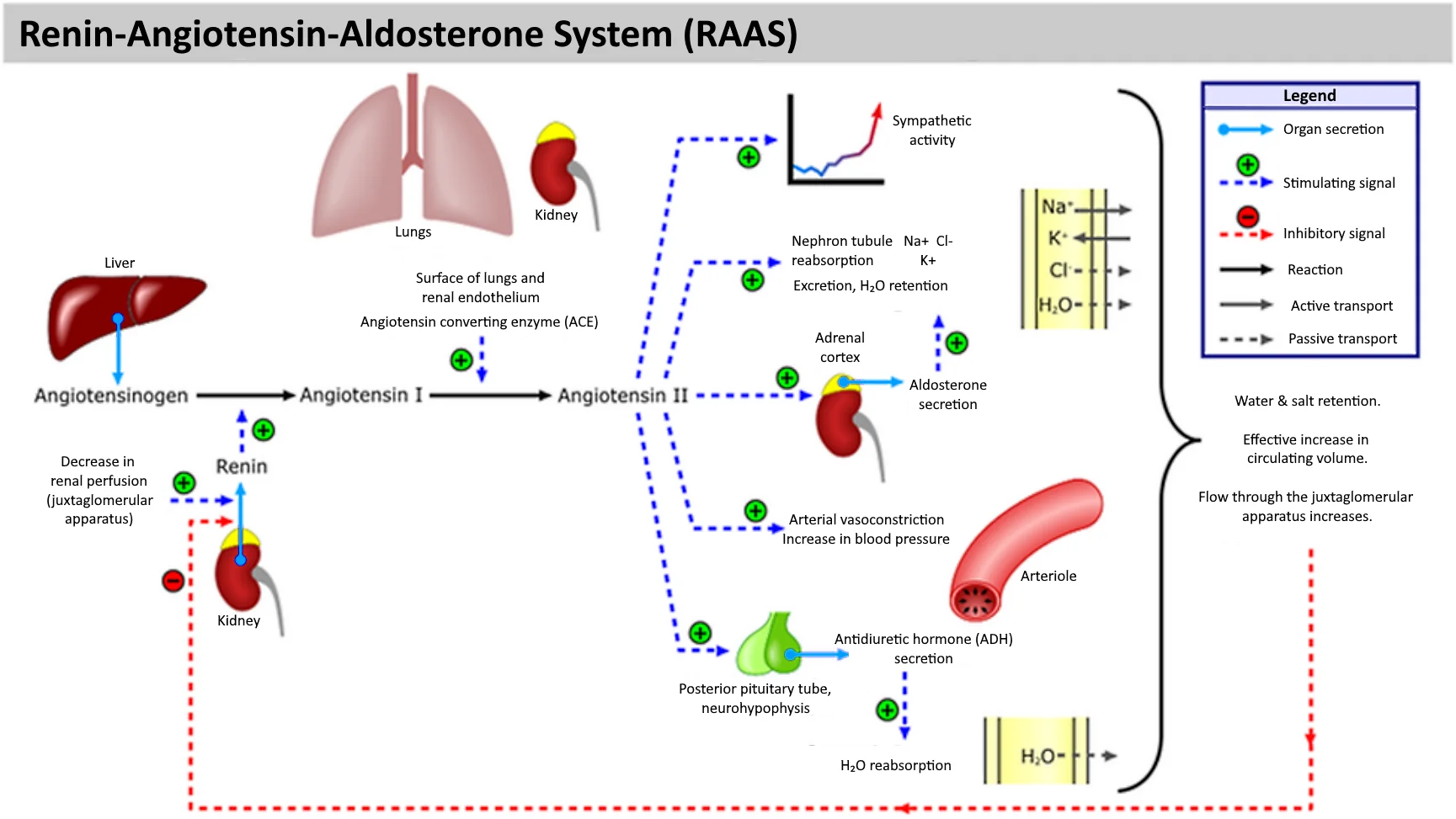
The RAAS cascade. Focus on: low BP → renin (from JG cells) → angiotensinogen → angiotensin I → (ACE in lungs) → angiotensin II → vasoconstriction + aldosterone (Na+ reabsorption) + ADH (water reabsorption) + thirst. ACE inhibitors and ARBs are common drug targets. Credit: Wikimedia Commons, CC BY-SA 4.0
Pharmacological Targets in the RAAS
The MCAT commonly describes drugs that target specific steps in the RAAS cascade:
ACE inhibitors - block the conversion of angiotensin I to angiotensin II. Less vasoconstriction, less aldosterone release, blood pressure drops. ACE also normally degrades bradykinin, so blocking ACE causes bradykinin accumulation - this explains the dry cough side effect.
ARBs (angiotensin receptor blockers) - block angiotensin II receptors. Same blood pressure-lowering effects as ACE inhibitors but without the cough (bradykinin is still degraded normally).
Negative Feedback in RAAS
The RAAS cascade has built-in negative feedback:
When blood pressure normalizes, the JG cells sense adequate pressure and stop releasing renin
Angiotensin II directly inhibits further renin release (short-loop feedback)
Elevated blood pressure activates baroreceptors that suppress sympathetic drive to the JG cells
ANP (released when blood volume is high) inhibits renin, aldosterone, and ADH
A patient takes an ACE inhibitor. What happens to renin, angiotensin I, angiotensin II, and aldosterone levels?
Click to reveal answer
Renin increases (the drop in angiotensin II removes negative feedback on JG cells). Angiotensin I increases (renin is still converting angiotensinogen, but ACE cannot convert AI to AII). Angiotensin II decreases (ACE is blocked). Aldosterone decreases (angiotensin II normally stimulates aldosterone release). Net effect: blood pressure drops.
Why does angiotensin II preferentially constrict the efferent arteriole? What is the physiological purpose?
Click to reveal answer
Efferent constriction maintains GFR even when systemic blood pressure is low. By constricting the "exit" of the glomerulus, blood backs up inside, maintaining glomerular hydrostatic pressure and filtration rate despite reduced renal blood flow. This ensures that waste filtration continues during hypovolemia. However, excessive efferent constriction (e.g., from chronic RAAS activation) can eventually damage the glomerulus.
Blood pH must stay between 7.35 and 7.45. A pH below 7.35 is acidosis; above 7.45 is alkalosis. Either extreme can be fatal. Two organ systems share the job of maintaining this narrow range: the lungs handle the fast response (seconds to minutes), and the kidneys handle the slow but powerful response (hours to days).
The MCAT tests acid-base physiology frequently and expects you to identify the disorder, determine the cause, and predict the compensation.
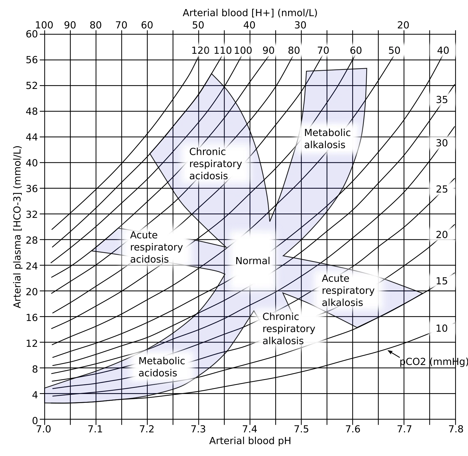
Acid-base nomogram showing all four disorders. Focus on: identify the primary disorder from pH (acidosis < 7.35, alkalosis > 7.45), then determine if it's respiratory (CO2 abnormal) or metabolic (HCO3- abnormal). The body compensates by adjusting the other system. Credit: Wikimedia Commons, Public Domain
The Bicarbonate Buffer System
The body’s most important extracellular buffer is the bicarbonate system. The chemistry behind this buffer (Henderson-Hasselbalch equation) is covered in general chemistry - here we focus on how the body regulates it:
How the Kidneys Regulate pH
The kidneys have three mechanisms for acid-base regulation:
1. Bicarbonate reabsorption (PCT)
The PCT reabsorbs ~80-90% of filtered HCO3-
This is not direct reabsorption - HCO3- cannot cross the apical membrane
Instead: H+ is secreted into the lumen (via Na+/H+ exchangers), combines with filtered HCO3- to form CO2 + H2O (catalyzed by carbonic anhydrase on the brush border)
CO2 diffuses into the PCT cell, is reconverted to HCO3- (intracellular carbonic anhydrase), and HCO3- exits on the basolateral side into the blood
Net effect: each H+ secreted “reclaims” one HCO3-
2. H+ secretion (collecting duct)
Type A intercalated cells actively secrete H+ via H+ ATPase and H+/K+ ATPase
This generates NEW bicarbonate (not just reclaiming filtered HCO3-)
Each H+ secreted generates one new HCO3- that enters the blood
This is the kidney’s mechanism for correcting acidosis
3. Ammonium (NH4+) excretion
The PCT produces NH3 (ammonia) from glutamine
NH3 diffuses into the tubular lumen and combines with H+ to form NH4+ (ammonium)
NH4+ is trapped in the lumen (charged, cannot diffuse back) and excreted
This allows the kidney to excrete large amounts of H+ without dropping urine pH below ~4.5 (NH3 acts as a urinary buffer)
The Four Acid-Base Disorders
Disorder
Primary Problem
pH
Primary Change
Compensation
Metabolic acidosis
Excess H+ or loss of HCO3-
< 7.35
HCO3- low
Lungs hyperventilate (blow off CO2)
Metabolic alkalosis
Loss of H+ or excess HCO3-
> 7.45
HCO3- high
Lungs hypoventilate (retain CO2)
Respiratory acidosis
CO2 retention (hypoventilation)
< 7.35
CO2 high
Kidneys retain HCO3-, excrete H+
Respiratory alkalosis
CO2 loss (hyperventilation)
> 7.45
CO2 low
Kidneys excrete HCO3-, retain H+
How to Read an Arterial Blood Gas (ABG)
The MCAT may give you ABG values and ask you to identify the disorder:
Step 1: Look at pH. Is it acidotic (< 7.35) or alkalotic (> 7.45)?
Step 2: Determine the primary cause.
If CO2 is abnormal and matches the pH direction, it is respiratory
If HCO3- is abnormal and matches the pH direction, it is metabolic
Step 3: Check for compensation.
Is the other value changing in the expected compensatory direction?
Normal values:
pH: 7.35-7.45
pCO2: 35-45 mmHg
HCO3-: 22-26 mEq/L
Example: pH 7.30, pCO2 55 mmHg, HCO3- 28 mEq/L
pH is low (acidosis)
CO2 is high (respiratory cause - hypoventilation)
HCO3- is slightly elevated (renal compensation - kidneys retaining bicarbonate)
Diagnosis: respiratory acidosis with partial renal compensation
Common Causes of Each Disorder
Metabolic acidosis:
Diabetic ketoacidosis (excess ketone production)
Lactic acidosis (anaerobic metabolism)
Renal failure (cannot excrete H+ or regenerate HCO3-)
Severe diarrhea (loss of HCO3- in stool)
Metabolic alkalosis:
Prolonged vomiting (loss of HCl from stomach)
Excessive antacid use
Hyperaldosteronism (excess H+ secretion in collecting duct)
Respiratory acidosis:
COPD, pneumonia, or any cause of hypoventilation
Opioid overdose (suppresses respiratory drive)
Respiratory alkalosis:
Hyperventilation (anxiety, pain, high altitude)
A patient has pH 7.50, pCO2 48 mmHg, HCO3- 36 mEq/L. Identify the primary disorder and the compensation.
Click to reveal answer
Primary disorder: metabolic alkalosis (pH is high, HCO3- is high - the bicarbonate excess is driving the alkalosis). Compensation: respiratory (CO2 is elevated because the lungs are hypoventilating to retain CO2 and lower pH). The compensation is partial because pH has not returned to normal range. A common cause would be prolonged vomiting (loss of gastric HCl).
Why can't the lungs fully compensate for a metabolic acid-base disorder?
Click to reveal answer
Respiratory compensation is self-limiting. In metabolic acidosis, the lungs hyperventilate to blow off CO2. But excessive hyperventilation reduces CO2 so much that the low CO2 itself begins to inhibit respiratory drive (the body will not voluntarily suffocate). Similarly, in metabolic alkalosis, hypoventilation raises CO2, but hypoxia eventually forces breathing to resume. Only the kidneys can fully correct the primary problem by adjusting HCO3- directly.
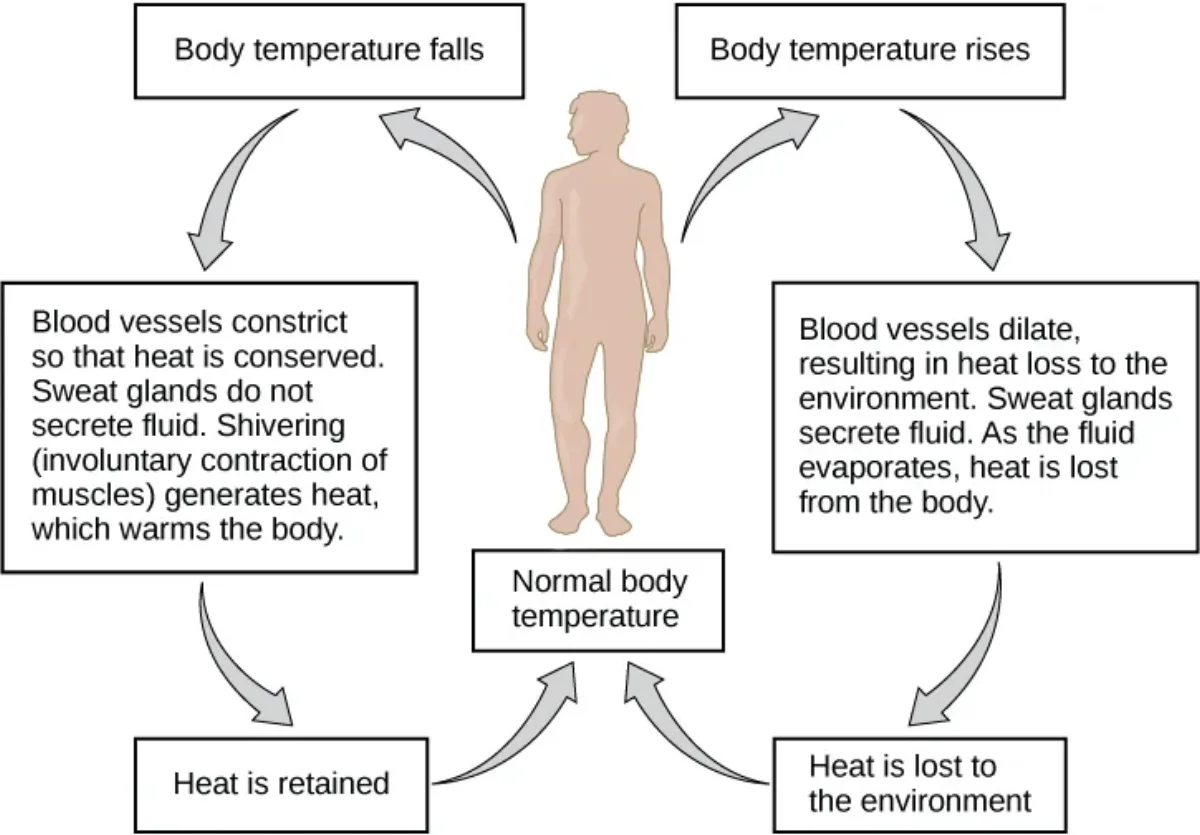
Humans are endotherms - we generate our own body heat and maintain a stable core temperature regardless of the environment. The hypothalamus acts as the body’s thermostat, integrating temperature information from peripheral and central thermoreceptors and coordinating the appropriate heating or cooling response.
Thermoregulation is a classic example of negative feedback, and the MCAT frequently tests your ability to trace the loop from stimulus to response.
The Hypothalamic Thermostat
The hypothalamus receives temperature input from:
Central thermoreceptors in the hypothalamus itself (monitor blood temperature directly)
Peripheral thermoreceptors in the skin (detect environmental temperature changes early)
The hypothalamus compares incoming data to the set point (~37 degrees C) and activates the appropriate effectors.
Cooling Mechanisms (When Body Temp Is Too High)
1. Cutaneous vasodilation - arterioles in the skin dilate, increasing blood flow to the body surface. Heat radiates from the blood through the skin to the environment. This is why you look flushed when hot.
2. Sweating - eccrine sweat glands secrete water and electrolytes onto the skin surface. As sweat evaporates, it removes heat (evaporative cooling). This is the most effective cooling mechanism in humans. In high humidity, sweating is less effective because the air is already saturated with water vapor.
3. Behavioral responses - seeking shade, removing clothing, reducing activity. These are voluntary but highly effective.
Heating Mechanisms (When Body Temp Is Too Low)
1. Cutaneous vasoconstriction - arterioles in the skin constrict, reducing blood flow to the surface. Heat is retained in the core. This is why you look pale when cold.
2. Shivering - involuntary rapid contraction of skeletal muscles generates heat as a byproduct of ATP hydrolysis. Shivering can increase heat production by 5-fold.
3. Non-shivering thermogenesis - brown adipose tissue generates heat by uncoupling oxidative phosphorylation. Uncoupling proteins in the inner mitochondrial membrane allow H+ to flow back across the membrane without passing through ATP synthase - the energy is released as heat instead of making ATP. Infants have significant brown fat; adults retain smaller amounts.
4. Piloerection - contraction of arrector pili muscles at the base of hair follicles. In furred animals, this traps an insulating layer of air. In humans, it produces goosebumps - a vestigial response with minimal insulating value.
5. Thyroid hormone - chronic cold exposure stimulates thyroid hormone production (T3/T4), which increases basal metabolic rate and heat generation over days to weeks.
Heat moves between your body and the environment by four physical mechanisms:
Mechanism
Definition
Example
Radiation
Heat transferred as infrared electromagnetic waves
Feeling warmth from a fire without touching it
Conduction
Heat transferred by direct contact with a surface
Sitting on a cold metal bench
Convection
Heat carried away by moving air or fluid
Wind chill making a cold day feel colder
Evaporation
Heat lost when liquid converts to gas
Sweating; stepping out of a pool and feeling cold
The four mechanisms of heat transfer between the body and the environment. Evaporation is the only mechanism that always removes heat from the body. Credit: OpenStax Biology 2e, CC BY 4.0
Countercurrent Heat Exchange
In the extremities (arms, legs), arteries and veins run parallel and close together. Warm arterial blood heading to the fingers transfers heat to the cooler venous blood returning to the core. This means:
Less heat reaches the extremities (conserving core temperature)
The returning venous blood is pre-warmed before reaching the core
This is the same countercurrent principle as the loop of Henle, but for heat instead of solutes. It is especially important in cold environments and in aquatic mammals (whales, penguins) that have highly developed countercurrent heat exchangers in their flippers.
Fever: A Shifted Set Point
Fever is NOT a failure of thermoregulation. It is the hypothalamus deliberately raising its set point.
Pyrogens stimulate the hypothalamus to produce prostaglandin E2 (PGE2)
PGE2 raises the hypothalamic set point (e.g., from 37 degrees C to 39 degrees C)
The body now “thinks” it is too cold and activates warming mechanisms (shivering, vasoconstriction)
Body temperature rises to the new set point and stabilizes
When the fever breaks:
The infection is controlled, pyrogen levels drop
PGE2 decreases, and the set point returns to 37 degrees C
The body now “thinks” it is too hot and activates cooling mechanisms (sweating, vasodilation)
Temperature falls back to normal
Anti-inflammatory drugs reduce fever by inhibiting cyclooxygenase (COX), which blocks PGE2 synthesis, lowering the set point back toward normal.
Endotherms vs. Ectotherms
Feature
Endotherms (mammals, birds)
Ectotherms (reptiles, fish, amphibians)
Heat source
Internal metabolism
External environment
Body temperature
Stable (homeothermic)
Variable (poikilothermic)
Metabolic rate
High
Low
Caloric requirement
High
Low
Activity in cold
Maintained
Reduced
Examples
Humans, dogs, eagles
Lizards, frogs, snakes
A patient has a fever of 39.5 degrees C. They are shivering. Explain this seemingly paradoxical response.
Click to reveal answer
The hypothalamic set point has been raised to 39.5 degrees C by pyrogens and PGE2. At the patient's current body temperature (still climbing toward 39.5), the hypothalamus registers the body as "too cold" relative to the new set point. It activates warming mechanisms (shivering, vasoconstriction) to drive body temperature UP to the new target. Shivering during fever = the body is still "climbing" to the elevated set point.
On a hot, humid day, a runner becomes hyperthermic despite sweating profusely. Why is sweating less effective in high humidity?
Click to reveal answer
Evaporative cooling requires sweat to evaporate from the skin surface. In high humidity, the air is already saturated with water vapor, reducing the rate of evaporation. Sweat forms on the skin but does not evaporate efficiently, so less heat is removed. The body's most effective cooling mechanism is compromised, leading to heat accumulation and potentially dangerous hyperthermia (heat stroke).
Osmoregulation is the maintenance of blood osmolarity within a narrow range (~285-295 mOsm/L). Because water moves freely across cell membranes by osmosis, changes in extracellular osmolarity directly affect cell volume. If blood becomes too dilute, cells swell. If blood becomes too concentrated, cells shrink. Either extreme disrupts cellular function - especially in the brain, where swelling or shrinking can be rapidly fatal.
The kidneys, working with ADH and the thirst mechanism, are the primary regulators of osmolarity.
Body Fluid Compartments
Total body water makes up approximately 60% of body weight in an average adult. This water is distributed between two main compartments:
Intracellular fluid (ICF) - ~32 of total body water. The fluid inside all cells. The major intracellular cation is K+.
Extracellular fluid (ECF) - ~31 of total body water. Subdivided into:
Plasma (~41 of ECF) - the fluid portion of blood within blood vessels
Interstitial fluid (~43 of ECF) - the fluid between cells, outside blood vessels
The major extracellular cation is Na+. Because Na+ is the primary extracellular solute, sodium levels largely determine ECF osmolarity and volume.
Osmolarity vs. Tonicity
These terms are related but not identical:
Osmolarity - the total concentration of all solutes in a solution (mOsm/L). Includes ALL solutes, whether or not they can cross the membrane.
Tonicity - the effective osmotic pressure of a solution relative to a cell. Only counts non-penetrating solutes (those that cannot cross the membrane). Tonicity determines whether cells will swell, shrink, or stay the same.
Urea can cross cell membranes freely, so it contributes to osmolarity but NOT to tonicity. A solution with high urea concentration may be hyperosmolar but isotonic (cells will not change volume because urea equilibrates across both sides).
Solution Type
Effect on Cell
Non-penetrating Solute Concentration
Hypotonic
Cell swells (water enters)
Lower than inside the cell
Isotonic
No change (water balanced)
Equal to inside the cell
Hypertonic
Cell shrinks (water leaves)
Higher than inside the cell
The Osmoregulation Feedback Loop
When blood osmolarity rises (dehydration, salt intake):
Osmoreceptors in the hypothalamus detect the increase
Two responses are triggered simultaneously:
ADH release from the posterior pituitary - increases water reabsorption in collecting ducts
Thirst - drives water intake (behavioral response)
Blood osmolarity decreases back toward normal
As osmolarity normalizes, ADH secretion and thirst decrease
When blood osmolarity falls (excess water intake):
Osmoreceptors detect the decrease
ADH release is suppressed
Collecting ducts remain impermeable to water
Large volumes of dilute urine are produced
Blood osmolarity increases back toward normal
Sodium and Volume Regulation
Because Na+ is the dominant extracellular solute, sodium balance is tightly linked to ECF volume:
More Na+ in the ECF -> water follows by osmosis -> ECF volume increases -> blood pressure rises
Less Na+ in the ECF -> water leaves by osmosis -> ECF volume decreases -> blood pressure falls
This is why aldosterone (which reabsorbs Na+) increases blood volume, and why ANP (which excretes Na+) decreases blood volume. It is also why excessive salt intake contributes to hypertension.
Edema: When Fluid Leaves the Vessels
Edema is the accumulation of excess fluid in the interstitial space (swelling). It occurs when the balance of Starling forces at the capillary level is disrupted:
Causes of edema:
Increased capillary hydrostatic pressure - CHF, venous obstruction, standing for long periods
Decreased plasma oncotic pressure - liver disease (low albumin production), kidney disease (protein lost in urine), malnutrition (kwashiorkor)
Lymphatic obstruction - lymphedema from surgery, infection (elephantiasis), or tumor
Electrolyte Disorders: Clinical Connections
Disorder
Definition
Key Cause
Key Symptom
Hyponatremia
Na+ < 135 mEq/L
SIADH, water intoxication
Confusion, seizures (brain swelling)
Hypernatremia
Na+ > 145 mEq/L
Dehydration, diabetes insipidus
Thirst, confusion (brain shrinking)
Hypokalemia
K+ < 3.5 mEq/L
Diuretics, vomiting, aldosterone excess
Muscle weakness, cardiac arrhythmia
Hyperkalemia
K+ > 5.0 mEq/L
Renal failure, K+-sparing diuretics, Addison’s
Cardiac arrhythmia (potentially fatal)
A patient drinks 8 liters of water in a short period. Predict the changes in blood osmolarity, ADH levels, and urine characteristics.
Click to reveal answer
Blood osmolarity decreases (dilutional effect from excess water). ADH levels drop (osmoreceptors sense low osmolarity and suppress ADH release). Urine becomes very dilute and high-volume (collecting ducts are impermeable without ADH, so water passes through as dilute urine). Severe cases cause hyponatremia and can lead to cerebral edema (water intoxication).
A solution contains 300 mOsm/L of urea. Is this solution hyperosmotic, isosmotic, or hyposmotic compared to plasma? Will cells placed in this solution swell, shrink, or stay the same?
Click to reveal answer
Isosmotic (300 mOsm/L matches plasma osmolarity). However, the solution is effectively hypotonic because urea freely crosses cell membranes. Urea enters the cell and equilibrates, contributing nothing to the osmotic gradient. The cell behaves as if it is in pure water - cells will swell. This illustrates the difference between osmolarity (total solute) and tonicity (only non-penetrating solutes).
When your body breaks down amino acids and nucleic acids, the nitrogen-containing groups must be removed and excreted. Nitrogen cannot simply be stored - its waste products are toxic. Different organisms have evolved different strategies for handling this nitrogen, and understanding these strategies reveals important principles about water conservation, toxicity, and evolution.
For the MCAT, the key concepts are: what nitrogenous wastes are, how the urea cycle works, and how the kidney excretes them.
The Three Nitrogenous Waste Products
1. Ammonia (NH3)
Produced directly from amino acid deamination
Extremely toxic to the nervous system (even at low concentrations)
Very water-soluble
Must be diluted in large volumes of water for safe excretion
Organisms that excrete ammonia directly are called ammonotelic (most aquatic animals - fish, aquatic invertebrates)
2. Urea
Produced in the liver via the urea cycle (from two ammonia molecules + one CO2)
Much less toxic than ammonia (~100,000x less toxic)
Water-soluble
Requires moderate water for excretion
Organisms that excrete urea are called ureotelic (mammals, adult amphibians, sharks)
3. Uric acid
Produced from purine (adenine, guanine) metabolism
Least toxic of the three
Poorly water-soluble (semi-solid paste)
Requires very little water for excretion
Organisms that excrete uric acid are called uricotelic (birds, reptiles, insects)
The Urea Cycle (Liver)
The urea cycle converts toxic ammonia into urea in the liver. This is the primary detoxification pathway for nitrogen in humans.
Key points:
Occurs partly in the mitochondria and partly in the cytoplasm of hepatocytes
Combines 2 NH3 (actually one free NH4+ and one from aspartate) + 1 CO2 to produce 1 urea molecule
Consumes 3 ATP equivalents per cycle
Urea is released into the blood, filtered at the glomerulus, and partially reabsorbed (about 50% in the PCT, with some recycled in the inner medulla)
Clinical Markers of Kidney Function
The MCAT often tests how kidney function is assessed:
Blood urea nitrogen (BUN) - measures urea in the blood. Elevated BUN suggests reduced kidney function (the kidneys are not clearing urea efficiently) or increased protein catabolism (more urea is being produced).
Serum creatinine - creatinine is a waste product of creatine phosphate metabolism in skeletal muscle. It is freely filtered at the glomerulus and NOT reabsorbed (making it an excellent marker of GFR). Elevated creatinine = reduced GFR = kidney impairment.
BUN/Creatinine ratio - helps distinguish the cause of elevated BUN:
Normal ratio: ~10-20:1
High ratio (>20:1): suggests prerenal cause (dehydration, CHF - urea is reabsorbed more because flow through tubules is slow)
Normal ratio with both elevated: suggests intrinsic renal disease (both accumulate equally)
Comparative Nitrogenous Waste Excretion
Feature
Ammonotelic
Ureotelic
Uricotelic
Primary waste
Ammonia (NH3)
Urea
Uric acid
Toxicity
Very high
Low
Very low
Water requirement
Very high
Moderate
Very low
Energy cost
Low (no conversion)
Moderate (3 ATP/urea cycle)
High
Typical organisms
Fish, aquatic invertebrates
Mammals, adult amphibians
Birds, reptiles, insects
Excretion form
Dissolved in water
Dissolved in urine
Semi-solid paste
What Happens When Waste Excretion Fails
When kidney function declines significantly, waste products accumulate in the blood:
Uremia - elevated urea and other waste products cause fatigue, nausea, confusion, and eventually coma
Metabolic acidosis - kidneys cannot excrete H+ or regenerate HCO3-
Hyperkalemia - kidneys cannot excrete K+
Fluid overload - kidneys cannot excrete excess water
Similarly, if the liver fails, ammonia from amino acid catabolism cannot be converted to urea. The resulting hyperammonemia is toxic to the brain because ammonia crosses the blood-brain barrier and disrupts neurotransmission.
A patient with liver cirrhosis develops confusion and elevated blood ammonia. Explain the pathophysiology using what you know about the urea cycle.
Click to reveal answer
The damaged liver cannot run the urea cycle efficiently. Ammonia from amino acid catabolism accumulates in the blood instead of being converted to urea. Ammonia crosses the blood-brain barrier and is toxic to neurons, causing confusion and altered mental status. This connection between liver function and nitrogen waste processing is a common MCAT passage topic.
Why is creatinine a better marker of GFR than urea?
Click to reveal answer
Creatinine is freely filtered and NOT reabsorbed by the tubules, so its clearance rate directly reflects GFR. Urea, on the other hand, is partially reabsorbed (~50% in the PCT and more in the collecting duct under ADH influence). This means BUN can change due to factors other than GFR - dehydration increases urea reabsorption, and high-protein diets increase urea production. Creatinine production is relatively constant (proportional to muscle mass), making it a more reliable GFR indicator.