
Glomerular Filtration
Filtration is the first step of urine formation. It is a bulk, nonselective process - the glomerulus does not “choose” what to filter. Instead, it acts like a sieve: anything small enough passes through, and anything too large stays behind. The selectivity comes later, in the tubules, where the kidney carefully reclaims what the body needs.
Understanding what drives filtration, what determines how much gets filtered, and what the filtration barrier looks like is critical for MCAT success.
The Filtration Membrane
The filtration barrier between blood and Bowman’s capsule has three layers:
1. Fenestrated endothelium of glomerular capillaries - has pores (fenestrations) that allow most plasma components to pass but block blood cells.
2. Basement membrane - a gel-like layer of glycoproteins with a net negative charge. This charge repels most plasma proteins (which are also negatively charged at physiological pH, especially albumin).
3. Podocytes - specialized cells that wrap around the capillaries with foot processes (pedicels). The gaps between the foot processes are called filtration slits. These provide the final size barrier.
What Gets Filtered vs. What Stays Behind
| Freely Filtered (passes into Bowman’s space) | NOT Filtered (stays in blood) |
|---|---|
| Water | Red blood cells |
| Glucose | White blood cells |
| Amino acids | Platelets |
| Urea, creatinine, uric acid | Plasma proteins (albumin, globulins) |
| Electrolytes (Na+, K+, Cl-, Ca2+, HCO3-) | Protein-bound substances |
| Small peptide hormones | Large molecules (>70 kDa) |
Clinical connection: If protein or blood appears in the urine, something is wrong with the filtration membrane. Proteinuria (protein in urine) indicates damage to the basement membrane or podocytes. Hematuria (blood in urine) indicates damage to the capillary endothelium.
Starling Forces: What Drives Filtration
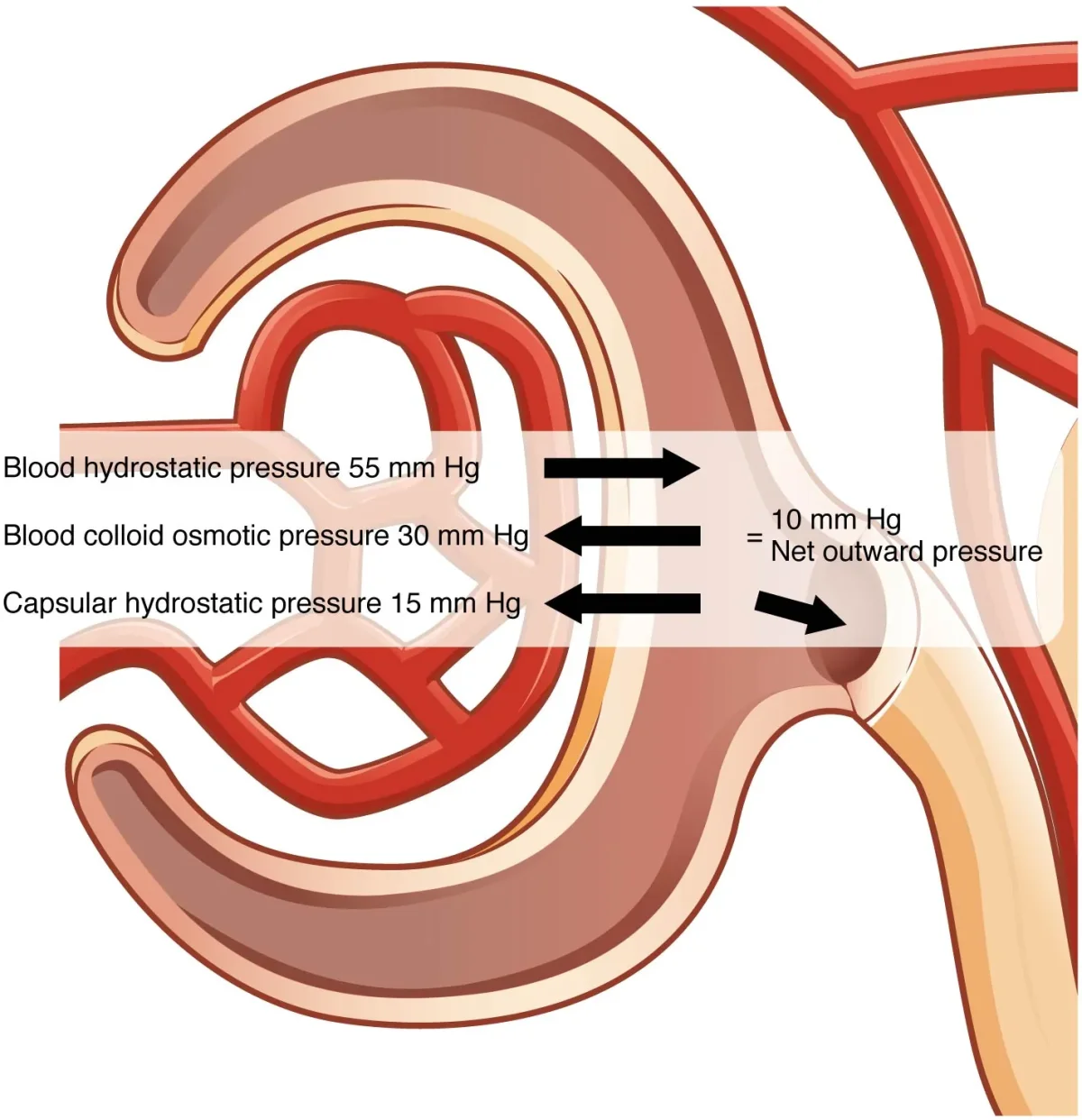
Filtration is driven by pressure. Specifically, the net filtration pressure (NFP) is determined by the balance of four forces - collectively called Starling forces:
Forces FAVORING filtration (pushing fluid out of capillaries):
- Glomerular hydrostatic pressure - the blood pressure inside the glomerular capillaries. This is the main driving force and is much higher than in most capillary beds.
Forces OPPOSING filtration (pushing fluid back or keeping it in):
- Bowman’s capsule hydrostatic pressure - the pressure of fluid already in Bowman’s space pushing back.
- Glomerular oncotic pressure - the osmotic pull of plasma proteins in the blood (proteins cannot cross, so they pull water back).
The net filtration pressure is the balance of these forces. Under normal conditions, hydrostatic pressure greatly exceeds the opposing forces, so the net flow is always from blood into Bowman’s space.
Glomerular Filtration Rate (GFR)
GFR is the volume of filtrate produced per minute by all glomeruli in both kidneys.
Normal GFR: ~125 mL/min = ~180 L/day
That is an enormous volume - far more than your total blood volume. You only excrete ~1-2 L as urine because the tubules reabsorb 99% of the filtrate.
Factors that increase GFR:
- Increased glomerular hydrostatic pressure (higher blood pressure)
- Afferent arteriole dilation (more blood enters)
- Efferent arteriole constriction (blood backs up, raising pressure)
- Decreased plasma protein concentration (less oncotic opposition)
Factors that decrease GFR:
- Decreased blood pressure (shock, hemorrhage)
- Afferent arteriole constriction (less blood enters)
- Efferent arteriole dilation (less backup, lower pressure)
- Increased plasma protein (dehydration concentrates proteins)
- Urinary obstruction (increases Bowman’s capsule pressure)
Filtration Fraction
Measuring GFR: Clearance
You cannot watch filtrate form, so GFR is estimated by measuring how fast the kidneys clear a substance from plasma. A substance that is freely filtered, not reabsorbed, and not secreted has a clearance that equals GFR.
Afferent vs Efferent: Why Direction Matters
Because the glomerulus sits between two arterioles, the kidney can control GFR with precision. Constriction at either end has opposite effects:
- Afferent constriction -> less blood arrives -> glomerular pressure falls -> GFR drops.
- Efferent constriction -> blood backs up in the glomerulus -> glomerular pressure rises -> GFR rises (up to a point).
Angiotensin II preferentially constricts the efferent arteriole, which protects GFR when renal perfusion is low. This is why ACE inhibitors and ARBs can drop GFR in patients with already-compromised renal blood flow: remove angiotensin II, and the efferent arteriole relaxes, dropping glomerular pressure.
Autoregulation of GFR
The kidneys maintain a stable GFR even when systemic blood pressure fluctuates (within the range of ~80-180 mmHg MAP). Two mechanisms accomplish this:
1. Myogenic mechanism - when blood pressure rises, the increased stretch on afferent arteriole walls triggers them to constrict (smooth muscle response). This prevents the pressure increase from reaching the glomerulus. When pressure drops, the arteriole relaxes.
2. Tubuloglomerular feedback (TGF) - the macula densa cells in the DCT (part of the juxtaglomerular apparatus) monitor NaCl concentration in the filtrate. If GFR increases, more NaCl reaches the macula densa. The macula densa signals the afferent arteriole to constrict, reducing blood flow and lowering GFR back to normal.
The Juxtaglomerular Apparatus (JGA)
The JGA is where the DCT brushes against its own afferent arteriole. Two cell types do the work:
- JG cells in the afferent arteriole wall secrete renin when blood pressure drops or sympathetic nerves fire.
- Macula densa cells in the DCT wall sense tubular NaCl and signal the JG cells.
That physical adjacency is the point - a local sensor (macula densa) sitting next to a local effector (JG cells), no hormones required for the feedback.