
Kidney Anatomy
Before you can understand how the kidney works, you need a clear mental map of what it looks like. The kidney’s structure is not random - every anatomical feature exists because of the function it serves. The cortex is where filtration begins. The medulla is where urine gets concentrated. The pelvis is where urine collects before leaving. Once you connect structure to function, the physiology becomes much easier.
Gross Anatomy: The Kidney
Each person has two kidneys, bean-shaped organs roughly the size of a fist, located retroperitoneally (behind the peritoneum) against the posterior abdominal wall. The right kidney sits slightly lower than the left because the liver pushes it down.
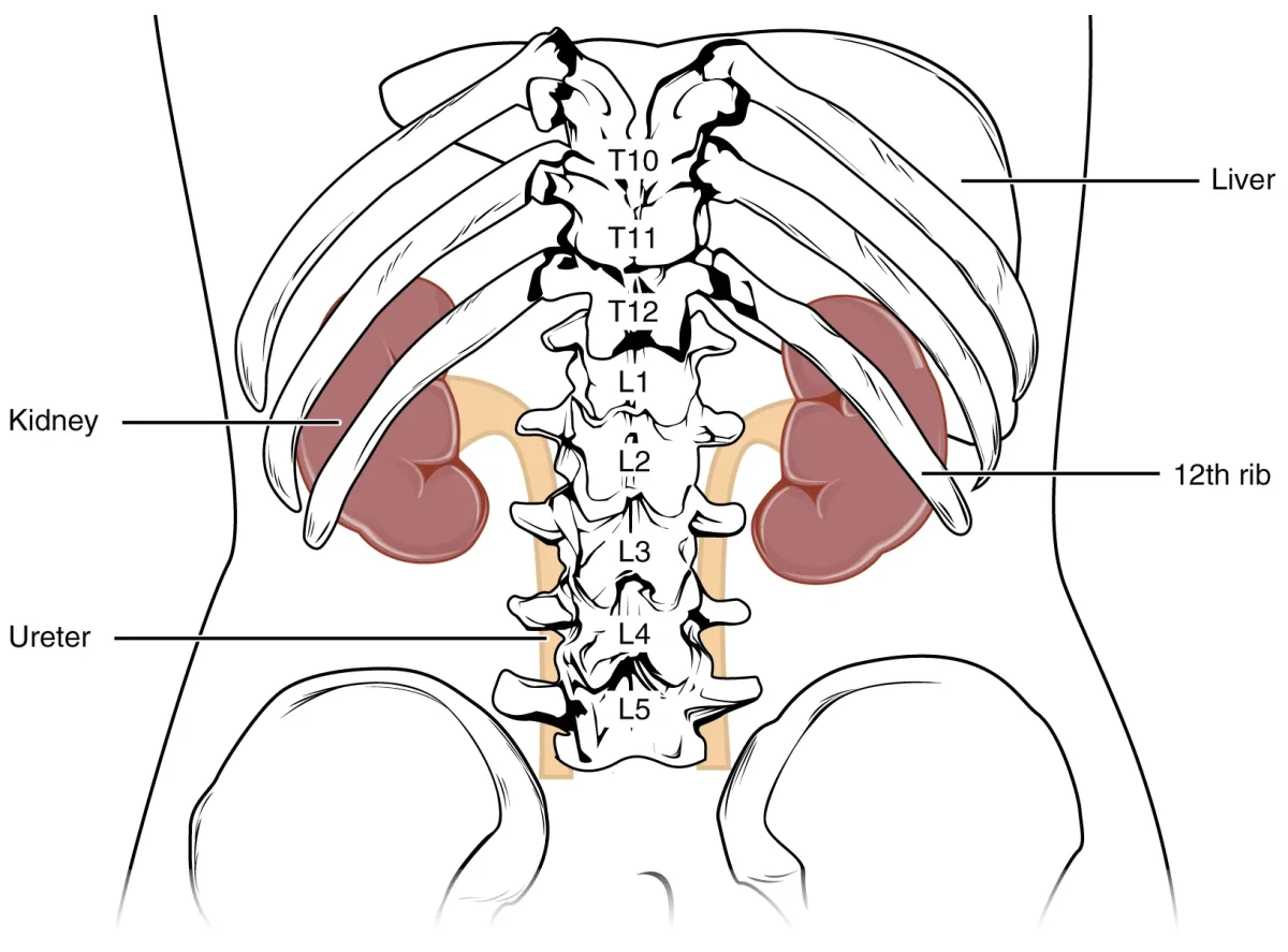
Three regions of the kidney:
Renal cortex - the outer layer. Contains the glomeruli, Bowman’s capsules, proximal convoluted tubules (PCT), and distal convoluted tubules (DCT). This is where blood filtration begins and where most reabsorption occurs.
Renal medulla - the inner region. Contains the loops of Henle and collecting ducts, organized into cone-shaped structures called renal pyramids. The medulla creates the osmotic gradient that allows urine concentration. The tips of the pyramids (renal papillae) point inward toward the renal pelvis.
Renal pelvis - the funnel-shaped collecting area at the center. Urine drains from the collecting ducts through the papillae into minor calyces, which merge into major calyces, which empty into the renal pelvis. From there, urine flows into the ureter.
The Urinary Tract: From Kidney to Exit
Once urine leaves the renal pelvis, it follows a simple path:
Renal pelvis -> Ureter -> Urinary bladder -> Urethra -> outside the body
- Ureters - muscular tubes (~25-30 cm long) that use peristalsis to move urine to the bladder. One from each kidney.
- Urinary bladder - hollow muscular organ lined with transitional epithelium (stretches as it fills). The detrusor muscle (smooth muscle) contracts during urination.
- Urethra - final passage to the exterior. In females, it is short (~4 cm) and carries only urine. In males, it is longer (~20 cm) and carries both urine and semen (at different times).
Two sphincters control urine release:
- Internal urethral sphincter - smooth muscle, involuntary (autonomic control)
- External urethral sphincter - skeletal muscle, voluntary (somatic control)
The Nephron: Functional Unit of the Kidney
Each kidney contains over 1 million nephrons. The nephron is where all the action happens - filtration, reabsorption, secretion, and excretion. Understanding the nephron’s anatomy is essential for every renal question on the MCAT.
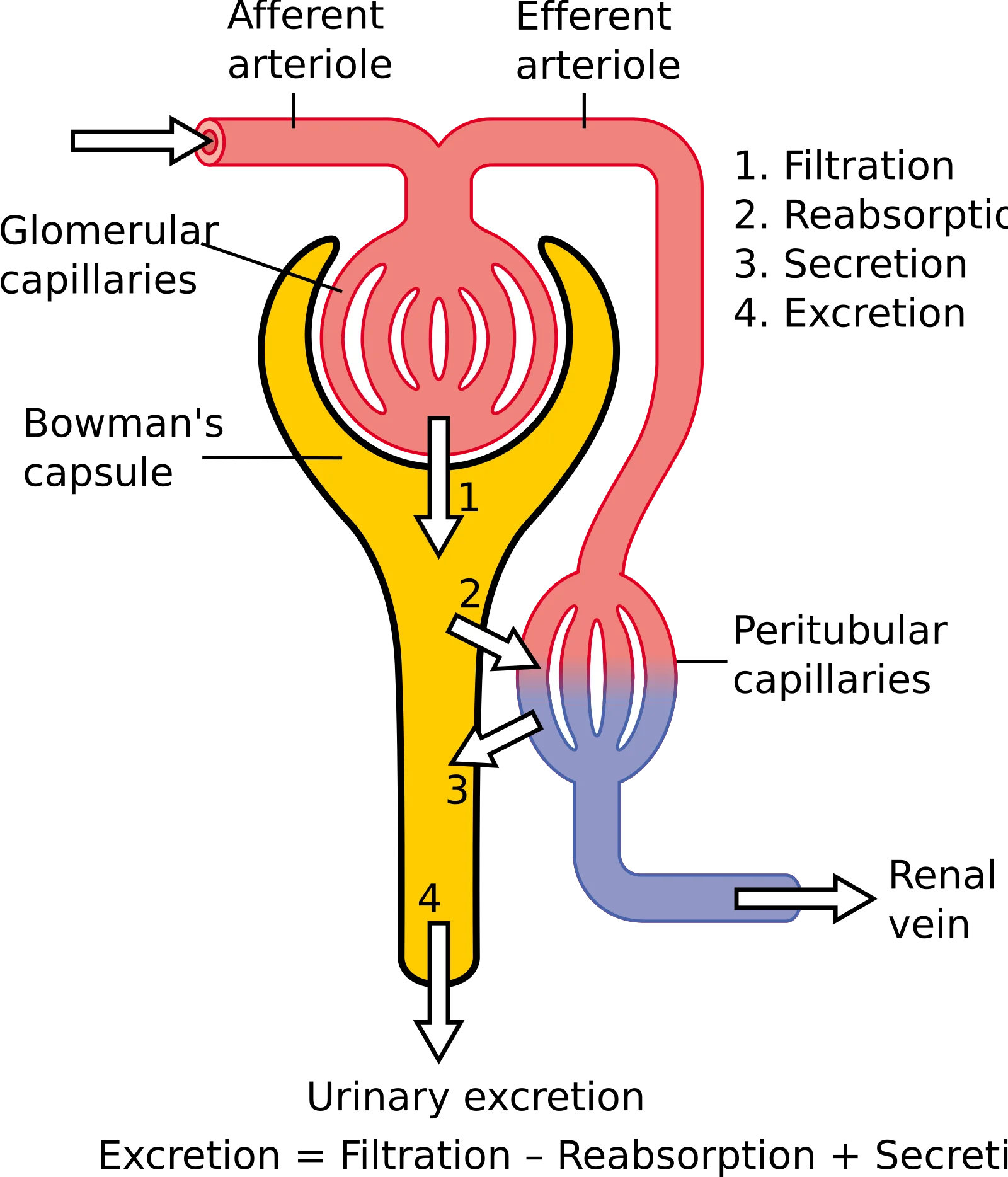
Walk through the nephron, segment by segment:
1. Renal corpuscle (in the cortex)
- Glomerulus - a ball of fenestrated capillaries where blood is filtered. High pressure forces fluid and small solutes out of the blood.
- Bowman’s capsule - a cup that surrounds the glomerulus and collects the filtrate. Lined with podocytes whose foot processes form filtration slits.
2. Proximal convoluted tubule (PCT) (in the cortex)
- The workhorse of reabsorption. Reabsorbs ~65% of filtered Na+, water, all glucose, all amino acids, and most bicarbonate.
- Cells have a brush border (microvilli) to maximize surface area.
3. Loop of Henle (dips into the medulla)
- Descending limb - permeable to water, impermeable to solutes. Water leaves by osmosis as the filtrate descends into the increasingly salty medulla.
- Ascending limb - impermeable to water, actively pumps out NaCl (especially the thick ascending limb). This is what creates the medullary osmotic gradient.
4. Distal convoluted tubule (DCT) (in the cortex)
- Fine-tuning station. Responds to aldosterone (reabsorbs Na+, secretes K+) and PTH (reabsorbs Ca2+). Site of additional H+ secretion for acid-base balance.
5. Collecting duct (runs from cortex through medulla)
- Receives filtrate from multiple nephrons. Permeability to water is controlled by ADH - more ADH means more aquaporin channels, more water reabsorbed, more concentrated urine.
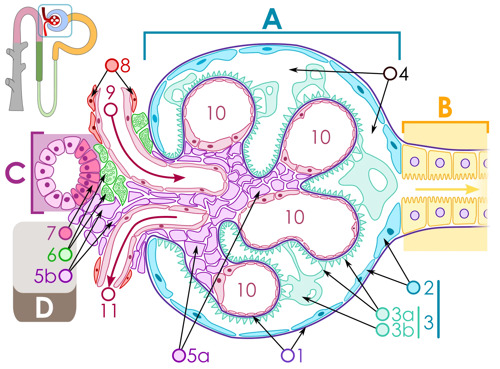
Nephron Vasculature
The kidney’s blood supply is unusual and highly testable. Pay close attention to the arrangement:
Renal artery -> segmental arteries -> interlobar arteries -> arcuate arteries -> cortical radiate (interlobular) arteries -> afferent arteriole -> glomerulus -> efferent arteriole -> peritubular capillaries (or vasa recta for juxtamedullary nephrons) -> venous system
Key features:
- Afferent arteriole - brings blood TO the glomerulus. “Afferent” = arriving.
- Efferent arteriole - carries blood AWAY from the glomerulus. “Efferent” = exiting.
- The glomerulus is a capillary bed sandwiched between TWO arterioles (not an arteriole and a venule). This is unique in the body and allows precise pressure control.
- Peritubular capillaries - surround the PCT and DCT in cortical nephrons. Low-pressure, high-surface-area vessels that reabsorb solutes and water from the tubular cells back into the blood.
- Vasa recta - long, straight capillaries that run alongside the loops of Henle in juxtamedullary nephrons. They maintain the medullary osmotic gradient by acting as countercurrent exchangers.
Two Types of Nephrons
| Feature | Cortical Nephrons (~85%) | Juxtamedullary Nephrons (~15%) |
|---|---|---|
| Location of glomerulus | Outer cortex | Near cortex-medulla border |
| Loop of Henle | Short, barely enters medulla | Long, extends deep into medulla |
| Capillary network | Peritubular capillaries | Vasa recta |
| Primary role | Routine filtration and reabsorption | Urine concentration (creates medullary gradient) |