
Cardiac Cycle
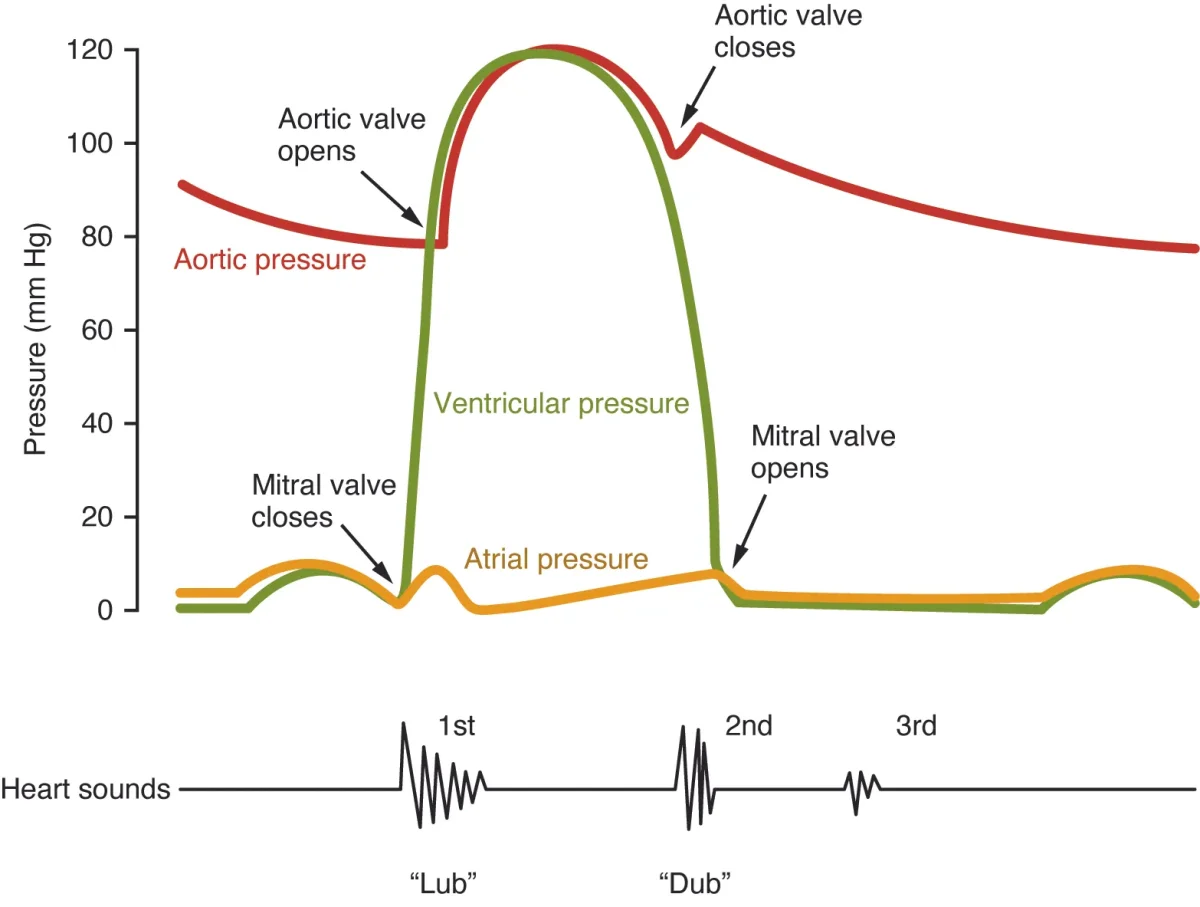
Every heartbeat is a precisely timed sequence of contraction and relaxation. The entire cycle - from the beginning of one heartbeat to the beginning of the next - takes about 0.8 seconds at a resting heart rate of 75 beats per minute. Understanding the cardiac cycle means understanding what is opening, what is closing, and what is moving at every moment.
The Two Phases
The cardiac cycle has two main phases:
Systole = contraction. The ventricles squeeze, ejecting blood into the arteries. AV valves are closed, semilunar valves are open.
Diastole = relaxation. The ventricles relax and fill with blood from the atria. Semilunar valves are closed, AV valves are open.
The Five Sub-Phases
Breaking it down further, the cardiac cycle has five distinct sub-phases:
1. Atrial systole (late diastole) - The atria contract, pushing the last 20-30% of blood into the ventricles. This “atrial kick” tops off ventricular filling. The AV valves are open.
2. Isovolumetric contraction - The ventricles begin to contract. Pressure rises rapidly, slamming the AV valves shut (this produces the S1 “lub” sound). But the semilunar valves have not opened yet because ventricular pressure has not exceeded arterial pressure. All valves are closed. Volume does not change.
3. Ventricular ejection - Ventricular pressure exceeds arterial pressure, forcing the semilunar valves open. Blood is ejected into the pulmonary artery and aorta. Volume drops rapidly.
4. Isovolumetric relaxation - The ventricles relax. Pressure falls below arterial pressure, causing the semilunar valves to snap shut (S2 “dub” sound). But the AV valves have not opened yet because ventricular pressure still exceeds atrial pressure. All valves are closed again. Volume does not change.
5. Ventricular filling (passive) - Ventricular pressure drops below atrial pressure, and the AV valves open. Blood flows passively from the atria into the ventricles, driven by the pressure gradient. This accounts for about 70-80% of filling.
Heart Sounds
You can hear the cardiac cycle with a stethoscope. The two main heart sounds are caused by valve closures, not by muscle contraction:
| Sound | Cause | When | Character |
|---|---|---|---|
| S1 (“lub”) | AV valves close | Start of systole | Lower pitch, longer duration |
| S2 (“dub”) | Semilunar valves close | End of systole | Higher pitch, shorter, “snappier” |
A heart murmur is an abnormal whooshing sound caused by turbulent blood flow, usually from a leaky valve (regurgitation) or a narrowed valve (stenosis). Murmurs are characterized by when they occur: a systolic murmur happens between S1 and S2, a diastolic murmur happens between S2 and the next S1.
Pressure-Volume (PV) Loops
The cardiac cycle can also be represented as a pressure-volume loop for the left ventricle. This graph plots ventricular pressure (y-axis) against ventricular volume (x-axis) and traces a rectangular loop:
- Bottom right → top right (vertical line going up): Isovolumetric contraction. Volume constant, pressure rises.
- Top right → top left (curve going left): Ejection. Volume decreases as blood is expelled.
- Top left → bottom left (vertical line going down): Isovolumetric relaxation. Volume constant, pressure falls.
- Bottom left → bottom right (curve going right): Filling. Volume increases as blood enters.
The width of the loop represents stroke volume (the difference between end-diastolic volume and end-systolic volume). The area inside the loop represents the work done by the ventricle during one beat.