
Conduction System
Most muscles in your body wait for a nerve signal before they contract. Cardiac muscle does not. The heart generates its own electrical impulses and conducts them through a built-in wiring system. Cut every nerve to the heart, and it will keep beating. This property is called autorhythmicity (or myogenic activity), and it is one of the most distinctive features of cardiac tissue.
The Conduction Pathway
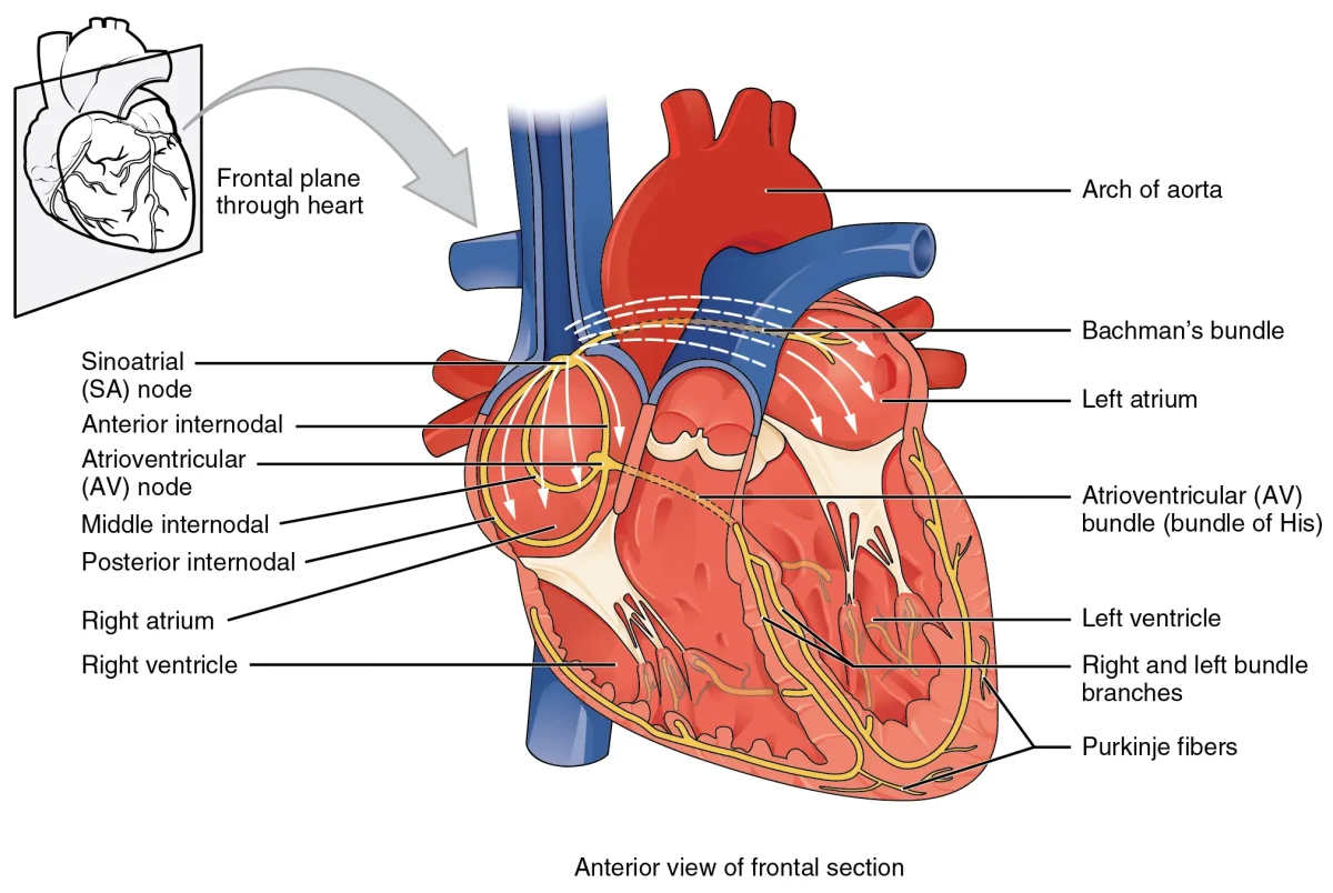
The heart’s electrical system follows a precise pathway that ensures the atria contract before the ventricles and that the ventricles contract from the bottom up (so blood is squeezed upward toward the arteries).
Step 1: SA Node (Sinoatrial Node) - Located in the wall of the right atrium near the superior vena cava. This is the heart’s primary pacemaker. It spontaneously depolarizes at a rate of about 60-100 beats per minute. When it fires, the electrical impulse spreads across both atria via gap junctions, causing atrial contraction.
Step 2: AV Node (Atrioventricular Node) - Located at the junction between the atria and ventricles, near the interatrial septum. The AV node introduces a critical 0.1-second delay. This pause ensures the atria finish contracting and emptying blood into the ventricles before the ventricles begin their own contraction.
Step 3: Bundle of His - The impulse travels from the AV node into the interventricular septum via the Bundle of His, which splits into the left and right bundle branches running down either side of the septum.
Step 4: Purkinje Fibers - The bundle branches fan out into the Purkinje fibers, which spread the impulse rapidly across the ventricular walls from apex to base. This ensures the ventricles contract from the bottom upward, efficiently squeezing blood toward the semilunar valves.
Intrinsic Rates
Each part of the conduction system can generate its own rhythm, but they fire at different rates. The fastest pacemaker wins:
| Structure | Intrinsic Rate | Role |
|---|---|---|
| SA node | 60-100 bpm | Primary pacemaker (normally controls heart rate) |
| AV node | 40-60 bpm | Backup pacemaker if SA node fails |
| Purkinje fibers | 20-40 bpm | Last-resort pacemaker |
If the SA node fails, the AV node takes over at a slower rate. If both fail, the Purkinje fibers can keep the ventricles beating - slowly and barely enough to sustain life. This is why a complete heart block (where the AV node stops conducting) produces a dangerously slow heart rate.
Intercalated Discs and Gap Junctions
Cardiac muscle cells are physically connected by intercalated discs - specialized junctions that contain:
- Gap junctions - allow ions to flow directly between adjacent cells, spreading the electrical impulse rapidly. This makes cardiac muscle behave as a functional syncytium (one cell fires, they all fire).
- Desmosomes - anchor cells together mechanically so they do not tear apart during contraction.
This is why the heart contracts as a unified wave rather than as individual cells twitching randomly. The atria form one functional syncytium; the ventricles form another. The AV node is the only electrical connection between them.
Autonomic Modulation
Although the heart generates its own rhythm, the autonomic nervous system modulates the rate:
| Division | Nerve | Effect on Heart Rate | Mechanism |
|---|---|---|---|
| Parasympathetic | Vagus nerve (CN X) | Decreases rate | Releases ACh → slows SA node depolarization |
| Sympathetic | Cardiac accelerator nerves | Increases rate and force | Releases norepinephrine → speeds SA node, increases contractility |
At rest, the vagus nerve is dominant. The SA node’s intrinsic rate is about 100 bpm, but tonic vagal input slows it to the normal resting rate of ~70 bpm. This is why cutting the vagus nerve (vagotomy) causes heart rate to jump to ~100 bpm.