
Smooth and Cardiac Muscle
Skeletal muscle gets most of the attention, but two other muscle types are equally important - and they work by different rules. Smooth muscle lines your blood vessels, digestive tract, and airways. Cardiac muscle pumps blood through your heart every second of your life. Both are involuntary, meaning you do not consciously control them.
The Three Muscle Types at a Glance
| Feature | Skeletal | Smooth | Cardiac |
|---|---|---|---|
| Location | Attached to bones | Walls of hollow organs, blood vessels, airways | Heart (myocardium) |
| Control | Voluntary (somatic nervous system) | Involuntary (autonomic NS, hormones) | Involuntary (autorhythmic, modulated by autonomic NS) |
| Striations | Yes | No | Yes |
| Nuclei | Multinucleated (peripheral) | Single nucleus (central) | 1-2 nuclei (central) |
| Sarcomeres | Yes (organized) | No (dense bodies instead of Z lines) | Yes (organized) |
| T-tubules | Yes | No (caveolae instead) | Yes (but wider, fewer) |
| SR development | Extensive | Minimal | Moderate |
| Ca²⁺ source | SR (internal) | SR + extracellular (both) | SR + extracellular (CICR) |
| Troponin/tropomyosin | Yes | No (uses calmodulin-MLCK) | Yes |
| Contraction speed | Fast | Slowest | Intermediate |
| Fatigue | Fatigues | Resistant to fatigue | Resistant to fatigue |
| Regeneration | Limited (satellite cells) | Can divide | Cannot divide (permanent G0) |
| Gap junctions | No | Yes (single-unit) | Yes (intercalated discs) |
Smooth Muscle
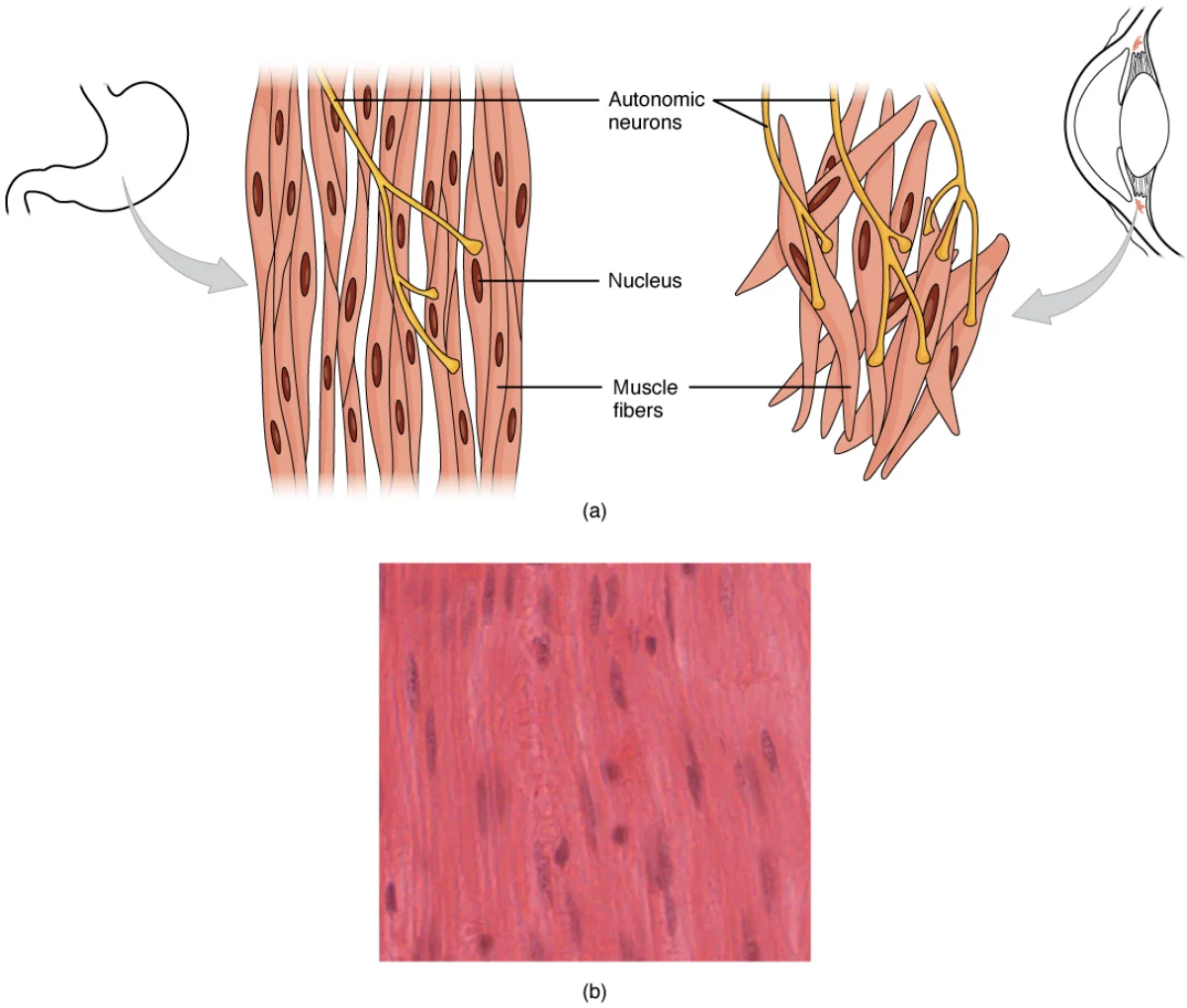
Smooth muscle gets its name from its lack of visible striations under the microscope. It does NOT have organized sarcomeres. Instead, contractile filaments are arranged in a criss-cross pattern anchored to structures called dense bodies (functionally similar to Z lines but scattered throughout the cell).
Smooth Muscle Contraction Mechanism
Smooth muscle uses a fundamentally different calcium-signaling pathway than skeletal muscle:
- Ca²⁺ enters the cell from both the SR and the extracellular space
- Ca²⁺ binds to calmodulin (NOT troponin - smooth muscle lacks troponin)
- The Ca²⁺-calmodulin complex activates myosin light chain kinase (MLCK)
- MLCK phosphorylates the myosin light chain, which activates the myosin ATPase
- Phosphorylated myosin can now bind actin and perform cross-bridge cycling
- Relaxation occurs when the myosin light chain is dephosphorylated
Two Types of Smooth Muscle
| Feature | Single-Unit (Visceral) | Multi-Unit |
|---|---|---|
| Location | GI tract, uterus, ureter, bladder | Large airways, large arteries, iris, ciliary body, arrector pili |
| Gap junctions | Yes - cells connected electrically | No - each cell independently innervated |
| Contraction | Synchronized (cells contract as a sheet) | Independent (fine control) |
| Pacemaker activity | Yes (spontaneous depolarization) | No (needs nerve/hormone stimulation) |
| Stretch response | Contracts when stretched (myogenic response) | Minimal stretch response |
Cardiac Muscle
Cardiac muscle shares features with both skeletal and smooth muscle. Like skeletal muscle, it has sarcomeres and striations. Like smooth muscle, it is involuntary and fatigue-resistant.
Unique Features of Cardiac Muscle
Intercalated discs are the defining structural feature of cardiac muscle. These are specialized junctions at the end-to-end connections between adjacent cardiac muscle cells. Each intercalated disc contains:
- Gap junctions - allow ions to flow directly between cells, enabling the entire heart to contract as a synchronized unit (functional syncytium). When one cell depolarizes, the signal spreads to all connected cells.
- Desmosomes - anchor cells together mechanically, preventing them from pulling apart during contraction
Cardiac E-C Coupling: Calcium-Induced Calcium Release (CICR)
Cardiac muscle uses a modified version of excitation-contraction coupling called calcium-induced calcium release (CICR):
- Action potential propagates along the sarcolemma and into T-tubules
- Calcium channels on T-tubules open and allow extracellular Ca²⁺ to enter the cell (unlike skeletal muscle, where the T-tubule acts as a mechanical sensor only)
- This small influx of Ca²⁺ triggers calcium release channels on the SR to open
- A much larger release of Ca²⁺ floods out of the SR (the “calcium-induced” part)
- Ca²⁺ binds troponin C → cross-bridge cycling → contraction
The key difference from skeletal muscle: in cardiac muscle, extracellular Ca²⁺ is required. Block the calcium channels on the T-tubule, and the heart weakens its contraction.
Cardiac Muscle Cannot Tetanize
Skeletal muscle can sustain maximal contraction (tetanus) through rapid, repeated stimulation. Cardiac muscle CANNOT tetanize because it has a long refractory period that nearly spans the entire contraction. By the time the muscle is ready to respond to another stimulus, it has already relaxed. This is a critical safety mechanism - if the heart could tetanize, it would stop pumping blood and you would die.