The heart is a muscular organ roughly the size of your fist, located slightly left of center in the thoracic cavity between the lungs. Its sole job is deceptively simple: pump blood. But the engineering behind that job is anything but simple. The heart must simultaneously send deoxygenated blood to the lungs and oxygenated blood to the entire body, and it must do this without ever letting the two streams mix.
Four Chambers, Two Pumps
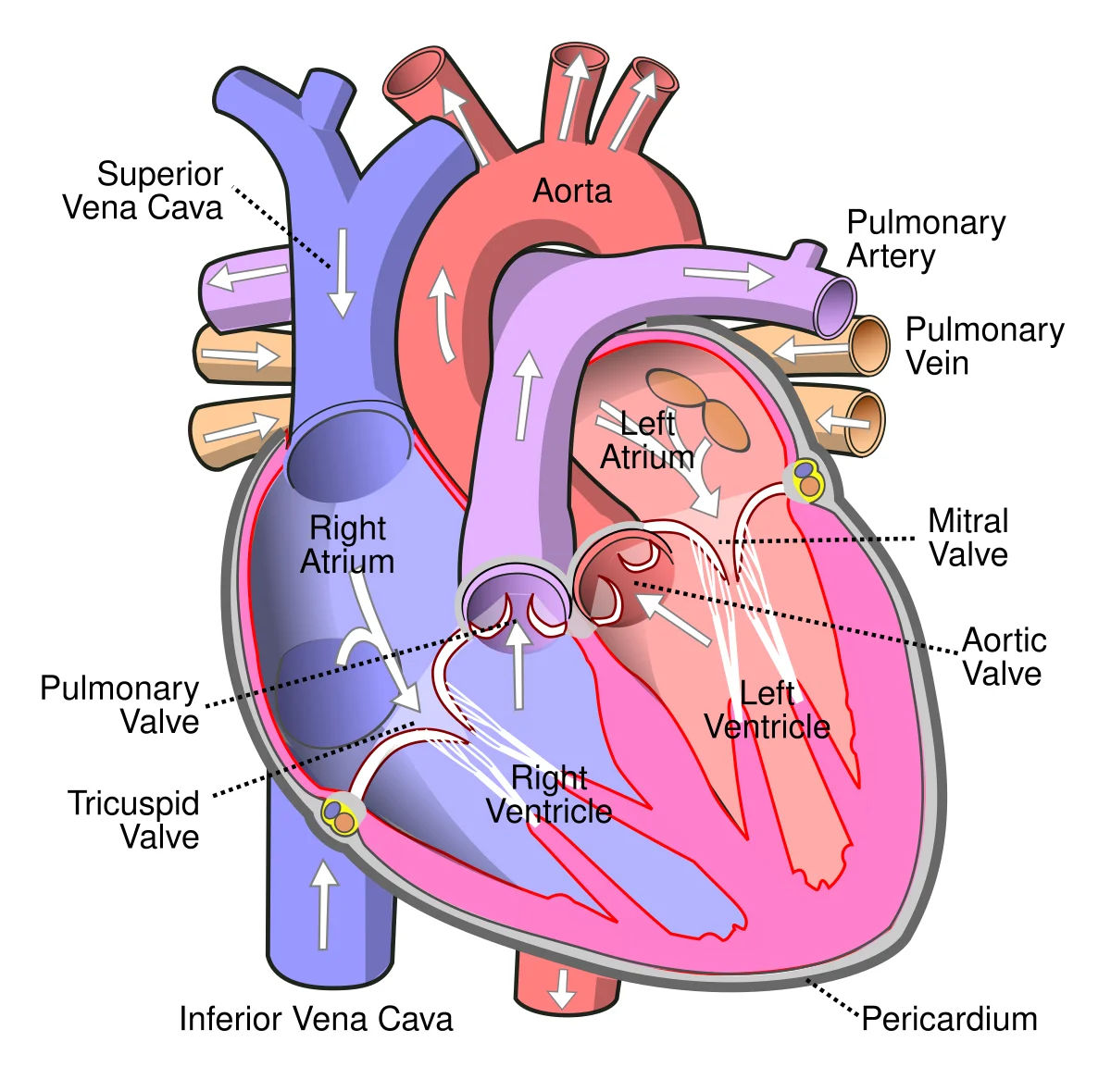
The heart has four chambers arranged as two side-by-side pumps.
The right atrium receives deoxygenated blood from the body via two large veins: the superior vena cava (draining the upper body) and the inferior vena cava (draining the lower body). The coronary sinus also empties deoxygenated blood from the heart muscle itself into the right atrium.
The right ventricle receives blood from the right atrium and pumps it to the lungs via the pulmonary trunk, which splits into the left and right pulmonary arteries. This is the only place in the body where arteries carry deoxygenated blood.
The left atrium receives oxygenated blood returning from the lungs via four pulmonary veins. These are the only veins in the body that carry oxygenated blood.
The left ventricle is the powerhouse. It receives blood from the left atrium and pumps it into the aorta, which distributes blood to every organ in the body. Because it must generate enough pressure to reach your toes, the left ventricular wall is approximately three times thicker than the right.
The four chambers, four valves, and great vessels of the heart. Blue = deoxygenated blood (right side, heading to lungs), red/pink = oxygenated blood (left side, heading to body). Arrows show the direction of flow. Credit: Wikimedia Commons (Wapcaplet), CC BY-SA 3.0
Interactive 3D Heart. Drag to rotate, scroll to zoom. See the internal chambers, valves, and great vessels from any angle.Credit: Haiqa Arif via Sketchfab, CC BY
The Four Valves
Valves ensure blood flows in only one direction. They open passively when pressure pushes blood forward and snap shut when blood tries to flow backward.
There are two types of valves:
Atrioventricular (AV) valves sit between atria and ventricles:
Tricuspid valve - right side, three leaflets
Bicuspid (mitral) valve - left side, two leaflets
Semilunar valves sit at the exits of the ventricles:
Pulmonary valve - exit of right ventricle, opens into pulmonary trunk
Aortic valve - exit of left ventricle, opens into aorta
Heart Wall and Support Structures
The heart wall is composed predominantly of cardiac muscle (myocardium), which generates the force for contraction. The myocardium is much thicker in the left ventricle than the right, reflecting the greater workload of pumping blood through the systemic circulation.
The entire heart sits inside the pericardial sac (pericardium), a double-walled membrane filled with serous fluid that reduces friction as the heart beats.
The AV valves are anchored by chordae tendineae - tough cords connected to papillary muscles in the ventricular walls. These prevent the valve leaflets from being blown back into the atria during ventricular contraction.
The Three Circulations
Pulmonary circulation: Right ventricle → pulmonary arteries → lungs (gas exchange) → pulmonary veins → left atrium. This is a low-pressure circuit (~825 mmHg).
Systemic circulation: Left ventricle → aorta → arteries → capillaries (nutrient/gas exchange) → veins → vena cavae → right atrium. This is a high-pressure circuit (~80120 mmHg).
Coronary circulation: The heart feeds itself first. The left and right coronary arteries branch off the aorta immediately above the aortic valve. After delivering oxygen to the myocardium, coronary veins drain into the coronary sinus, which empties into the right atrium.
Portal Systems
In most circulations, blood passes through one capillary bed before returning to the heart. In a portal system, blood passes through two capillary beds in series before returning. There are three portal systems to know:
Allows reabsorption of filtered substances back into the blood
Fetal Circulation
Before birth, the lungs are collapsed and non-functional. The fetus receives oxygen from the placenta, not from breathing. Three temporary shunts redirect blood away from the lungs:
Shunt
Connects
Purpose
Closes after birth
Foramen ovale
Right atrium → left atrium
Bypasses right ventricle/lungs
Becomes fossa ovalis
Ductus arteriosus
Pulmonary artery → aorta
Diverts blood past the lungs
Becomes ligamentum arteriosum
Ductus venosus
Umbilical vein → inferior vena cava
Bypasses the liver
Becomes ligamentum venosum
All three shunts close shortly after birth when the newborn takes its first breaths and pulmonary resistance drops dramatically.
Why is the left ventricular wall thicker than the right ventricular wall?
Click to reveal answer
The left ventricle must generate much higher pressure to pump blood through systemic circulation (to every organ in the body), while the right ventricle only needs to push blood the short distance to the lungs (pulmonary circulation). Higher workload requires thicker muscle.
A fetus has a patent foramen ovale. What happens to blood flow as a result of this opening?
Click to reveal answer
Blood flows from the right atrium directly to the left atrium, bypassing the lungs. This is normal in fetal circulation because the fetus receives oxygen from the placenta, not from breathing. The foramen ovale typically closes after birth when left atrial pressure exceeds right atrial pressure due to increased pulmonary blood flow.
Every heartbeat is a precisely timed sequence of contraction and relaxation. The entire cycle - from the beginning of one heartbeat to the beginning of the next - takes about 0.8 seconds at a resting heart rate of 75 beats per minute. Understanding the cardiac cycle means understanding what is opening, what is closing, and what is moving at every moment.
The Two Phases
The cardiac cycle has two main phases:
Systole = contraction. The ventricles squeeze, ejecting blood into the arteries. AV valves are closed, semilunar valves are open.
Diastole = relaxation. The ventricles relax and fill with blood from the atria. Semilunar valves are closed, AV valves are open.
The Five Sub-Phases
Breaking it down further, the cardiac cycle has five distinct sub-phases:
1. Atrial systole (late diastole) - The atria contract, pushing the last 20-30% of blood into the ventricles. This “atrial kick” tops off ventricular filling. The AV valves are open.
2. Isovolumetric contraction - The ventricles begin to contract. Pressure rises rapidly, slamming the AV valves shut (this produces the S1 “lub” sound). But the semilunar valves have not opened yet because ventricular pressure has not exceeded arterial pressure. All valves are closed. Volume does not change.
3. Ventricular ejection - Ventricular pressure exceeds arterial pressure, forcing the semilunar valves open. Blood is ejected into the pulmonary artery and aorta. Volume drops rapidly.
4. Isovolumetric relaxation - The ventricles relax. Pressure falls below arterial pressure, causing the semilunar valves to snap shut (S2 “dub” sound). But the AV valves have not opened yet because ventricular pressure still exceeds atrial pressure. All valves are closed again. Volume does not change.
5. Ventricular filling (passive) - Ventricular pressure drops below atrial pressure, and the AV valves open. Blood flows passively from the atria into the ventricles, driven by the pressure gradient. This accounts for about 70-80% of filling.
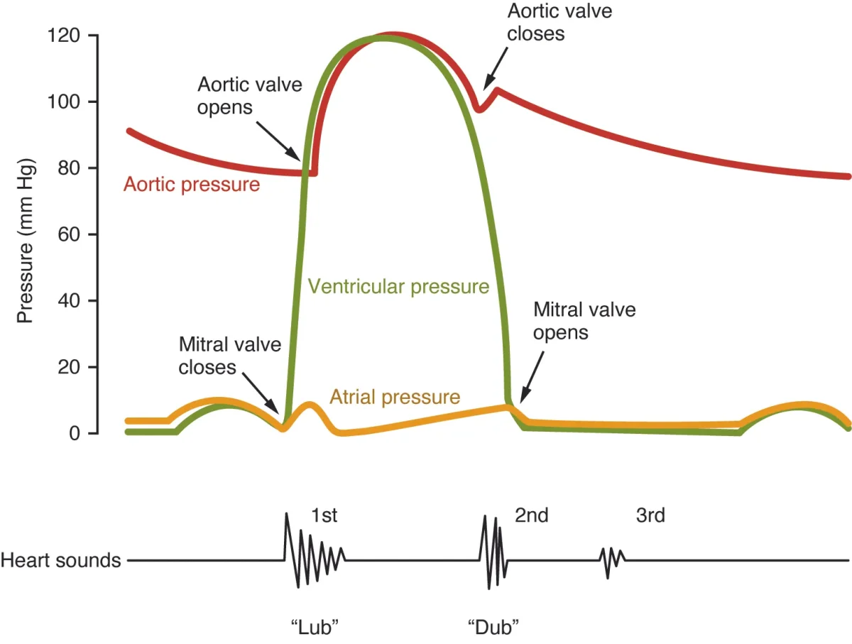
Pressure changes in the aorta, left ventricle, and left atrium during one cardiac cycle, correlated with valve events and heart sounds (S1 "lub" and S2 "dub"). Credit: OpenStax Anatomy and Physiology 2e, CC BY 4.0
Heart Sounds
You can hear the cardiac cycle with a stethoscope. The two main heart sounds are caused by valve closures, not by muscle contraction:
Sound
Cause
When
Character
S1 (“lub”)
AV valves close
Start of systole
Lower pitch, longer duration
S2 (“dub”)
Semilunar valves close
End of systole
Higher pitch, shorter, “snappier”
A heart murmur is an abnormal whooshing sound caused by turbulent blood flow, usually from a leaky valve (regurgitation) or a narrowed valve (stenosis). Murmurs are characterized by when they occur: a systolic murmur happens between S1 and S2, a diastolic murmur happens between S2 and the next S1.
Pressure-Volume (PV) Loops
The cardiac cycle can also be represented as a pressure-volume loop for the left ventricle. This graph plots ventricular pressure (y-axis) against ventricular volume (x-axis) and traces a rectangular loop:
Bottom right → top right (vertical line going up): Isovolumetric contraction. Volume constant, pressure rises.
Top right → top left (curve going left): Ejection. Volume decreases as blood is expelled.
Top left → bottom left (vertical line going down): Isovolumetric relaxation. Volume constant, pressure falls.
Bottom left → bottom right (curve going right): Filling. Volume increases as blood enters.
The width of the loop represents stroke volume (the difference between end-diastolic volume and end-systolic volume). The area inside the loop represents the work done by the ventricle during one beat.
During isovolumetric contraction, what is the status of all four heart valves, and why?
Click to reveal answer
All four valves are closed. The AV valves just closed (producing S1) because ventricular pressure exceeded atrial pressure. The semilunar valves have not yet opened because ventricular pressure has not yet exceeded arterial pressure. The ventricle is contracting in a completely sealed chamber, building pressure without changing volume.
Which phase of the cardiac cycle accounts for the majority of ventricular filling - atrial contraction or passive filling?
Click to reveal answer
Passive filling accounts for 70-80% of ventricular filling. When the AV valves open, blood flows down its pressure gradient from the atria into the ventricles without any atrial contraction. Atrial systole only adds the final 20-30% (the "atrial kick"). This is why patients with atrial fibrillation (where atria quiver instead of contracting) can still maintain reasonable cardiac output - they just lose the atrial kick.
Most muscles in your body wait for a nerve signal before they contract. Cardiac muscle does not. The heart generates its own electrical impulses and conducts them through a built-in wiring system. Cut every nerve to the heart, and it will keep beating. This property is called autorhythmicity (or myogenic activity), and it is one of the most distinctive features of cardiac tissue.
The Conduction Pathway
The heart’s electrical system follows a precise pathway that ensures the atria contract before the ventricles and that the ventricles contract from the bottom up (so blood is squeezed upward toward the arteries).
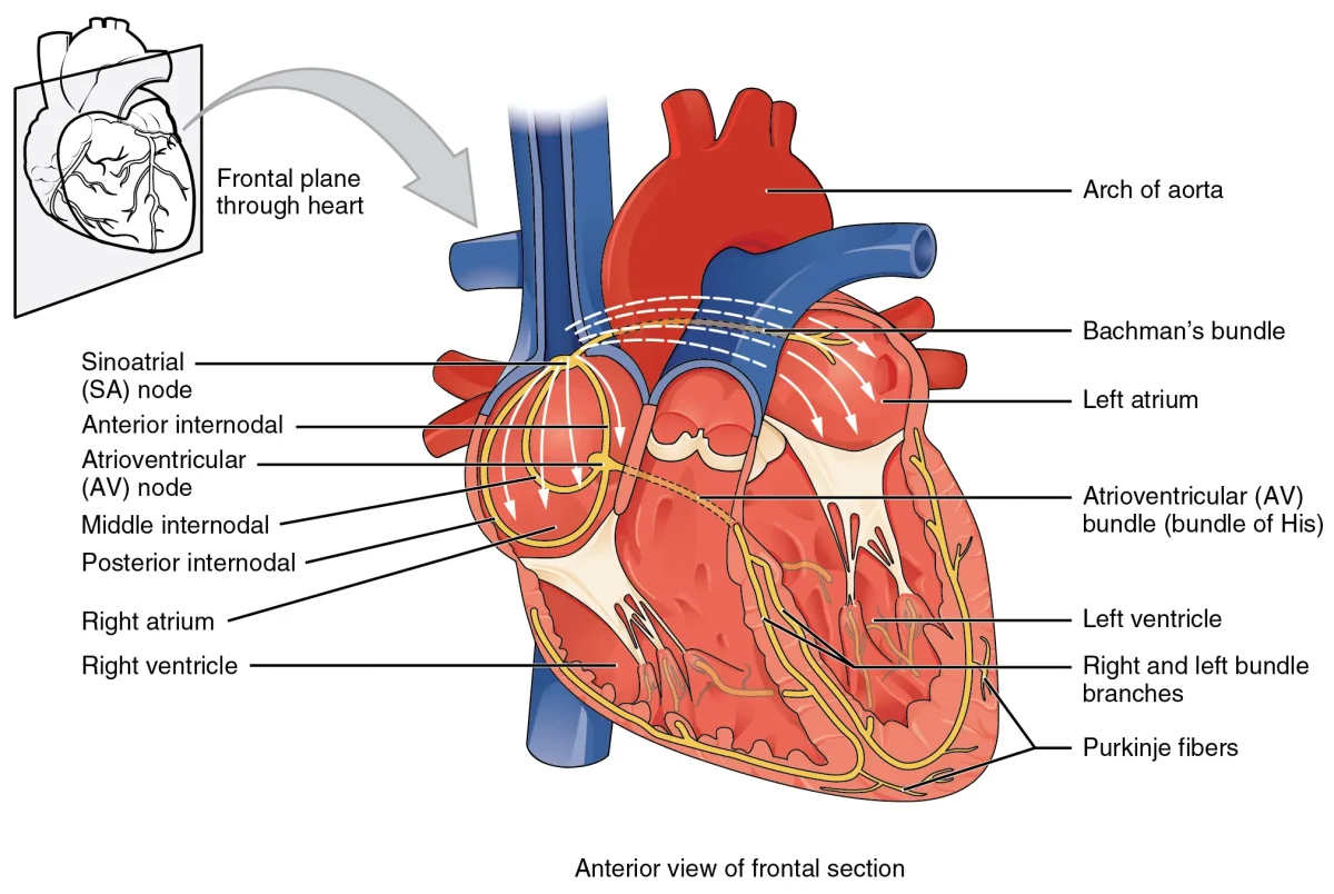
Step 1: SA Node (Sinoatrial Node) - Located in the wall of the right atrium near the superior vena cava. This is the heart’s primary pacemaker. It spontaneously depolarizes at a rate of about 60-100 beats per minute. When it fires, the electrical impulse spreads across both atria via gap junctions, causing atrial contraction.
Step 2: AV Node (Atrioventricular Node) - Located at the junction between the atria and ventricles, near the interatrial septum. The AV node introduces a critical 0.1-second delay. This pause ensures the atria finish contracting and emptying blood into the ventricles before the ventricles begin their own contraction.
Step 3: Bundle of His - The impulse travels from the AV node into the interventricular septum via the Bundle of His, which splits into the left and right bundle branches running down either side of the septum.
Step 4: Purkinje Fibers - The bundle branches fan out into the Purkinje fibers, which spread the impulse rapidly across the ventricular walls from apex to base. This ensures the ventricles contract from the bottom upward, efficiently squeezing blood toward the semilunar valves.
The cardiac conduction pathway ensures coordinated, sequential contraction of the atria followed by the ventricles. Credit: OpenStax Anatomy and Physiology 2e, CC BY 4.0
Intrinsic Rates
Each part of the conduction system can generate its own rhythm, but they fire at different rates. The fastest pacemaker wins:
Structure
Intrinsic Rate
Role
SA node
60-100 bpm
Primary pacemaker (normally controls heart rate)
AV node
40-60 bpm
Backup pacemaker if SA node fails
Purkinje fibers
20-40 bpm
Last-resort pacemaker
If the SA node fails, the AV node takes over at a slower rate. If both fail, the Purkinje fibers can keep the ventricles beating - slowly and barely enough to sustain life. This is why a complete heart block (where the AV node stops conducting) produces a dangerously slow heart rate.
Intercalated Discs and Gap Junctions
Cardiac muscle cells are physically connected by intercalated discs - specialized junctions that contain:
Gap junctions - allow ions to flow directly between adjacent cells, spreading the electrical impulse rapidly. This makes cardiac muscle behave as a functional syncytium (one cell fires, they all fire).
Desmosomes - anchor cells together mechanically so they do not tear apart during contraction.
This is why the heart contracts as a unified wave rather than as individual cells twitching randomly. The atria form one functional syncytium; the ventricles form another. The AV node is the only electrical connection between them.
Autonomic Modulation
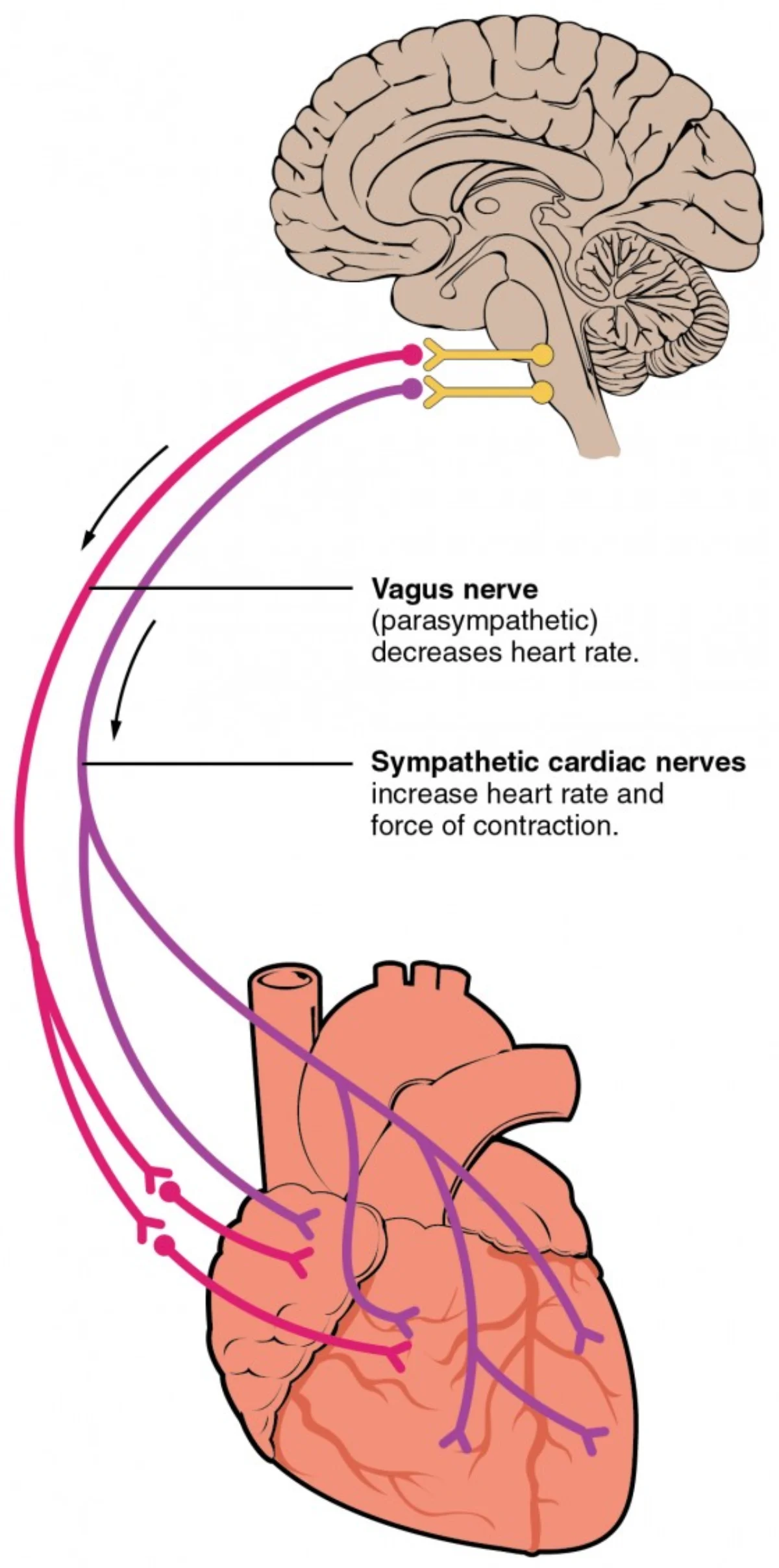
Although the heart generates its own rhythm, the autonomic nervous system modulates the rate:
Division
Nerve
Effect on Heart Rate
Mechanism
Parasympathetic
Vagus nerve (CN X)
Decreases rate
Releases ACh → slows SA node depolarization
Sympathetic
Cardiac accelerator nerves
Increases rate and force
Releases norepinephrine → speeds SA node, increases contractility
At rest, the vagus nerve is dominant. The SA node’s intrinsic rate is about 100 bpm, but tonic vagal input slows it to the normal resting rate of ~70 bpm. This is why cutting the vagus nerve (vagotomy) causes heart rate to jump to ~100 bpm.
What is the purpose of the 0.1-second delay at the AV node?
Click to reveal answer
The delay ensures the atria finish contracting before the ventricles begin. Without this pause, the atria and ventricles would contract simultaneously, and the atrial kick (the final 20-30% of ventricular filling) would be lost. The delay allows complete emptying of atrial blood into the ventricles before ventricular ejection begins.
If a drug blocks all sympathetic input to the heart but leaves parasympathetic input intact, what happens to resting heart rate?
Click to reveal answer
Heart rate decreases slightly below normal resting rate. At rest, parasympathetic (vagal) tone is dominant anyway, so blocking sympathetic input removes only a small excitatory influence. The heart rate would drop modestly. If you blocked parasympathetic input instead, heart rate would jump to ~100 bpm (the SA node's intrinsic rate) because the dominant vagal brake is removed.
An electrocardiogram (ECG or EKG) is a recording of the heart’s electrical activity detected by electrodes placed on the skin. It does not measure contraction directly - it measures the electrical events that trigger contraction. Think of it as listening to the commands, not watching the soldiers march.
The MCAT expects you to know what each wave represents and to reason through how changes in conduction would alter the tracing. You will not be asked to diagnose specific arrhythmias, but you may see an abnormal ECG described in a passage and need to connect it to the underlying physiology.
The Normal ECG Tracing
A single heartbeat produces three recognizable waveforms:
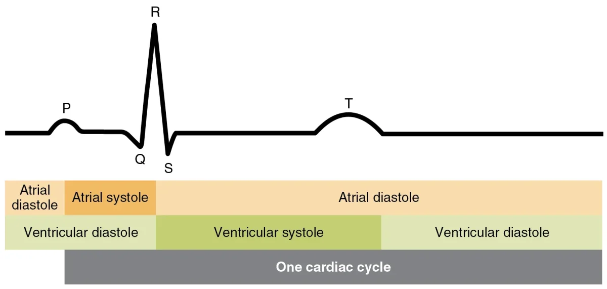
A normal ECG tracing with the P wave, QRS complex, and T wave labeled, correlated with the phases of atrial and ventricular systole and diastole. Credit: OpenStax Anatomy and Physiology 2e, CC BY 4.0
Wave/Segment
Electrical Event
Mechanical Event
P wave
Atrial depolarization
Atria contract (atrial systole)
PR interval
Impulse travels through AV node (delay)
Ventricles finish filling
QRS complex
Ventricular depolarization
Ventricles contract (ventricular systole)
ST segment
Ventricles fully depolarized (plateau phase)
Blood being ejected
T wave
Ventricular repolarization
Ventricles begin to relax
Key Points for the MCAT
Why is the QRS complex larger than the P wave? The ventricles have much more muscle mass than the atria, so ventricular depolarization produces a much larger electrical signal.
Where is atrial repolarization? It occurs during the QRS complex but is completely masked by the much stronger ventricular depolarization signal. You cannot see it on a normal ECG.
What does the PR interval represent? The time from the beginning of atrial depolarization to the beginning of ventricular depolarization - essentially, the AV node delay. If the AV node conducts slowly, the PR interval lengthens.
What does the QT interval represent? The time from the start of ventricular depolarization to the end of ventricular repolarization - the total duration of ventricular electrical activity.
Connecting the ECG to the Conduction System
Each part of the ECG tracing maps directly to the conduction pathway you learned in the previous section:
P wave = SA node fires, impulse spreads across atria
PR interval (flat line) = impulse pauses at the AV node (the critical delay that lets atria finish emptying)
QRS complex = impulse travels through Bundle of His and Purkinje fibers, depolarizing the ventricles
T wave = ventricles repolarize and prepare for the next cycle
If any part of the conduction system fails - for example, the AV node stops conducting - the relationship between these waves changes. A passage might describe P waves and QRS complexes occurring at different rates or with abnormal spacing. Your job is to identify which part of the conduction pathway is affected based on what you know about each wave.
Why is the QRS complex so much taller than the P wave on an ECG?
Click to reveal answer
The ventricles have far more muscle mass than the atria. The amplitude of an ECG waveform reflects the amount of tissue being depolarized. Since the ventricular myocardium is much thicker and more massive, its depolarization produces a much larger electrical signal than atrial depolarization.
A researcher records an ECG and notices the PR interval is significantly longer than normal. Which structure in the conduction pathway is most likely affected?
Click to reveal answer
The AV node. The PR interval represents the time for the impulse to travel from the SA node (start of P wave) through the AV node to the ventricles (start of QRS). A prolonged PR interval means the AV node is conducting the impulse more slowly than normal, delaying ventricular depolarization.
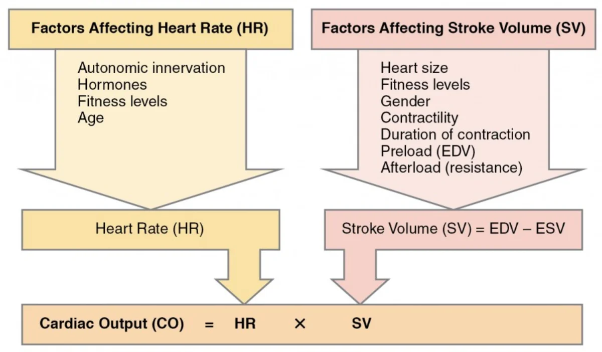
Cardiac output (CO) answers a simple question: how much blood does the heart pump per minute? It is the single most important measure of the heart’s performance, and the MCAT tests it relentlessly - both the formula itself and the physiological factors that change it. For the physics of blood flow through vessels (Poiseuille’s law, Bernoulli’s equation), see fluid dynamics in physics.
The Core Formula
Think of it this way: the total water flowing out of a pump per minute depends on how much water per squeeze (stroke volume) multiplied by how many squeezes per minute (heart rate). Increase either one, and cardiac output goes up.
Stroke Volume
Stroke volume is the amount of blood ejected from the left ventricle with each heartbeat. It is calculated as:
Ejection fraction (EF) is the percentage of blood ejected per beat: EF = SV/EDV. A normal EF is about 55-70%. An EF below 40% indicates heart failure - the ventricle is not emptying effectively.
The Three Determinants of Stroke Volume
Stroke volume is controlled by three factors. You must know all three:
The major factors influencing cardiac output. Stroke volume depends on preload, contractility, and afterload. Credit: Lumen Learning / OpenStax Anatomy and Physiology, CC BY 4.0
1. Preload - the degree of stretch on the ventricular wall at the end of diastole. More blood returning to the heart (increased venous return) = more stretch = more preload. Preload is essentially EDV.
2. Afterload - the resistance the ventricle must overcome to eject blood. In the left ventricle, afterload is determined primarily by aortic pressure (or total peripheral resistance). Higher blood pressure = higher afterload = harder for the ventricle to eject = decreased stroke volume.
3. Contractility (inotropy) - the intrinsic strength of contraction, independent of preload. Sympathetic stimulation and epinephrine increase contractility. Contractility increases stroke volume by reducing ESV (the ventricle squeezes more completely).
The Frank-Starling Mechanism
This is arguably the most important concept in cardiovascular physiology for the MCAT.
The Frank-Starling law states: the more the ventricle fills with blood during diastole (greater preload/EDV), the more forcefully it contracts during systole, ejecting a greater stroke volume.
Why? When cardiac muscle fibers are stretched more, actin and myosin filaments reach a more optimal overlap, generating stronger cross-bridge cycling. Up to a point, more stretch = more force.
The physiological power of Frank-Starling: it automatically matches the output of the right and left ventricles. If the right ventricle suddenly pumps more blood to the lungs, more blood returns to the left atrium, increasing left ventricular preload, which increases left ventricular stroke volume. The system is self-balancing.
Autonomic Regulation of Heart Rate
Factor
Effect on HR
Effect on Contractility
Mechanism
Sympathetic stimulation
Increases (chronotropy)
Increases (inotropy)
Norepinephrine on beta-1 receptors
Parasympathetic (vagus)
Decreases
Minimal effect on ventricles
ACh on muscarinic receptors at SA/AV nodes
Epinephrine (adrenal)
Increases
Increases
Circulating catecholamine on beta-1 receptors
Autonomic innervation of the heart. The vagus nerve (parasympathetic) slows the heart; sympathetic nerves speed it up and increase contractility. Credit: Lumen Learning / OpenStax Anatomy and Physiology, CC BY 4.0
A patient loses 1 liter of blood. Using the Frank-Starling mechanism, explain what happens to stroke volume and why.
Click to reveal answer
Stroke volume decreases. Blood loss reduces total blood volume, which decreases venous return to the heart. Less venous return means less ventricular filling (decreased preload/EDV). By the Frank-Starling mechanism, less stretch on the ventricular wall means weaker contraction and reduced stroke volume.
A patient's cardiac output is 4,900 mL/min and their heart rate is 70 bpm. What is their stroke volume? If their EDV is 130 mL, what is their ejection fraction?
Click to reveal answer
SV = CO / HR = 4,900 / 70 = 70 mL per beat. ESV = EDV - SV = 130 - 70 = 60 mL. EF = SV / EDV = 70 / 130 = 53.8%. This is at the low end of normal (55-70%), suggesting mildly reduced systolic function.
Blood pressure is the force that blood exerts on the walls of blood vessels. Without adequate pressure, blood cannot reach the brain, kidneys, or any other organ. Too much pressure damages vessel walls and leads to heart attacks and strokes. The body maintains blood pressure within a narrow range using a combination of rapid neural reflexes and slower hormonal systems.
Measuring Blood Pressure
Blood pressure is recorded as systolic/diastolic (e.g., 80120 mmHg).
Systolic pressure (~120 mmHg) - the peak pressure during ventricular contraction
Diastolic pressure (~80 mmHg) - the lowest pressure during ventricular relaxation
The pulse you feel in your wrist is the difference between these two values - the pulse pressure (systolic minus diastolic = 40 mmHg).
The Blood Pressure Drop Across the Circulation
Blood pressure is not the same everywhere. It drops progressively as blood moves further from the heart: highest in the aorta (~120 mmHg systolic), lower in the arteries, sharply lower across the arterioles (the biggest single drop), low in the capillaries (~35 → 15 mmHg), and near zero in the large veins.
The biggest pressure drop occurs across the arterioles - these small, muscular vessels are the primary site of resistance in the circulatory system. This is why arterioles are called the “resistance vessels.” By constricting or dilating, they control both blood pressure and blood flow distribution to individual organs.
Rapid Regulation: The Baroreceptor Reflex
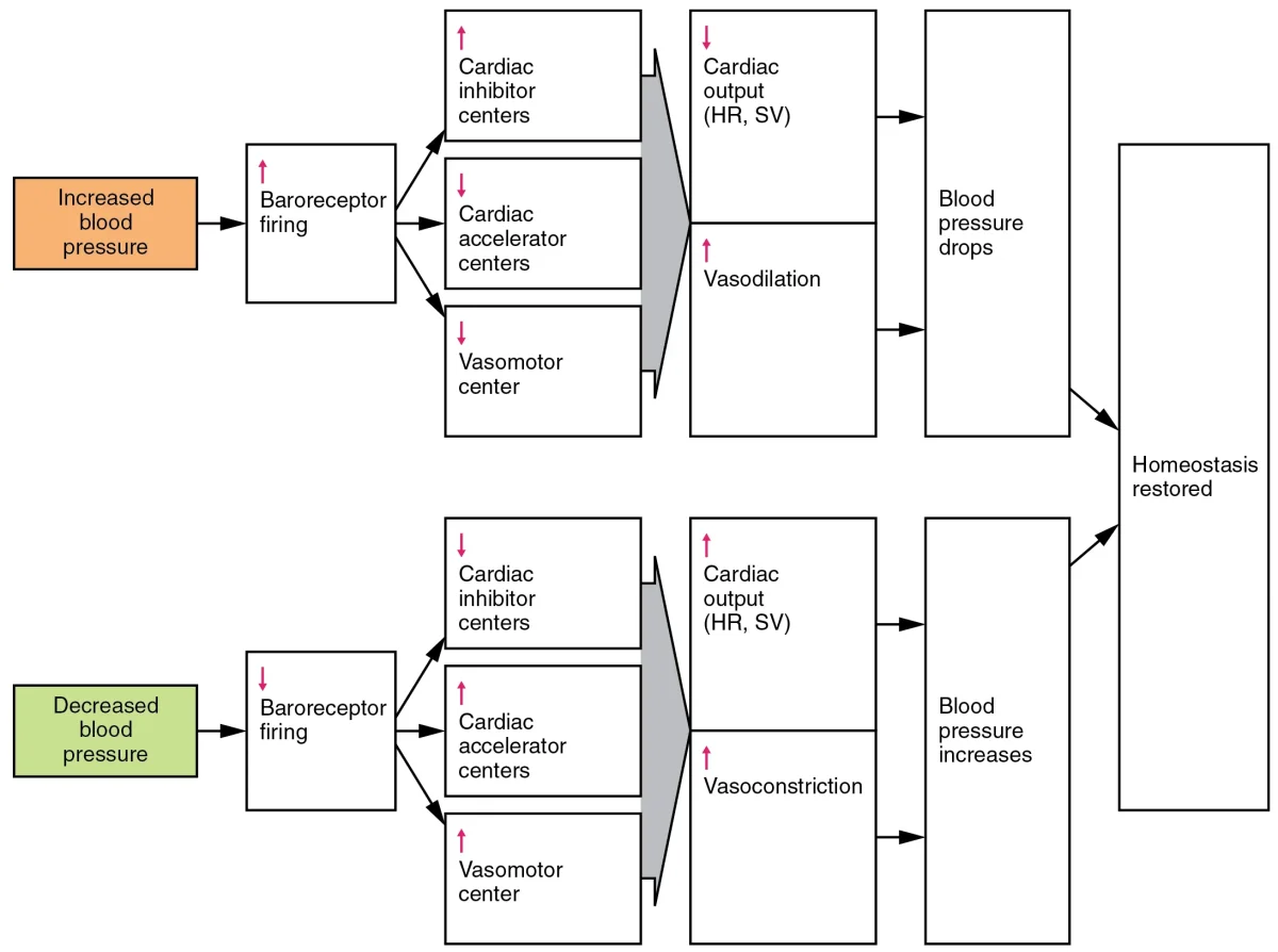
The body’s fastest blood pressure correction system is the baroreceptor reflex, a neural feedback loop that operates in seconds.
Baroreceptors are stretch-sensitive nerve endings located in the walls of the carotid sinus (at the bifurcation of the common carotid artery) and the aortic arch. When blood pressure rises, the vessel walls stretch more, and baroreceptors fire more frequently. When blood pressure falls, they fire less.
The baroreceptor reflex adjusts heart rate and vascular resistance in response to blood pressure changes. Credit: OpenStax Anatomy and Physiology 2e, CC BY 4.0
When blood pressure drops:
Baroreceptors detect decreased stretch → fire less
Cardiovascular center in the medulla increases sympathetic output and decreases parasympathetic output
Renin converts angiotensinogen (from the liver) → angiotensin I
ACE (angiotensin-converting enzyme, in the lungs) converts angiotensin I → angiotensin II
Angiotensin II causes vasoconstriction (raises TPR) and stimulates aldosterone release from the adrenal cortex
Aldosterone increases Na+ and water reabsorption in the kidneys → increases blood volume → increases CO → raises BP
A patient stands up quickly and feels dizzy. Trace the reflex pathway that corrects this blood pressure drop.
Click to reveal answer
Standing → blood pools in legs → decreased venous return → decreased CO → decreased BP → baroreceptors in carotid sinus/aortic arch detect decreased stretch → fire less → medulla increases sympathetic output, decreases parasympathetic → increased HR, increased contractility, vasoconstriction → BP restored. If this reflex fails (e.g., from dehydration or autonomic neuropathy), the patient experiences orthostatic hypotension.
A patient is given an ACE inhibitor. What happens to angiotensin II levels, aldosterone levels, and blood pressure?
Click to reveal answer
Angiotensin II decreases (ACE can no longer convert angiotensin I to angiotensin II). Aldosterone decreases (angiotensin II normally stimulates aldosterone release). Blood pressure decreases because there is less vasoconstriction (from reduced angiotensin II) and less sodium/water retention (from reduced aldosterone).
The circulatory system uses five types of blood vessels, each engineered for a specific job. Understanding their structural differences is the key to understanding why blood flows the way it does - and why it fails in disease.
The Three Layers of Blood Vessels
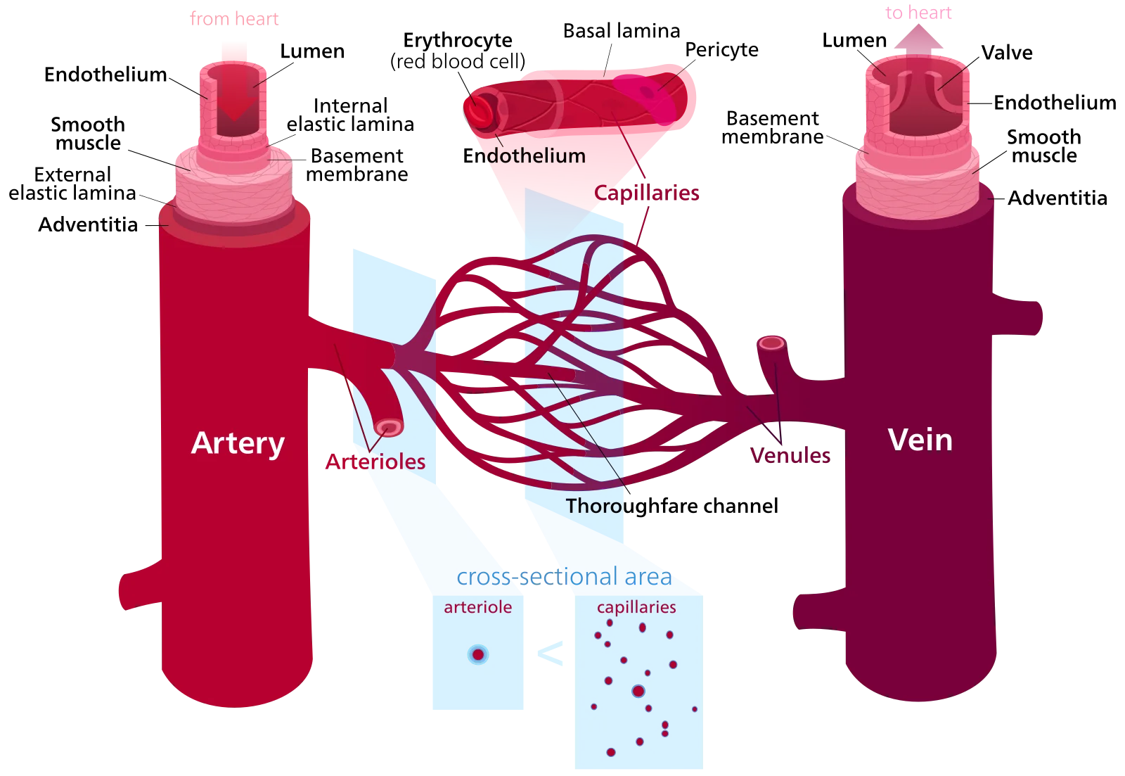
Most blood vessels share a common three-layer wall structure:
Comparison of artery, vein, and capillary. Focus on: arteries have thick walls (more smooth muscle) to handle high pressure, veins have thinner walls with valves to prevent backflow, and capillaries are one cell thick for gas exchange. Credit: Wikimedia Commons, CC BY-SA 3.0
Layer
Composition
Function
Tunica intima (innermost)
Single layer of endothelial cells
Smooth, non-thrombogenic surface for blood flow; regulates permeability
Tunica media (middle)
Smooth muscle + elastic fibers
Vasoconstriction/vasodilation; maintains vessel tone
Tunica externa (outermost)
Connective tissue (collagen)
Anchors vessel to surrounding tissue; structural support
Arteries
Arteries carry blood away from the heart under high pressure. They have thick walls with abundant smooth muscle and elastic fibers in the tunica media.
Elastic (conducting) arteries - the largest arteries (aorta, pulmonary trunk, carotid arteries). Their walls are rich in elastic fibers that stretch during systole and recoil during diastole, smoothing out the pulsatile flow from the heart. This is why you still have blood flow during diastole - the elastic recoil of these arteries acts like a secondary pump.
Muscular (distributing) arteries - medium-sized arteries (brachial, femoral, renal arteries). More smooth muscle, less elastic tissue. They distribute blood to specific organs and can constrict or dilate to redirect flow.
Arterioles: The Resistance Vessels
Arterioles are the smallest arteries (diameter: 10-100 micrometers) and the primary regulators of blood pressure and blood flow distribution. Their smooth muscle walls can constrict (vasoconstriction) or relax (vasodilation) to change resistance dramatically.
A small change in arteriole radius produces a massive change in resistance. This relationship follows Poiseuille’s law: resistance is inversely proportional to the fourth power of the radius. Halve the radius and resistance increases 16-fold.
Capillaries: The Exchange Vessels
Capillaries are the thinnest vessels (diameter: 5-10 micrometers, just wide enough for a single red blood cell). Their walls are only one endothelial cell thick - no tunica media, no tunica externa. This thinness is essential for diffusion of gases, nutrients, and waste.
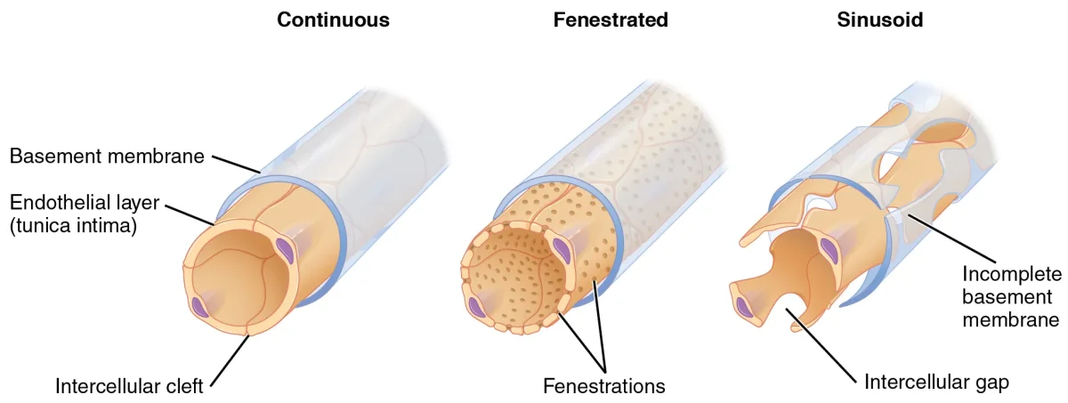
There are three types of capillaries:
The three types of capillaries differ in how permeable they are. Continuous capillaries are the most restrictive; sinusoidal are the most permeable. Credit: OpenStax Anatomy and Physiology 2e, CC BY 4.0
Type
Structure
Where Found
What Passes Through
Continuous
Tight junctions, no pores
Muscle, skin, lungs, brain (BBB)
Small molecules, water, ions only
Fenestrated
Small pores (60-80 nm) in endothelial cells
Kidneys, intestines, endocrine glands
Small proteins, filtered fluids
Sinusoidal
Large gaps (30-40 micrometers)
Liver, spleen, bone marrow
RBCs, WBCs, large proteins - almost everything
Velocity and Cross-Sectional Area
Blood flows fastest in the aorta and slowest in the capillaries. This seems counterintuitive - the capillaries are tiny, so shouldn’t flow speed up? No, because while each individual capillary is narrow, the total cross-sectional area of all capillaries combined is enormous (about 600 times the area of the aorta).
By the continuity equation (A1v1 = A2v2), when total cross-sectional area increases, velocity must decrease. This slow flow in capillaries is essential - it gives time for gas and nutrient exchange.
Veins: The Capacitance Vessels
Veins return blood to the heart under low pressure. Their walls are thinner and more compliant (stretchable) than arteries. Veins hold about 60-65% of total blood volume at any time, which is why they are called capacitance vessels.
Because venous pressure is so low (~5-10 mmHg), veins use several mechanisms to push blood back to the heart:
Venous valves - one-way flaps that prevent backflow, especially in the legs where blood must travel against gravity
Respiratory pump - during inhalation, decreased thoracic pressure creates a pressure gradient that pulls venous blood toward the heart
Vessel Comparison Summary
Feature
Arteries
Arterioles
Capillaries
Venules
Veins
Wall thickness
Thickest
Thick
Thinnest (1 cell)
Thin
Moderate
Lumen diameter
Large
Small
Tiny (5-10 um)
Small
Large
Blood pressure
Highest
High
Low
Very low
Lowest
Flow velocity
Fast
Moderate
Slowest
Slow
Moderate
Special features
Elastic recoil
Resistance control
Gas exchange
WBC migration
Valves, high volume
Why does blood flow slowest in the capillaries, even though they are the narrowest individual vessels?
Click to reveal answer
The total cross-sectional area of all capillaries combined is far greater than any other vessel type. By the continuity equation (A x v = constant), when total area increases, velocity must decrease. Each capillary is tiny, but there are billions of them, so their combined area is enormous. This slow flow maximizes time for gas and nutrient exchange.
A person stands motionless for a long time and starts to feel lightheaded. Which venous return mechanisms are being compromised?
Click to reveal answer
The skeletal muscle pump is inactive. Without leg muscle contractions, blood pools in the lower extremity veins. Reduced venous return decreases preload (Frank-Starling), which decreases stroke volume and cardiac output, leading to reduced cerebral perfusion and lightheadedness. This is why soldiers standing at attention sometimes faint.
Every cell in your body needs oxygen and nutrients delivered and waste products removed. This exchange does not happen in arteries or veins - it happens exclusively at the capillary level. Understanding how fluid and solutes move across capillary walls is essential for the MCAT and for understanding conditions like edema (swelling).
The Tug-of-War at the Capillary Wall
Fluid movement across capillary walls is governed by two opposing forces:
The Four Starling Forces
There are actually four pressures at play, though two dominate:
Force
Direction
Typical Value
Description
Capillary hydrostatic pressure (Pc)
Pushes fluid OUT
~35 mmHg (arteriole end) → ~15 mmHg (venule end)
Blood pressure pushing fluid through the capillary wall
Interstitial hydrostatic pressure (Pi)
Pushes fluid IN (opposes filtration)
~0 mmHg (often negligible)
Pressure of fluid already in the tissue space
Capillary oncotic pressure (πc)
Pulls fluid IN
~25 mmHg (relatively constant)
Osmotic pull of plasma proteins (mainly albumin)
Interstitial oncotic pressure (πi)
Pulls fluid OUT
~1 mmHg (usually small)
Osmotic pull of proteins in the interstitial fluid
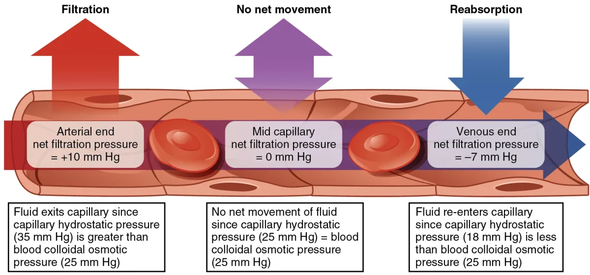
Fluid dynamics across a capillary bed. At the arteriole end, hydrostatic pressure exceeds oncotic pressure (net filtration). At the venule end, oncotic pressure exceeds hydrostatic pressure (net reabsorption). Credit: OpenStax Anatomy and Physiology 2e, CC BY 4.0
Net reabsorption - fluid moves back INTO the capillary
This picks up CO2, urea, and other waste products
About 85% of the filtered fluid is reabsorbed at the venule end. The remaining 15% (~3 liters per day) is picked up by the lymphatic system and returned to the venous circulation.
Edema: When the Balance Breaks
Edema (tissue swelling) occurs when more fluid leaves capillaries than returns. There are four main causes, each linked to a Starling force:
Liver failure or nephrotic syndrome (low albumin production or loss)
Increased capillary permeability
Proteins leak out, pulling fluid with them
Inflammation, burns, allergic reactions
Blocked lymphatic drainage
Filtered fluid cannot return
Lymphedema after lymph node removal, parasitic infection (elephantiasis)
Bulk Flow vs. Diffusion
Do not confuse bulk flow (Starling forces) with diffusion:
Bulk flow moves large volumes of fluid across capillary walls. It is driven by pressure gradients (hydrostatic and oncotic). This is what Starling forces describe.
Diffusion moves individual molecules (O2, CO2, glucose) down their concentration gradients. It is the primary mechanism for gas and nutrient exchange. O2 diffuses from blood (high PO2) to tissues (low PO2). CO2 diffuses the opposite direction.
Both processes happen simultaneously at the capillary level, but they are driven by different forces.
A patient with nephrotic syndrome is losing large amounts of albumin in their urine. Why do they develop edema?
Click to reveal answer
Loss of albumin decreases plasma oncotic pressure. Albumin is the primary protein responsible for pulling fluid back into capillaries (oncotic pressure). When albumin drops, less fluid is reabsorbed at the venule end. The excess fluid accumulates in the interstitial space, causing edema. The Starling force affected is decreased capillary oncotic pressure.
At the arteriole end of a capillary, capillary hydrostatic pressure is 35 mmHg and capillary oncotic pressure is 25 mmHg. What is the net filtration pressure, and in which direction does fluid move?
Click to reveal answer
Net filtration pressure = 35 - 25 = +10 mmHg, favoring filtration (fluid moves OUT of the capillary). The positive value means hydrostatic pressure exceeds oncotic pressure, pushing fluid into the interstitial space. At the venule end, the math reverses: 15 - 25 = -10 mmHg, favoring reabsorption (fluid moves back IN).
Blood is not just a red liquid. It is a complex tissue made up of cells suspended in a protein-rich fluid. An average adult has about 5 liters of blood, and every component has a specific job. Understanding blood composition is essential for MCAT questions on gas transport, immunity, clotting, and transfusion compatibility.
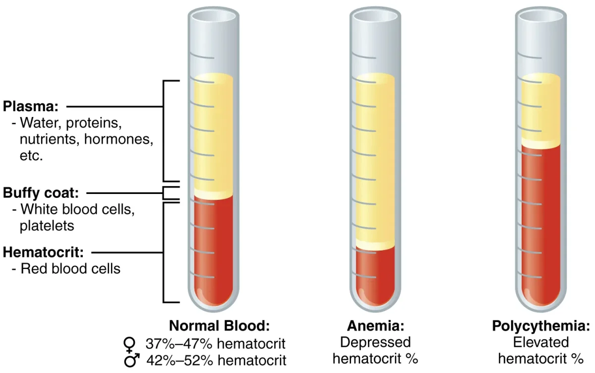
When blood is centrifuged, it separates into three layers: plasma (top), the buffy coat of WBCs and platelets (middle), and packed red blood cells (bottom). Credit: OpenStax Anatomy and Physiology 2e, CC BY 4.0
Plasma (~55% of Blood Volume)
Plasma is the liquid matrix of blood. It is about 90% water and 10% dissolved solutes:
Plasma proteins (~7%) - albumin (maintains oncotic pressure), globulins (antibodies and transport), fibrinogen (clotting precursor)
Electrolytes - Na+, K+, Ca2+, Cl-, HCO3-
Nutrients - glucose, amino acids, lipids
Waste products - urea, creatinine, bilirubin
Dissolved gases - O2, CO2
Hormones - transported to target organs
Serum is plasma minus the clotting factors (what you get after blood clots and the clot is removed).
Red Blood Cells (Erythrocytes)
RBCs are the most abundant cells in blood (~4.5-5.5 million per microliter). Their sole purpose is gas transport.
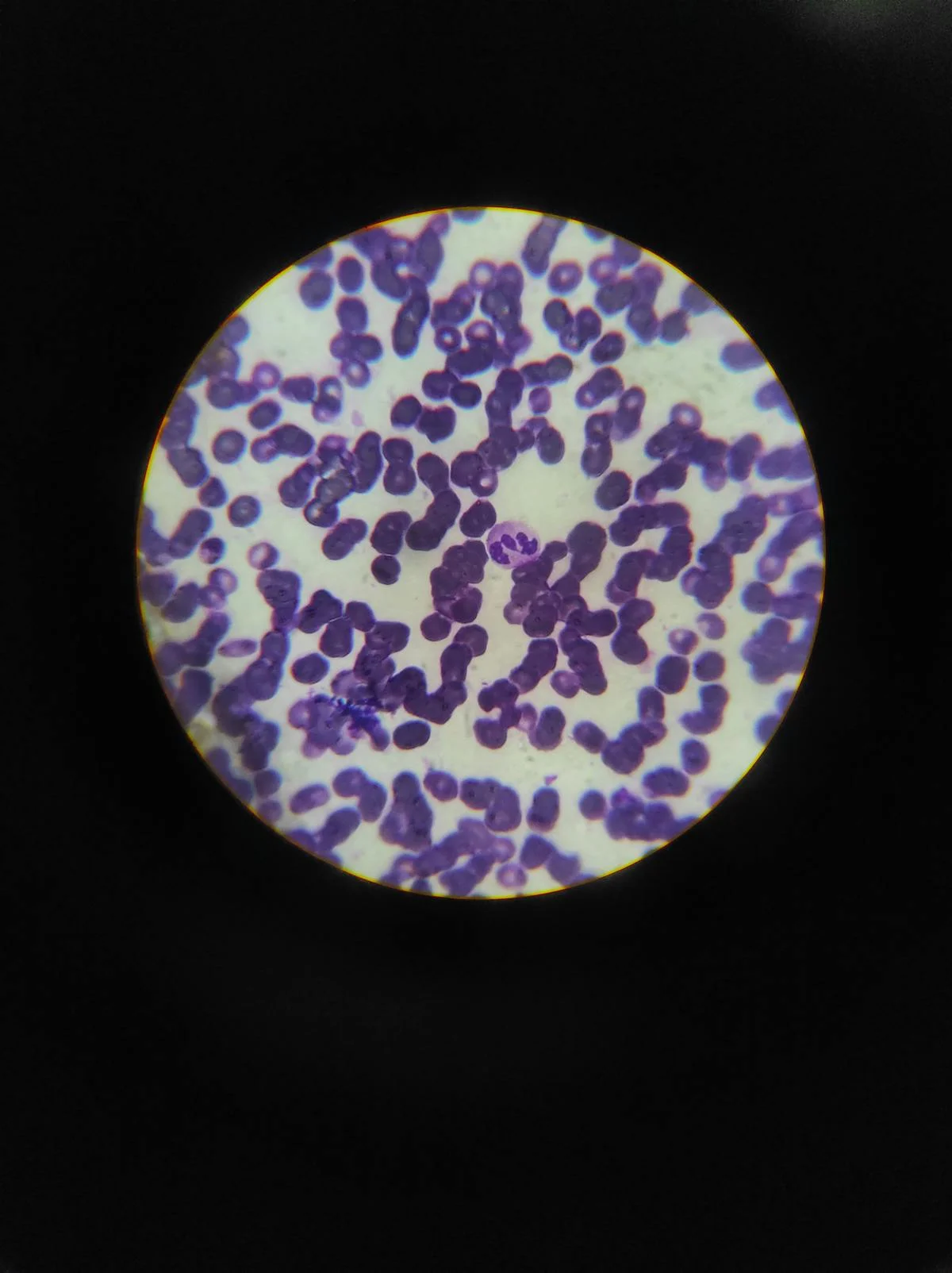
A Wright-stained blood smear viewed through a light microscope. This is the type of preparation used in clinical labs to count and identify blood cell types. Credit: Pexels, free to use
The proportion of blood volume occupied by RBCs is called the hematocrit (normally ~42-52% in males, ~37-47% in females). RBC production is stimulated by erythropoietin (EPO), a hormone released by the kidneys in response to low oxygen levels (hypoxia). This is why people living at high altitude develop higher hematocrit - their kidneys sense low O2 and ramp up RBC production.
Interactive 3D Hemoglobin. Rotate to see the four subunits (2 alpha, 2 beta) and the heme groups that bind oxygen.Credit: Nima via Sketchfab, CC BY
Key features of RBCs:
Biconcave disc shape - maximizes surface area for gas exchange and allows flexibility to squeeze through narrow capillaries
No nucleus or organelles - mature RBCs eject their nucleus during development, leaving more room for hemoglobin
Packed with hemoglobin - each RBC contains ~280 million hemoglobin molecules
No mitochondria - RBCs rely entirely on anaerobic glycolysis for their own energy (they cannot consume the oxygen they carry)
Lifespan: ~120 days - old RBCs are removed by macrophages in the spleen and liver
Hemoglobin is a tetrameric protein (two alpha and two beta subunits in adult hemoglobin, HbA), each containing a heme group with an iron (Fe2+) atom that binds one O2 molecule. Each hemoglobin molecule can carry 4 O2 molecules.
The Oxygen-Hemoglobin Dissociation Curve
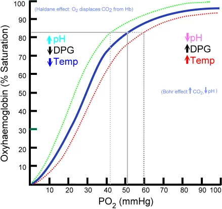
Hemoglobin does not bind oxygen all-or-nothing. As each O2 binds, the hemoglobin subunits shift toward a higher-affinity state, making the next O2 easier to bind. This positive cooperativity produces the classic sigmoid (S-shaped) dissociation curve when % saturation is plotted against PO2.
The sigmoid O2-Hb dissociation curve. The plateau at high PO2 (lungs) ensures near-complete loading; the steep portion at low PO2 (tissues) ensures rapid unloading. Rightward shifts promote unloading; leftward shifts promote loading. Credit: Wikimedia Commons, CC BY-SA 4.0
Two regions of the curve matter:
Plateau (high PO2, ~100 mmHg in lungs): Hemoglobin stays ~97% saturated even if PO2 drops somewhat. Loading is protected.
Steep portion (low PO2, ~40 mmHg in tissues): Small drops in PO2 cause large drops in saturation. Unloading is maximized where O2 is needed.
Right Shift = More Unloading
A rightward shift means hemoglobin releases O2 more readily at any given PO2 (lower affinity). This happens in metabolically active tissue, which is exactly where extra O2 is needed.
The Bohr effect is the specific case of ↑CO2 and ↑H+ lowering hemoglobin’s O2 affinity. Active muscle produces both, so the curve shifts right exactly where O2 needs to be delivered. A leftward shift (↓CO2, ↓H+, ↓temp, ↓2,3-BPG) means tighter O2 binding and less unloading.
Fetal Hemoglobin
Fetal hemoglobin (HbF) has two alpha and two gamma subunits instead of two alpha, two beta. The gamma subunits bind 2,3-BPG more weakly, so HbF has a higher O2 affinity than adult HbA. Its dissociation curve sits to the left of the adult curve, letting the fetus pull O2 off maternal hemoglobin at the placenta.
White Blood Cells (Leukocytes)
WBCs defend the body against infection. They are far less numerous than RBCs (~5,000-10,000 per microliter) but are larger and nucleated. The specific roles of neutrophils, macrophages, and other frontline defenders are covered in innate immune cells.
Cell Type
Function
Key Facts
Neutrophils
First responders; phagocytose bacteria
Most abundant WBC; multilobed nucleus; short-lived; pus = dead neutrophils
Lymphocytes
Adaptive immunity (B cells, T cells, NK cells)
Second most abundant; small cells with large nucleus
Monocytes
Become macrophages in tissue; phagocytosis + antigen presentation
Largest WBC; kidney-shaped nucleus
Eosinophils
Combat parasites; modulate allergic response
Bilobed nucleus; red-staining granules
Basophils
Release histamine and heparin; involved in allergic reactions
Rarest WBC; large purple/blue granules; similar to mast cells
Platelets (Thrombocytes)
Platelets are not true cells - they are small, anucleate cell fragments (~150,000-400,000 per microliter) produced by megakaryocytes in the bone marrow. Platelet production is stimulated by thrombopoietin, a hormone from the liver and kidneys.
Platelets are essential for hemostasis (stopping bleeding). When a blood vessel is damaged, platelets adhere to exposed collagen, activate, change shape, and aggregate to form a temporary platelet plug. They also release chemicals that initiate the coagulation cascade.
Blood Types: ABO System
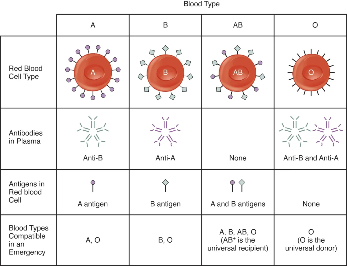
Blood type is determined by antigens on the surface of RBCs and antibodies in the plasma.
The ABO blood group system. Each blood type has specific antigens on RBCs and antibodies in the plasma against the antigens it lacks. Credit: OpenStax Anatomy and Physiology 2e, CC BY 4.0
Blood Type
Antigens on RBCs
Antibodies in Plasma
Can Donate To
Can Receive From
A
A antigen
Anti-B
A, AB
A, O
B
B antigen
Anti-A
B, AB
B, O
AB
Both A and B
Neither (universal recipient)
AB only
A, B, AB, O
O
Neither
Both anti-A and anti-B (universal donor)
A, B, AB, O
O only
The Rh Factor
The Rh system adds a second layer of compatibility. If you have the Rh antigen (Rh D protein) on your RBCs, you are Rh-positive (+). If not, you are Rh-negative (-).
Rh incompatibility in pregnancy: An Rh-negative mother carrying an Rh-positive fetus can develop anti-Rh antibodies if fetal blood enters her circulation (usually during delivery). These antibodies are harmless to the first baby but can attack the RBCs of subsequent Rh-positive fetuses, causing hemolytic disease of the newborn. Prevention: the mother receives Rh immunoglobulin injections to destroy any fetal Rh+ cells before her immune system can mount a response.
True universal donor: O-negative (no A, B, or Rh antigens). True universal recipient: AB-positive (no antibodies against any common antigens).
Why do mature red blood cells lack a nucleus and mitochondria? What is the advantage and disadvantage of this design?
Click to reveal answer
Advantage: More space for hemoglobin, maximizing oxygen-carrying capacity. Disadvantage: RBCs cannot repair themselves, synthesize new proteins, or undergo cell division. They rely on anaerobic glycolysis for energy (no mitochondria = no aerobic respiration). They wear out after ~120 days and must be replaced by new cells from the bone marrow.
A type B-negative patient urgently needs blood. No B-negative blood is available. What other blood type(s) can be safely transfused?
Click to reveal answer
O-negative. Type O blood has no A or B antigens (so the patient's anti-A antibodies will not react), and Rh-negative means no Rh antigen. O-negative is the universal donor. O-positive could also work in a life-threatening emergency (the Rh antigen is less immediately dangerous than ABO mismatches), but O-negative is the safest choice.
When a blood vessel is damaged, the body must stop the bleeding quickly without clotting the entire circulatory system. This process - hemostasis - involves three overlapping stages that work together like a construction crew sealing a broken pipe.
Stage 1: Vascular Spasm
Immediately after injury, the damaged vessel’s smooth muscle contracts, narrowing the lumen and reducing blood flow to the area. This reflex vasoconstriction buys time for the other stages to kick in.
Stage 2: Platelet Plug Formation
When the endothelial lining is breached, the underlying collagen is exposed. Platelets stick to exposed collagen via von Willebrand factor (vWF), a sticky glycoprotein that acts as molecular glue.
Once adhered, platelets activate and undergo several changes:
Change shape from smooth discs to spiky spheres with extending pseudopods
Release chemical signals (ADP, thromboxane A2) that recruit more platelets
Aggregate together, forming a platelet plug that temporarily seals the wound
This positive feedback loop (activated platelets recruit more platelets) ensures rapid plug formation.
Stage 3: The Coagulation Cascade
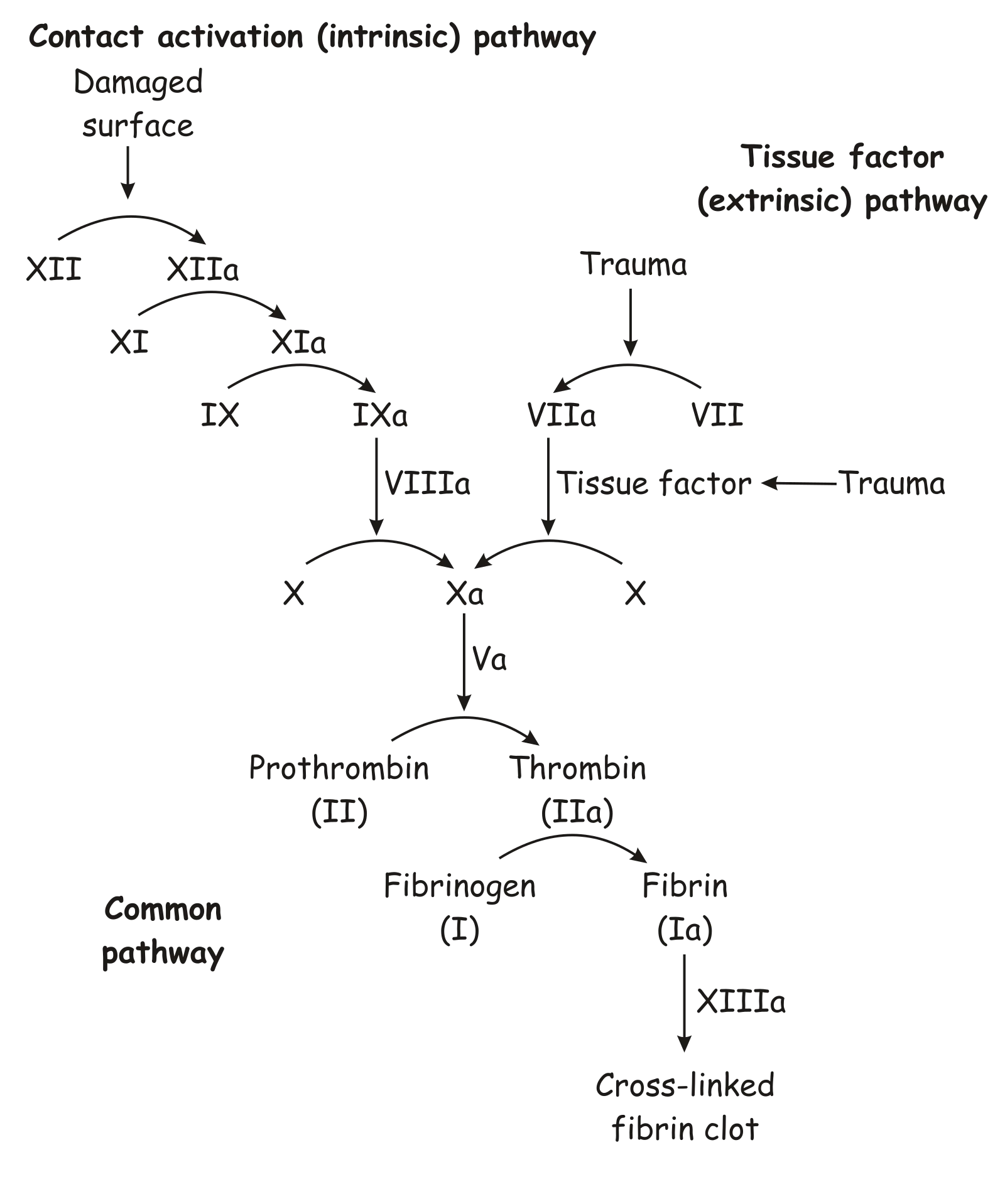
The coagulation cascade is a series of enzymatic reactions that converts soluble fibrinogen into insoluble fibrin threads, which weave through the platelet plug and harden it into a stable clot.
The coagulation cascade (simplified). Focus on: the intrinsic and extrinsic pathways converge on the common pathway, where prothrombin is converted to thrombin, which converts fibrinogen to fibrin. You do not need to memorize individual clotting factor numbers - just know the common pathway and that vitamin K is required. Credit: Wikimedia Commons, CC BY-SA 3.0
The key reaction to know is the common pathway:
A cascade of clotting factors (triggered by vessel damage) activates an enzyme that converts prothrombin → thrombin
Thrombin converts fibrinogen → fibrin
Fibrin polymerizes into a mesh that traps RBCs, platelets, and plasma, forming the stable clot
Vitamin K and Clotting
Vitamin K is essential for the liver to produce several functional clotting factors, including prothrombin. Without vitamin K, these factors cannot be properly activated, and the clotting cascade fails.
This is why newborns receive a vitamin K injection at birth - their gut flora, which normally produces vitamin K, has not yet been established.
Clot Dissolution
Once the vessel is repaired, the clot must be removed so it does not block normal blood flow. The enzyme plasmin breaks down fibrin threads, dissolving the clot. Plasmin is produced from its inactive precursor, plasminogen, which is embedded within the clot itself.
The body also has built-in safeguards to prevent clots from forming where they should not. Intact endothelial cells produce chemical signals (prostacyclin and nitric oxide) that inhibit platelet adhesion, keeping blood flowing smoothly through undamaged vessels.
What is the difference between the platelet plug and the fibrin clot? Which forms first?
Click to reveal answer
The platelet plug forms first. It is a temporary, loose aggregate of activated platelets that quickly seals the wound. The fibrin clot forms second through the coagulation cascade - fibrin threads weave through and reinforce the platelet plug, creating a stable, long-lasting clot. Think of the platelet plug as a quick patch and the fibrin clot as the permanent repair.
A patient has a vitamin K deficiency. Which stage of hemostasis is most directly impaired, and why?
Click to reveal answer
Stage 3 (the coagulation cascade) is most directly impaired. Vitamin K is required for the liver to produce functional clotting factors, including prothrombin. Without these factors, the cascade cannot convert fibrinogen to fibrin, so the permanent fibrin clot cannot form. Stages 1 (vascular spasm) and 2 (platelet plug) are unaffected because they do not depend on vitamin K.
Every day, about 20 liters of fluid filters out of your capillaries into the interstitial space. About 17 liters is reabsorbed back into the capillaries at the venule end (via Starling forces). That leaves ~3 liters per day stranded in the tissues. Without a way to return this fluid to the blood, your tissues would swell until the pressure shut down circulation entirely.
That recovery system is the lymphatic system. It is not a separate circulatory loop - it is a one-way drainage network that collects excess interstitial fluid, filters it for pathogens, and dumps it back into the venous bloodstream.
Lymphatic Vessels
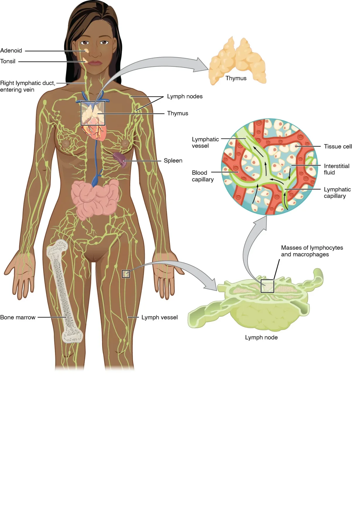
The lymphatic system collects interstitial fluid via a network of vessels and returns it to the venous circulation through the thoracic duct and right lymphatic duct. Credit: OpenStax Anatomy and Physiology 2e, CC BY 4.0
Lymph capillaries are tiny, blind-ended vessels found in nearly all tissues. They have overlapping endothelial cells that act as one-way flaps - interstitial fluid can push in, but cannot leak back out. They are more permeable than blood capillaries and can absorb large molecules, cell debris, and even bacteria.
Lymph capillaries merge into larger lymphatic vessels that resemble veins - they have thin walls, one-way valves, and rely on skeletal muscle contraction and respiratory movements to propel lymph forward (just like venous return).
All lymph eventually drains into the venous bloodstream at two points:
The thoracic duct (left lymphatic duct) - drains about 75% of the body (left side + everything below the diaphragm) into the left subclavian vein
The right lymphatic duct - drains the right arm, right side of the head, and right thorax into the right subclavian vein
Lymph Nodes
Lymph nodes are small, bean-shaped organs (1-25 mm) scattered along lymphatic vessels, clustered in the neck, axillae (armpits), and groin. Each node is packed with lymphocytes (B cells and T cells) and macrophages.
As lymph flows through a node, macrophages phagocytose pathogens and debris, and lymphocytes scan for foreign antigens. If an antigen is detected, an immune response is activated - beginning with the rapid, nonspecific defenses covered in innate immunity. This is why lymph nodes swell during infection - they are filling with proliferating immune cells.
Lymphoid Organs
Beyond lymph nodes, several organs play key roles in the lymphatic/immune system:
Site of T cell maturation; most active in childhood, involutes (shrinks) with age
Tonsils
Throat
First line of immune defense against ingested/inhaled pathogens
Peyer’s patches
Small intestine wall
Monitor intestinal bacteria; mount immune responses to gut pathogens
Bone marrow
Inside bones
Produces all blood cells (hematopoiesis); site of B cell maturation
Three Functions of the Lymphatic System
Fluid balance - returns ~3 liters/day of excess interstitial fluid to the blood, preventing edema
Immune surveillance - lymph nodes filter lymph for pathogens; lymphocytes mount adaptive immune responses
Fat absorption - specialized lymphatic vessels called lacteals in the small intestine absorb dietary fats (as chylomicrons) that are too large for blood capillaries. This is why lymph from the intestine looks milky and is called chyle.
Connection to the Cardiovascular System
The lymphatic system is the cardiovascular system’s essential partner:
Without lymphatic drainage, the 3 liters/day of unrecovered filtrate would accumulate, causing progressive edema and eventually circulatory collapse
Lymph returns to the blood at the subclavian veins, helping maintain blood volume and blood pressure
The spleen serves as a blood reservoir and filter, recycling iron from old RBCs back into new hemoglobin production
If lymphatic drainage is blocked (surgical removal of lymph nodes, parasitic infection like filariasis), the affected area develops severe, chronic swelling called lymphedema. In extreme cases (elephantiasis), a limb can swell to many times its normal size.
What would happen to the tissues if the lymphatic system stopped functioning?
Click to reveal answer
Progressive, severe edema. About 3 liters of fluid per day filters out of capillaries and is not reabsorbed by Starling forces alone. Without lymphatic drainage, this fluid accumulates in the interstitial space, causing tissue swelling that worsens over time. Eventually, the increased interstitial pressure could compress capillaries and compromise blood flow to tissues.
Why does the spleen filter blood while lymph nodes filter lymph? What is the functional significance of this difference?
Click to reveal answer
The spleen is positioned directly in the bloodstream (supplied by the splenic artery), so it filters blood for old/damaged RBCs, platelets, and blood-borne pathogens. Lymph nodes are positioned along lymphatic vessels, so they filter interstitial fluid (lymph) for pathogens and debris that entered the tissues. Together, they provide immune surveillance of both the blood and the tissue compartments.