Your body faces a constant stream of challenges - a hot stove, an approaching car, a change in blood pressure - and it needs a system that can respond in milliseconds. That system is the nervous system, the body’s rapid communication network. While the endocrine system handles slower, long-lasting signals through hormones, the nervous system uses electrical impulses and neurotransmitters to deliver near-instantaneous responses.
Understanding how the nervous system is organized is the foundation for every neuroscience topic on the MCAT, from action potentials to complex behaviors.
The Two Major Divisions: CNS and PNS
The nervous system is divided into two major structural components: the central nervous system (CNS) and the peripheral nervous system (PNS).
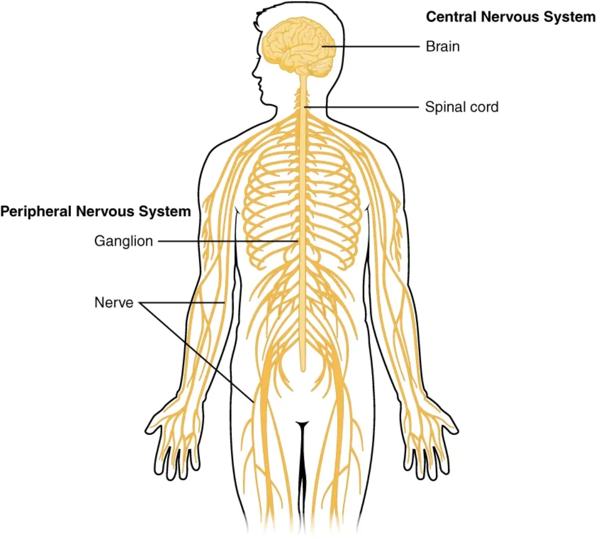
The CNS consists of the brain and spinal cord. It is the integration and command center of the body - where sensory information is processed, decisions are made, and motor commands originate. The CNS is protected by bone (skull and vertebral column), meninges (three protective membrane layers), and cerebrospinal fluid (CSF).
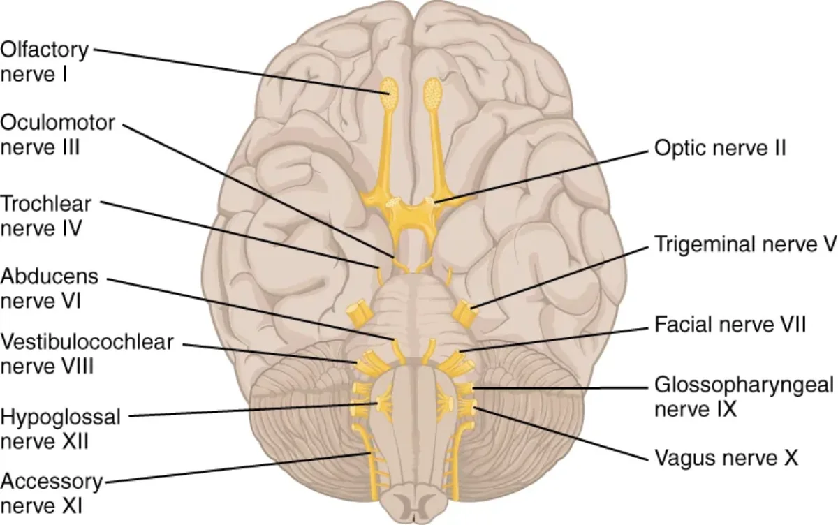
The PNS includes everything outside the brain and spinal cord. This means all 12 pairs of cranial nerves, 31 pairs of spinal nerves, ganglia (clusters of neuron cell bodies outside the CNS), and the extensive network of peripheral nerve fibers that reach every tissue in the body.
The CNS (brain and spinal cord) serves as the command center, while the PNS connects it to the rest of the body. Credit: OpenStax Anatomy & Physiology 2e, CC BY 4.0
The PNS is further divided into functional subdivisions based on what it controls.
The somatic nervous system controls voluntary movements. It carries motor commands to skeletal muscles and returns sensory information from the skin, muscles, and joints. When you decide to pick up a pen, the somatic nervous system executes that command.
The autonomic nervous system (ANS) controls involuntary functions - things your body handles without conscious thought. This includes heart rate, digestion, respiratory rate, and glandular secretion. The ANS is further divided into the sympathetic (“fight or flight”), parasympathetic (“rest and digest”), and enteric divisions.
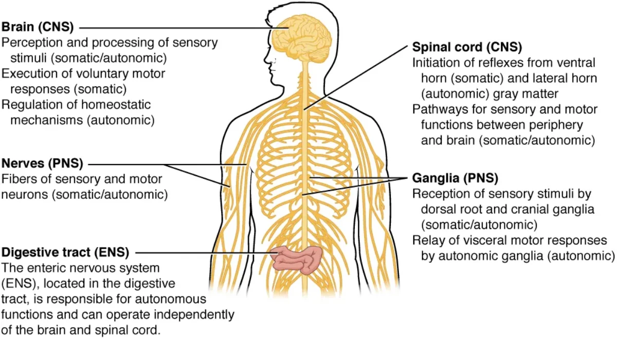
The enteric nervous system is sometimes classified separately because of its remarkable independence. It is a mesh-like network of neurons embedded in the walls of the gastrointestinal tract. It can coordinate digestion entirely on its own, which is why it is sometimes called the “second brain.” It contains roughly 100 million neurons - more than the spinal cord.
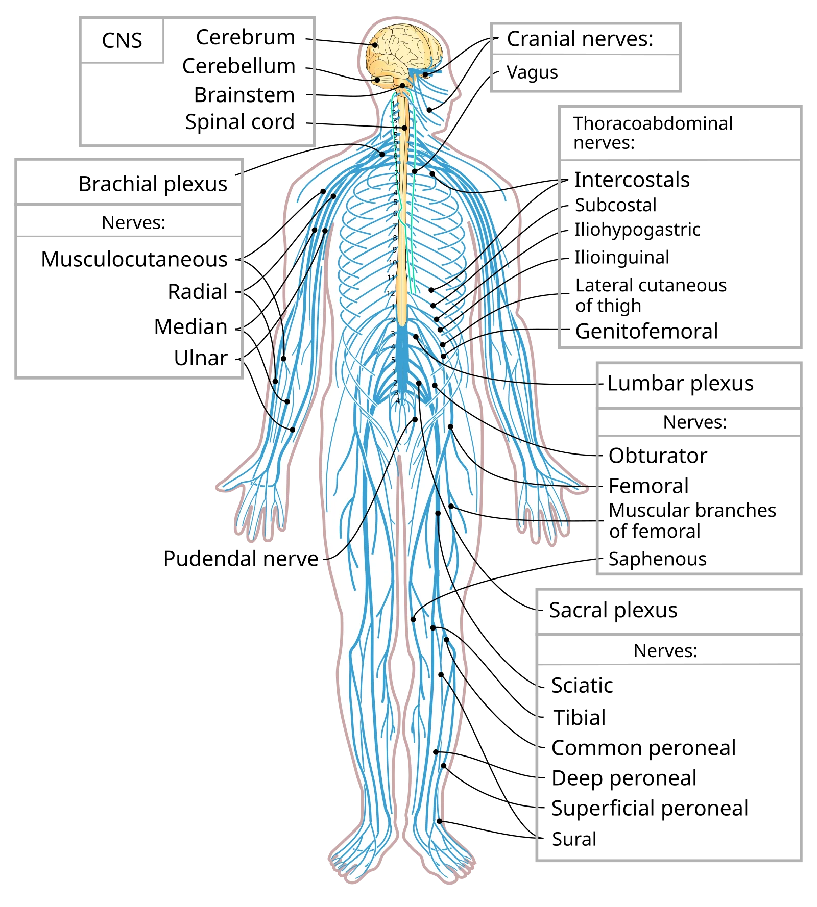
The PNS branches into somatic (voluntary), autonomic (involuntary), and enteric (gut-specific) divisions. Credit: OpenStax Anatomy & Physiology 2e, CC BY 4.0The nervous system extends from the brain and spinal cord to every region of the body through an extensive network of peripheral nerves. Credit: Wikimedia Commons, CC BY-SA 4.0
Afferent vs. Efferent Pathways
Regardless of which subdivision we are discussing, nerve signals travel in one of two directions:
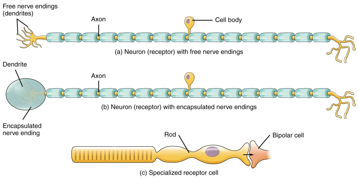
Afferent neurons (sensory neurons) carry information toward the CNS. They detect stimuli - light, pressure, temperature, pain - and transmit that data to the brain and spinal cord for processing.
Efferent neurons (motor neurons) carry commands away from the CNS to effector organs such as muscles and glands. They execute the responses the CNS has determined are appropriate.
There is a third category worth knowing: interneurons reside entirely within the CNS and connect afferent and efferent neurons. They are responsible for integration, analysis, and the formation of complex circuits. The vast majority of neurons in your body are interneurons.
Putting It All Together: The Reflex Arc
The simplest example of nervous system organization in action is the reflex arc. When you touch a hot stove:
Sensory receptors in your skin detect the heat.
An afferent neuron transmits the signal through a dorsal root into the spinal cord (CNS).
An interneuron in the spinal cord relays the signal directly to a motor neuron - no brain involvement needed.
An efferent neuron exits through a ventral root and stimulates your arm muscles to pull away.
This entire process takes a fraction of a second. The signal also travels up to the brain so you consciously register the pain, but the reflex withdrawal happens before you even “feel” it.
What are the two structural divisions of the nervous system, and what does each include?
Click to reveal answer
The CNS includes the brain and spinal cord. The PNS includes all neural tissue outside the CNS - cranial nerves (12 pairs), spinal nerves (31 pairs), and ganglia.
Using the SAME DAVE mnemonic, which type of neuron enters the dorsal root of the spinal cord, and which exits the ventral root?
Click to reveal answer
Afferent (sensory) neurons enter through the dorsal root (Dorsal = Afferent). Efferent (motor) neurons exit through the ventral root (Ventral = Efferent).
What are the three functional subdivisions of the PNS?
Click to reveal answer
Somatic (voluntary control of skeletal muscle), Autonomic (involuntary control of smooth muscle, cardiac muscle, and glands - divided into sympathetic, parasympathetic), and Enteric (semi-independent neural network of the GI tract).
Neurons are the functional units of the nervous system - specialized cells that generate and transmit electrical signals across long distances at remarkable speed. Although they come in many shapes and sizes, all neurons share the same basic architecture. Understanding each structural component and its role is essential for making sense of signal transmission, synaptic communication, and the clinical conditions the MCAT tests.
Interactive 3D Neuron. Trace the signal path: dendrites receive, soma integrates, axon hillock decides, myelinated axon transmits, synaptic terminals deliver.Credit: Nima via Sketchfab, CC BY
Dendrites: The Receivers
Dendrites are highly branched extensions that project from the neuron’s cell body. Their job is to receive incoming signals from other neurons, sensory receptors, or the environment. The more branches a dendrite has, the more connections it can make - some neurons in the brain have thousands of dendritic branches.
Dendrites contain receptors and ligand-gated ion channels on their surface that bind neurotransmitters released by neighboring neurons. When a neurotransmitter binds, it may produce a small local change in voltage called a graded potential. These graded potentials are not all-or-nothing; they vary in size and can be either excitatory or inhibitory.
Soma (Cell Body): The Integration Center
The soma is the metabolic heart of the neuron. It contains the nucleus, rough endoplasmic reticulum (called Nissl bodies in neurons), Golgi apparatus, and mitochondria. All the proteins and organelles the neuron needs are manufactured here and transported outward.
Critically, the soma integrates all the graded potentials arriving from the dendrites. Excitatory signals (EPSPs) and inhibitory signals (IPSPs) are summed together. If the net signal is strong enough when it reaches the next structure - the axon hillock - an action potential fires.
Axon Hillock: The Decision Point
The axon hillock is the cone-shaped region where the soma transitions into the axon. It has the highest density of voltage-gated sodium channels anywhere on the neuron, giving it the lowest threshold for firing an action potential.
This is where the all-or-nothing decision is made. If the summed graded potentials depolarize the membrane at the axon hillock to threshold (approximately -55 mV), an action potential is generated. If they do not reach threshold, nothing happens. There is no “partial” action potential.
Axon: The Transmission Line
The axon is a long, slender projection that carries the action potential away from the cell body toward the target cell. Axons can be extremely short (a fraction of a millimeter for interneurons in the brain) or remarkably long (over one meter for motor neurons extending from the spinal cord to the toes).
The cytoplasm of the axon is called axoplasm, and its membrane is called the axolemma. Axons do not contain rough ER or Golgi apparatus, so they depend on the soma for protein synthesis. Materials are moved along the axon by motor proteins: kinesin transports cargo toward the synaptic terminal (anterograde transport), while dynein transports cargo back toward the soma (retrograde transport).
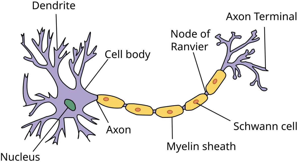
Neuron anatomy showing the signal path: dendrites receive input, the cell body integrates it, and the myelinated axon transmits the action potential to the axon terminal. Credit: Wikimedia Commons, CC BY-SA 3.0
Myelin Sheath and Nodes of Ranvier
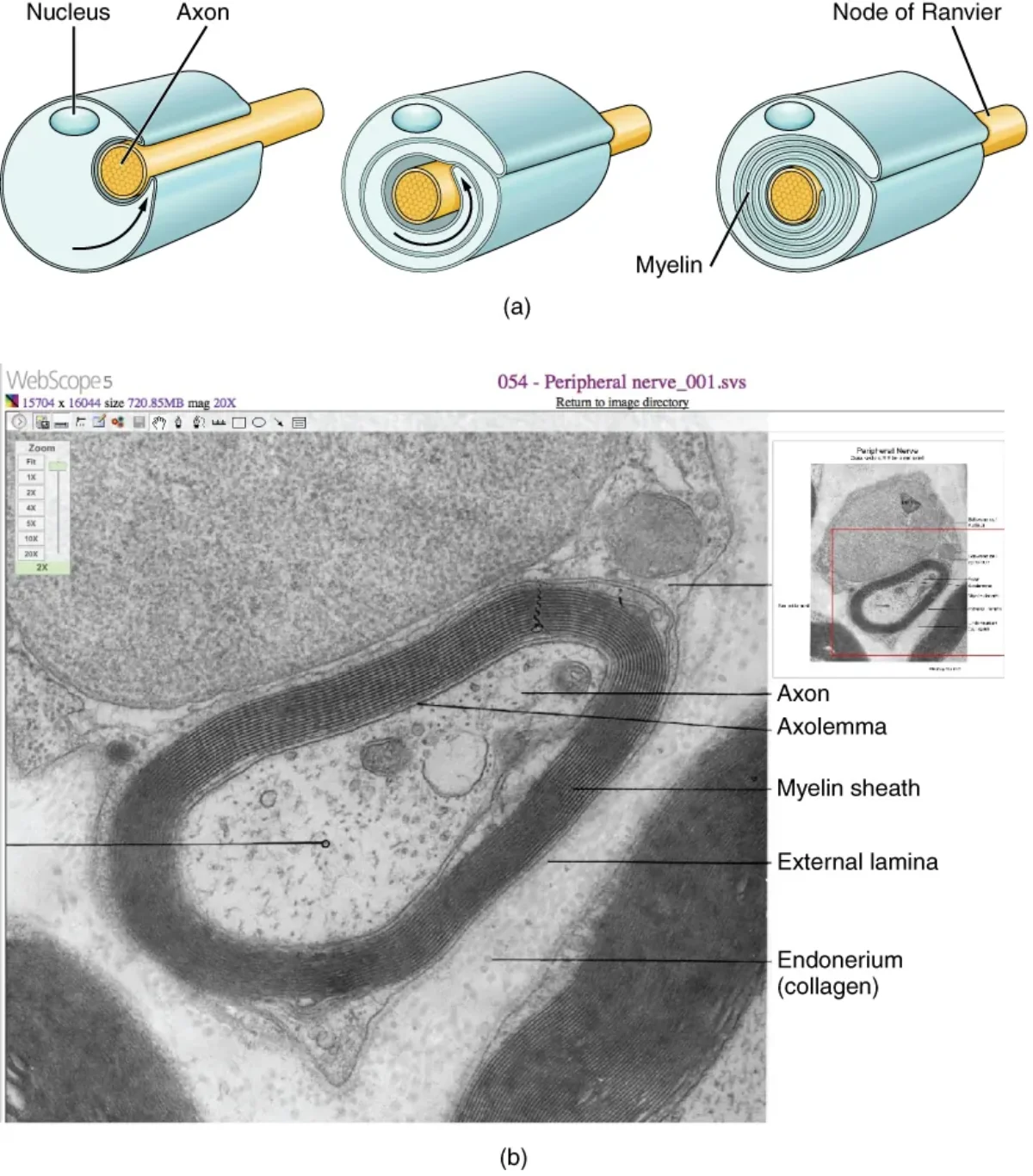
Many axons are wrapped in a fatty insulating layer called the myelin sheath. Myelin is composed of lipid-rich cell membrane wound tightly around the axon in concentric layers. It serves two critical functions: it insulates the axon to prevent current leakage, and it dramatically increases the speed of signal conduction.
The cells that produce myelin differ by location:
In the PNS, Schwann cells each wrap around a single segment of one axon.
In the CNS, oligodendrocytes extend multiple processes, each myelinating a segment of a different axon. A single oligodendrocyte can myelinate portions of up to 50 axons.
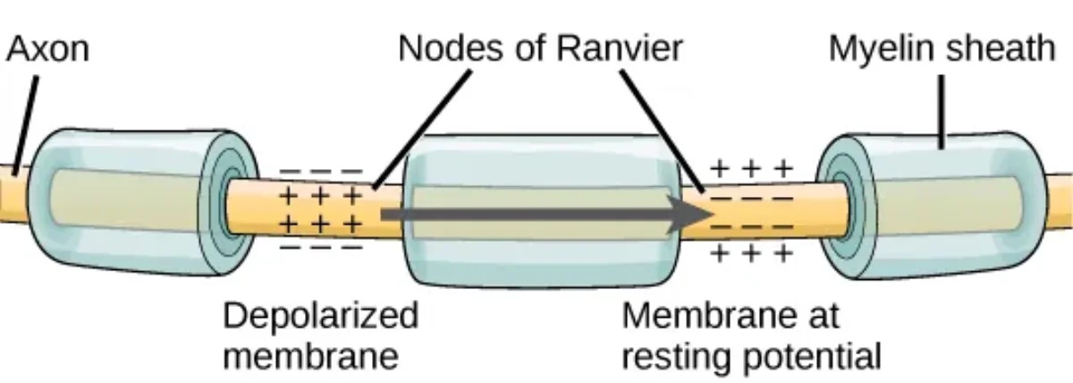
Between each myelinated segment are small gaps called nodes of Ranvier. These gaps are packed with voltage-gated sodium channels. Because the action potential effectively “jumps” from node to node rather than traveling continuously along the axon, myelinated neurons conduct signals much faster. This jumping conduction is called saltatory conduction (from the Latin “saltare,” meaning to jump).
Synaptic Terminals: The Output Zone
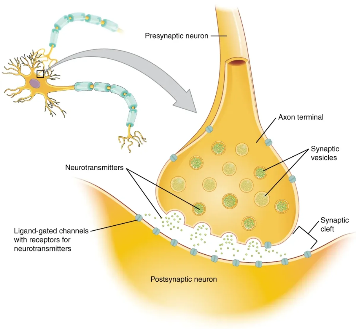
At its end, the axon branches into many fine extensions that terminate in synaptic terminals (also called terminal boutons or axon terminals). Each terminal contains synaptic vesicles loaded with neurotransmitter molecules.
When an action potential arrives at the synaptic terminal, voltage-gated calcium channels open. The influx of Ca2+ triggers the vesicles to fuse with the presynaptic membrane and release their neurotransmitter into the synaptic cleft - the tiny gap between the sending and receiving cells. This process is called exocytosis, and it converts the electrical signal back into a chemical one.
Structural Classification of Neurons
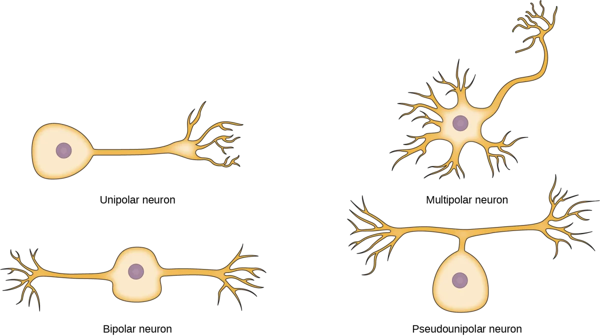
Neurons are classified by how many processes (extensions) emerge from the cell body.
Multipolar neurons have one axon and many dendrites. They are the most common type in the CNS and include most motor neurons and interneurons.
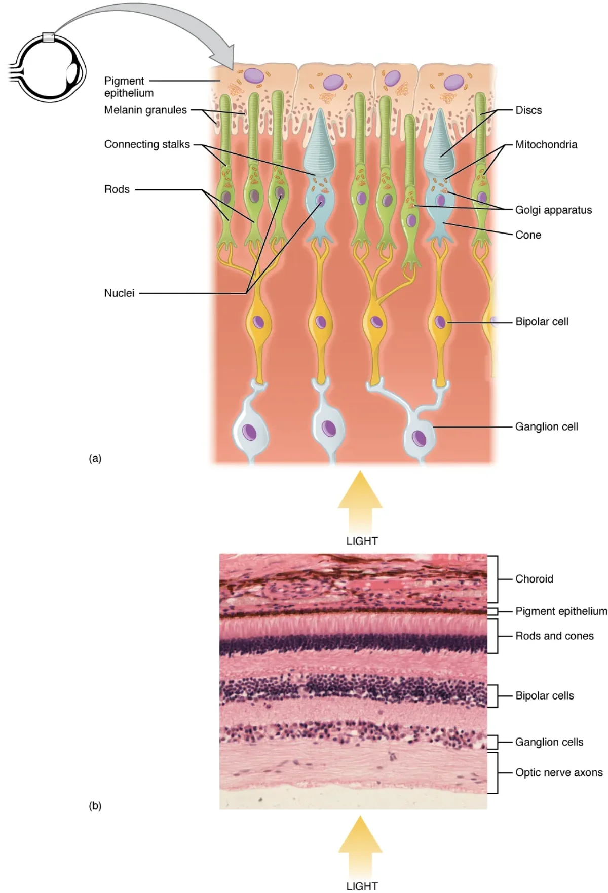
Bipolar neurons have one axon and one dendrite on opposite sides of the cell body. They are found in special sensory organs - the retina, olfactory epithelium, and inner ear.
Unipolar (pseudounipolar) neurons have a single process that splits into two branches - one heading toward the periphery and one toward the CNS. Most sensory neurons in the PNS are pseudounipolar. Their cell bodies sit in the dorsal root ganglia.
Structural classification of neurons based on the number of processes extending from the cell body. Credit: OpenStax Anatomy & Physiology 2e, CC BY 4.0
Functional Classification of Neurons
Neurons can also be classified by the direction they carry signals:
Sensory (afferent) neurons carry information from receptors toward the CNS. Most are pseudounipolar.
Motor (efferent) neurons carry commands from the CNS to muscles and glands. Most are multipolar.
Interneurons connect neurons within the CNS. They are exclusively multipolar and represent the vast majority of all neurons in the body.
What is the function of the axon hillock, and why is it uniquely suited for this role?
Click to reveal answer
The axon hillock is the "trigger zone" where the decision to fire an action potential is made. It has the highest concentration of voltage-gated sodium channels on the neuron, giving it the lowest threshold for firing. If summed graded potentials depolarize it to threshold (~-55 mV), an action potential is initiated.
What is saltatory conduction, and why does myelination increase conduction speed?
Click to reveal answer
Saltatory conduction is the "jumping" of an action potential from one node of Ranvier to the next along a myelinated axon. Myelin insulates the axon between nodes, preventing current leak. Voltage-gated sodium channels are concentrated at the nodes, so the signal regenerates only at these gaps - skipping the myelinated segments and dramatically increasing speed.
Compare Schwann cells and oligodendrocytes: where is each found, and how many axon segments does each myelinate?
Click to reveal answer
Schwann cells are in the PNS and each one wraps a single segment of one axon. Oligodendrocytes are in the CNS and each one can myelinate segments of up to 50 different axons by extending multiple processes.
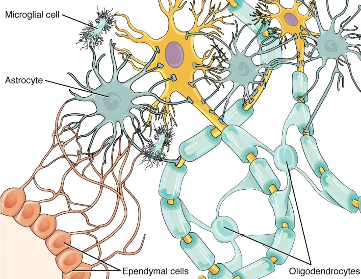
Neurons get all the attention, but they could not function for a second without glial cells. Glia (from the Greek word for “glue”) outnumber neurons by roughly 10:1 in certain regions and perform every essential support function the nervous system requires - from insulation and nutrition to immune defense and waste removal. Six major types of glial cells appear on the MCAT, four in the CNS and two in the PNS. Knowing each one’s location, function, and clinical relevance is non-negotiable for test day.
CNS Glial Cells
Astrocytes: The Multitaskers
Astrocytes are the most abundant glial cells in the CNS and arguably the most versatile. They are star-shaped cells with numerous processes that contact both neurons and blood vessels. Their functions include:
Blood-brain barrier (BBB) maintenance: Astrocyte foot processes (end-feet) wrap around CNS capillaries and help maintain the tight junctions that form the BBB. This barrier prevents most pathogens and large molecules in the blood from entering brain tissue.
Nutrient transfer: Astrocytes shuttle glucose from blood vessels to neurons and can convert glucose to lactate for neuronal fuel.
Neurotransmitter recycling: They take up excess neurotransmitters (especially glutamate) from the synaptic cleft, preventing excitotoxicity.
Ion homeostasis: Astrocytes buffer extracellular K+ concentrations, which is critical because even small changes in K+ levels alter neuronal excitability.
Structural support and repair: After CNS injury, astrocytes proliferate and form a glial scar (a process called reactive gliosis).
Oligodendrocytes: The CNS Insulators
Oligodendrocytes produce the myelin sheath in the CNS. Unlike Schwann cells, a single oligodendrocyte extends multiple flat, paddle-like processes that each wrap around a segment of a different axon. One oligodendrocyte can myelinate portions of up to 50 axons simultaneously.
This efficiency comes with a clinical downside: if a single oligodendrocyte is damaged, multiple axons lose their myelin at once.
Microglia: The Immune Defense
Microglia are the resident immune cells of the CNS. Unlike other glial cells, which derive from neural ectoderm, microglia originate from mesoderm (specifically, from yolk sac macrophage precursors that migrate into the brain during development). They function as the brain’s macrophages.
In their resting state, microglia extend long, thin processes that continuously survey the local environment. When they detect pathogens, debris, or damaged neurons, they retract their processes, become amoeboid, and phagocytose the threat. They also release pro-inflammatory cytokines to recruit additional immune responses.
Ependymal Cells: The Fluid Managers
Ependymal cells are ciliated epithelial cells that line the ventricles of the brain and the central canal of the spinal cord. Their beating cilia help circulate cerebrospinal fluid (CSF). Specialized ependymal cells in the choroid plexus actively produce CSF by filtering blood plasma.
CSF serves as a shock absorber for the brain, a medium for nutrient and waste exchange, and a source of buoyancy that reduces the effective weight of the brain from about 1,400 g to roughly 50 g.
The four glial cell types of the CNS, each performing a distinct support function. Credit: OpenStax Anatomy & Physiology 2e, CC BY 4.0
PNS Glial Cells
Schwann Cells: The PNS Insulators
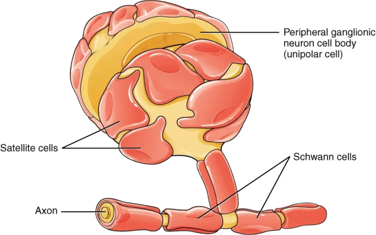
Schwann cells are the myelinating glia of the PNS. Each Schwann cell wraps around a single segment of a single axon, forming one internode of the myelin sheath. The gaps between adjacent Schwann cells are the nodes of Ranvier.
Schwann cells also play a crucial role in nerve regeneration. After PNS axon damage, Schwann cells form a regeneration tube that guides the regrowing axon back to its target. This is a major reason PNS nerves can regenerate while CNS axons generally cannot - the CNS lacks this guidance mechanism.
Not all Schwann cells produce myelin. Non-myelinating Schwann cells loosely envelop small-diameter axons without wrapping them in myelin layers. These unmyelinated fibers conduct signals more slowly.
Satellite Cells: The Ganglia Supporters
Satellite cells surround neuron cell bodies in PNS ganglia (such as dorsal root ganglia and autonomic ganglia). They provide structural support, regulate the chemical environment around the neuron, and may play a role in chronic pain signaling.
Think of satellite cells as the PNS counterpart to astrocytes - they nurture and protect neuronal cell bodies, just as astrocytes do in the CNS.
Schwann cells myelinate PNS axons while satellite cells support neuron cell bodies in ganglia. Credit: OpenStax Anatomy & Physiology 2e, CC BY 4.0
The Myelination Process
Whether performed by oligodendrocytes or Schwann cells, myelination follows the same basic principle: the glial cell membrane wraps concentrically around the axon, squeezing out cytoplasm to form a dense, lipid-rich sheath. The high lipid content (roughly 80% lipid, 20% protein) is what makes myelin such an effective electrical insulator.
Myelination involves the progressive wrapping of glial cell membrane around the axon. Credit: OpenStax Anatomy & Physiology 2e, CC BY 4.0
Myelinate CNS axons (one cell, many axon segments)
Neural ectoderm
Microglia
CNS
Immune defense, phagocytosis
Mesoderm
Ependymal cells
CNS
Line ventricles, produce and circulate CSF
Neural ectoderm
Schwann cells
PNS
Myelinate PNS axons (one cell, one axon segment), aid regeneration
Neural crest
Satellite cells
PNS
Support neuron cell bodies in ganglia
Neural crest
Which glial cell maintains the blood-brain barrier, and how does it do so?
Click to reveal answer
Astrocytes maintain the blood-brain barrier. Their foot processes (end-feet) wrap around CNS capillaries and help maintain the tight junctions between endothelial cells, preventing most pathogens and large molecules from crossing into brain tissue.
Why can PNS nerves regenerate after injury but CNS nerves generally cannot?
Click to reveal answer
After PNS injury, Schwann cells form a regeneration tube (band of Bungner) that physically guides the regrowing axon back to its target. The CNS lacks this mechanism - oligodendrocytes do not form regeneration tubes, and astrocytes form inhibitory glial scars that actively block regrowth.
Which glial cell type is derived from mesoderm rather than ectoderm, and what is its function?
Click to reveal answer
Microglia are derived from mesoderm (yolk sac macrophage precursors). They serve as the resident immune cells of the CNS, constantly surveying the environment and phagocytosing pathogens, debris, and damaged neurons when activated.
Every neuron in your body, even when completely “at rest” and not firing, maintains a voltage difference across its membrane of approximately -70 mV. This resting membrane potential is not a passive state - it is an actively maintained, energy-consuming condition that keeps the neuron primed and ready to fire at a moment’s notice. Understanding how this voltage is established, maintained, and calculated is one of the most tested topics in MCAT biology.
What Is Resting Membrane Potential?
The resting membrane potential is the voltage difference across the neuronal membrane when the neuron is not transmitting a signal. By convention, it is measured as the inside of the cell relative to the outside. At rest, the inside of a typical neuron sits at approximately -70 mV - meaning the interior is negatively charged compared to the extracellular fluid.
This negativity exists because of an unequal distribution of ions across the membrane and the selective permeability of the membrane to those ions.
Ion Distribution at Rest
The key ions involved are sodium (Na+), potassium (K+), chloride (Cl-), and large organic anions (A-, primarily proteins and nucleic acids that cannot cross the membrane).
At rest, the concentration gradients are:
Ion
Higher Concentration
Lower Concentration
Na+
Outside the cell (~145 mM)
Inside the cell (~12 mM)
K+
Inside the cell (~140 mM)
Outside the cell (~4 mM)
Cl-
Outside the cell (~120 mM)
Inside the cell (~4 mM)
Organic anions (A-)
Inside the cell
Cannot cross membrane
These gradients are not accidental. They are actively maintained by the Na+/K+ ATPase and represent a massive store of potential energy.
The Na+/K+ ATPase: The Master Pump
The sodium-potassium ATPase (Na+/K+ pump) is a transmembrane protein that uses the energy from one ATP molecule to pump 3 Na+ ions out of the cell and 2 K+ ions in. This 3-out, 2-in ratio is critical for two reasons:
It maintains the concentration gradients: Na+ is kept high outside and K+ is kept high inside.
It is electrogenic: Each cycle exports one more positive charge than it imports, creating a net loss of positive charge from the cell interior. This directly contributes about -3 to -5 mV to the resting potential.
The pump consumes a staggering amount of energy - neurons spend roughly 70% of their total ATP on Na+/K+ ATPase activity alone.
Leak Channels: Why K+ Dominates at Rest
While the Na+/K+ pump creates the concentration gradients, leak channels determine the resting potential. The neuronal membrane at rest is far more permeable to K+ than to Na+ because it contains many more K+ leak channels than Na+ leak channels (roughly 50-100 times more permeable to K+).
Here is what happens: K+ ions, which are concentrated inside the cell, flow outward through leak channels down their concentration gradient. As each positive K+ ion leaves, it leaves behind unmatched negative charges (the large organic anions that cannot cross the membrane). This outflow of positive charge makes the interior increasingly negative.
But this process does not continue indefinitely. As the inside becomes more negative, the growing electrical gradient begins to pull K+ back in. Eventually, the outward chemical driving force (concentration gradient pushing K+ out) is exactly balanced by the inward electrical driving force (negative interior pulling K+ back in). This balance point is the equilibrium potential for K+, approximately -90 mV.
The actual resting potential (-70 mV) is slightly less negative than the K+ equilibrium potential because the membrane is also slightly permeable to Na+. A small, steady leak of Na+ into the cell partially offsets the K+ effect. The resting potential thus sits between the K+ equilibrium potential (-90 mV) and the Na+ equilibrium potential (+60 mV), but much closer to K+ because the membrane is far more permeable to K+ at rest.
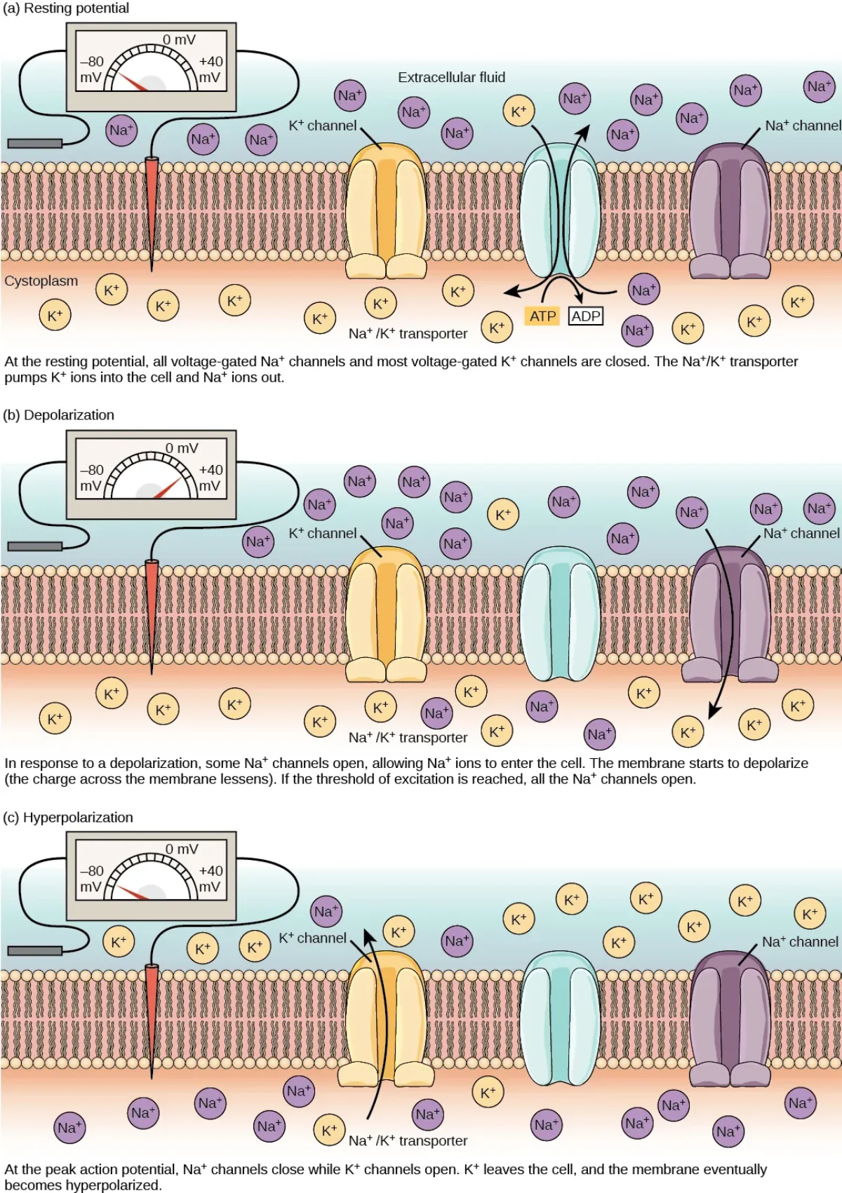
The resting membrane potential of -70 mV results from the Na+/K+ pump maintaining ion gradients and the membrane's high resting permeability to K+. Credit: OpenStax Anatomy & Physiology 2e, CC BY 4.0
The Nernst Equation: Equilibrium Potential for a Single Ion
The Nernst equation calculates the equilibrium potential for a single ion species - the membrane voltage at which there is no net movement of that particular ion across the membrane. This is the same Nernst equation from electrochemistry - learn it once, apply it in both subjects.
These calculations reveal why K+ equilibrium is deeply negative (K+ wants to leave the cell, making the inside negative) and why Na+ equilibrium is strongly positive (Na+ wants to enter, making the inside positive).
The Goldman Equation: The Full Picture
The Nernst equation only handles one ion at a time. In reality, the membrane is permeable to multiple ions simultaneously. The Goldman-Hodgkin-Katz (GHK) equation accounts for all permeable ions and their relative permeabilities to calculate the actual membrane potential.
The key conceptual takeaway: the Goldman equation weights each ion’s contribution by its permeability. At rest, PK is roughly 50-100 times larger than PNa, so the resting potential (-70 mV) is close to EK (-90 mV). During an action potential, Na+ channels open and PNa skyrockets - the membrane potential shifts toward ENa (+60 mV). This is depolarization.
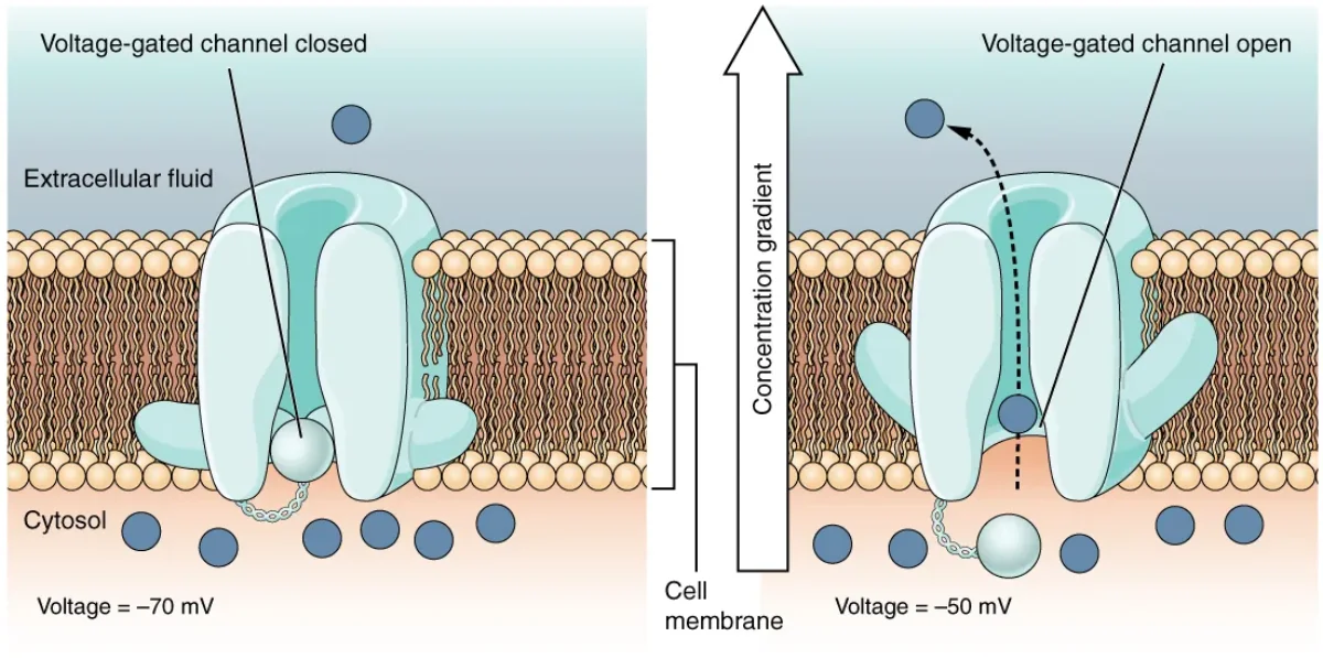
Voltage-gated ion channels open and close in response to changes in membrane potential, altering ion permeabilities. Credit: OpenStax Anatomy & Physiology 2e, CC BY 4.0
Factors That Alter Resting Membrane Potential
Several conditions can shift the resting potential, and the MCAT expects you to predict which direction:
Hyperkalemia (elevated extracellular K+): Reduces the K+ concentration gradient, so less K+ leaves the cell. The resting potential becomes less negative (depolarized), making the neuron more excitable and prone to spontaneous firing. This is why hyperkalemia is a medical emergency - it can cause fatal cardiac arrhythmias.
Hypokalemia (low extracellular K+): Increases the K+ gradient, so more K+ leaves the cell. The resting potential becomes more negative (hyperpolarized), making the neuron harder to excite.
Blocking the Na+/K+ ATPase: If the pump is inhibited, it stops maintaining gradients. Over time, Na+ accumulates inside and K+ leaks out, and the resting potential drifts toward 0 mV as concentration gradients dissipate.
The Na+/K+ ATPase pumps how many ions in each direction per ATP, and why is it called electrogenic?
Click to reveal answer
The pump moves 3 Na+ out and 2 K+ in per ATP hydrolyzed. It is electrogenic because it exports one more positive charge than it imports, creating a net outward positive current that directly contributes about -3 to -5 mV to the resting potential.
Why is the resting membrane potential (-70 mV) closer to EK (-90 mV) than to ENa (+60 mV)?
Click to reveal answer
At rest, the membrane is 50-100 times more permeable to K+ than to Na+ (due to abundant K+ leak channels). According to the Goldman equation, the membrane potential is weighted toward the equilibrium potential of the most permeable ion - so it sits near EK but is slightly less negative because of small Na+ leak inward.
How does hyperkalemia affect resting membrane potential and neuronal excitability?
Click to reveal answer
Hyperkalemia (high extracellular K+) reduces the K+ concentration gradient, so less K+ leaves the cell. The resting potential becomes less negative (depolarized, closer to threshold), making the neuron more excitable and prone to spontaneous firing. This can cause dangerous cardiac arrhythmias.
The resting membrane potential you learned in Section 4.4 is like a loaded spring - it stores energy, waiting for something to release it. That release is the action potential, a rapid, self-propagating electrical signal that travels the length of an axon without losing strength. Action potentials are the language of the nervous system: every thought, movement, and sensation depends on them.
This is one of the most heavily tested topics on the MCAT. You need to know each phase, every ion channel involved, and why the signal never fades as it travels.
Threshold: The Tipping Point
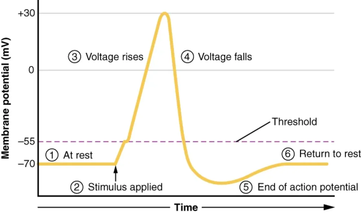
A neuron at rest sits at roughly -70 mV. Small depolarizations (graded potentials) can nudge the membrane potential upward, but nothing dramatic happens until the membrane reaches threshold, approximately -55 mV.
At threshold, a critical number of voltage-gated Na+ channels snap open, and the positive feedback loop begins: Na+ rushes in, depolarizing the membrane further, which opens even more Na+ channels. This is the point of no return. Any stimulus too weak to reach threshold will simply fade away - the neuron will not fire.
Phase 1: Depolarization
Once threshold is reached, voltage-gated Na+ channels open rapidly. Na+ floods into the cell down both its concentration gradient and electrical gradient. The membrane potential rockets from -55 mV all the way to approximately +30 mV in less than a millisecond.
This is the rising phase of the action potential. The interior of the cell briefly becomes positive relative to the outside - a complete reversal of the resting polarity.
Why does it stop at +30 mV? Two things happen almost simultaneously: (1) Na+ channels inactivate (a separate process from closing - more on this below), and (2) voltage-gated K+ channels finally open (they are slower to respond to voltage changes).
The stages of an action potential. Na+ channels open during depolarization, then inactivate as K+ channels open for repolarization. Credit: OpenStax Anatomy & Physiology 2e, CC BY 4.0
Phase 2: Repolarization
With Na+ channels inactivated and K+ channels now open, K+ rushes out of the cell. This outward flow of positive charge drives the membrane potential back down toward the resting value. The falling phase is repolarization.
Notice the key distinction: Na+ channels inactivate (a ball-and-chain mechanism physically blocks the channel pore), while K+ channels open. Both events contribute to repolarization, but they involve entirely different channel behaviors.
Phase 3: Hyperpolarization (The Undershoot)
Voltage-gated K+ channels are slow to open, and they are also slow to close. Even after the membrane potential returns to -70 mV, K+ continues to flow out. This drives the membrane potential briefly below the resting level, to about -80 to -90 mV. This dip is called hyperpolarization or the undershoot.
The Na+/K+ ATPase and K+ leak channels eventually restore the resting membrane potential to -70 mV, but the hyperpolarization phase has important consequences for refractory periods.
AnimationThe Action Potential
Key idea
An action potential is all-or-nothing: below −55 mV you get only a small graded depolarization that decays back to rest, and at or above −55 mV you get one spike of fixed size and duration. Stimulus strength is coded by firing frequency, never by spike amplitude.
Drag Stimulus: under 15 mV the membrane only bumps and decays, at or above it every spike is identical. Press Stimulate to try firing again inside the shaded refractory bands.
The All-or-Nothing Principle
An action potential is all-or-nothing. If threshold is reached, the neuron fires a full-strength action potential. If threshold is not reached, nothing happens. There is no such thing as a “half” action potential or a “strong” versus “weak” one.
This raises an important question: if all action potentials have the same amplitude, how does your nervous system distinguish between a light tap and a hard punch? The answer is frequency coding. A stronger stimulus causes a neuron to fire more action potentials per second, not bigger ones. It also recruits more neurons to fire simultaneously. The brain interprets higher frequency and greater neuron recruitment as a more intense stimulus.
The Three States of Na+ Channels
Understanding Na+ channel states is critical for understanding refractory periods:
Closed (resting): The channel is closed but capable of opening. This is the state at resting membrane potential.
Open (activated): The channel is open and Na+ flows through. This happens during depolarization.
Inactivated: A separate inactivation gate blocks the channel even though the activation gate is open. The channel CANNOT be opened again until it resets to the closed state. This requires repolarization.
The difference between “closed” and “inactivated” is the single most important detail for understanding refractory periods.
Absolute Refractory Period
During the absolute refractory period, it is impossible to fire another action potential regardless of how strong the stimulus is. This corresponds to the time when Na+ channels are either open or inactivated - they physically cannot be activated again.
The absolute refractory period spans from the moment threshold is reached through most of repolarization. Its purpose is to ensure that the action potential propagates in one direction only. The region of membrane that just fired cannot be re-excited, so the signal moves forward.
Relative Refractory Period
During the relative refractory period, a neuron can fire again, but only if it receives a stronger-than-normal stimulus. This corresponds to the hyperpolarization phase, when voltage-gated K+ channels are still open and the membrane potential is more negative than usual.
Because the membrane starts from a more negative baseline, a larger depolarization is needed to reach threshold. A normal stimulus will not do it, but a very strong one can.
Action Potential Propagation
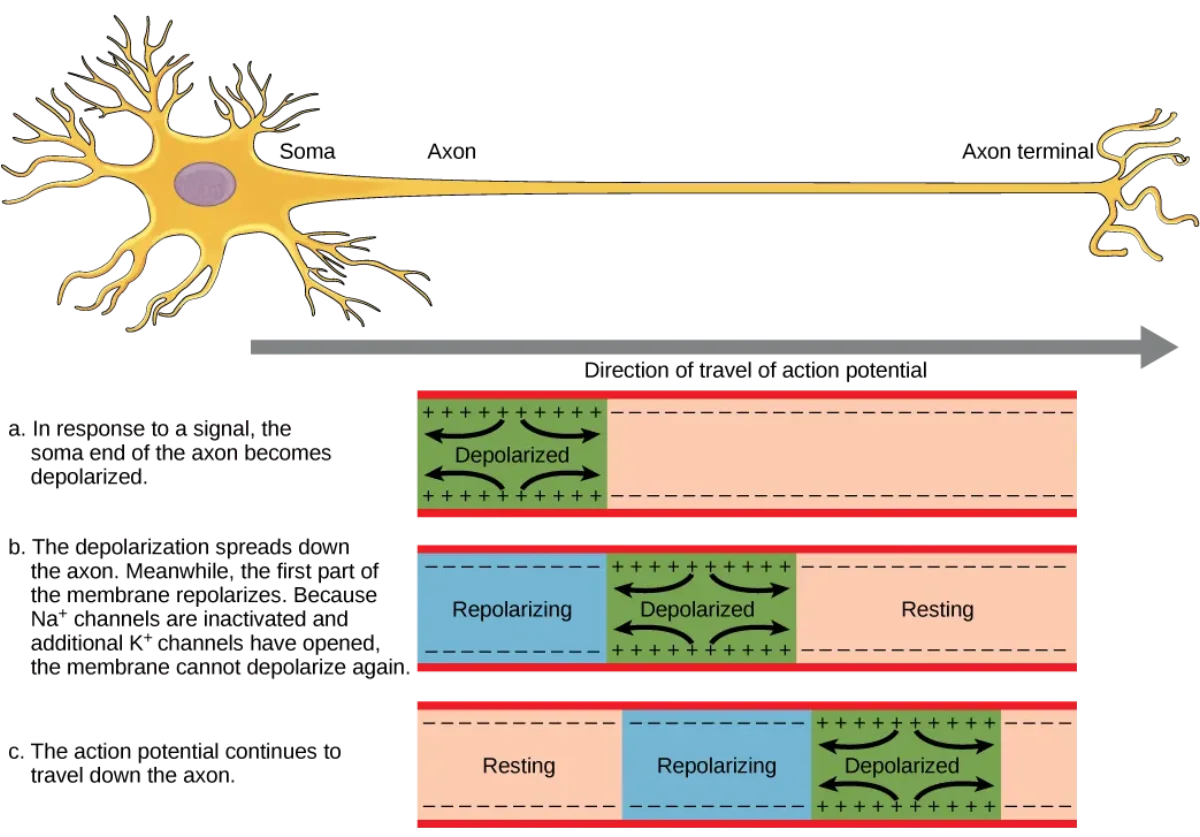
Once an action potential fires at one spot on the axon, it does not stay there. The influx of Na+ at one location creates a local current that depolarizes the adjacent membrane to threshold, triggering a new action potential there. This process repeats continuously down the length of the axon.
In unmyelinated fibers, this happens at every point along the membrane - continuous conduction. It works, but it is relatively slow because every patch of membrane must go through the full action potential cycle.
In myelinated fibers, the action potential appears to “jump” from one node of Ranvier to the next - saltatory conduction (covered in detail in Section 4.6). This is dramatically faster and more energy-efficient.
Action potential propagation. Local currents from the depolarized region spread to adjacent resting membrane, triggering the next action potential. Credit: OpenStax Anatomy & Physiology 2e, CC BY 4.0
Key Ions and Channels Summary
| Phase | Channel Activity | Ion Movement | Membrane Potential |
|---|---|---|---|
| Resting | K+ leak channels open, voltage-gated channels closed | K+ slowly leaks out | -70 mV |
| Depolarization | Voltage-gated Na+ channels open | Na+ rushes in | -55 mV to +30 mV |
| Repolarization | Na+ channels inactivate, voltage-gated K+ channels open | K+ rushes out | +30 mV to -70 mV |
| Hyperpolarization | K+ channels slow to close | K+ continues leaving | -70 mV to -90 mV |
| Return to rest | K+ channels close, Na+/K+ ATPase restores gradients | Na+ pumped out, K+ pumped in | Back to -70 mV |
What happens at threshold (-55 mV) that makes depolarization a “point of no return”?
Click to reveal answer
A critical number of voltage-gated Na+ channels open, and the resulting Na+ influx depolarizes the membrane further, opening even more Na+ channels. This positive feedback loop becomes self-sustaining, guaranteeing a full action potential.
What is the difference between the absolute and relative refractory periods?
Click to reveal answer
During the absolute refractory period, Na+ channels are inactivated and it is impossible to fire another action potential regardless of stimulus strength. During the relative refractory period, K+ channels are still open (hyperpolarization), so firing is possible but requires a stronger-than-normal stimulus to reach threshold.
If all action potentials have the same amplitude (all-or-nothing), how does the nervous system encode stimulus intensity?
Click to reveal answer
Stimulus intensity is encoded by frequency coding - stronger stimuli cause neurons to fire more action potentials per second and recruit more neurons to fire simultaneously. The brain interprets higher firing frequency and greater neuron recruitment as a stronger stimulus.
In Section 4.5 you saw that action potentials propagate by triggering the next patch of membrane to fire. In an unmyelinated axon, this happens at every point along the fiber - a slow, energy-expensive process. Evolution solved this problem with myelin, a fatty insulating sheath that transforms the way signals travel. The result is saltatory conduction, one of the most elegant speed upgrades in biology.
The word “saltatory” comes from the Latin saltare, meaning “to jump.” That is exactly what the action potential appears to do - it leaps from one gap in the myelin to the next, skipping over the insulated segments entirely.
Continuous Conduction: The Slow Lane
In unmyelinated fibers, voltage-gated Na+ and K+ channels are distributed along the entire length of the axon. When an action potential fires at one location, local currents depolarize the immediately adjacent membrane to threshold, and a new action potential is generated there. This repeats point by point.
This approach works, but it has two major drawbacks. First, it is slow - conduction velocities in unmyelinated fibers typically range from 0.5 to 2 m/s. Second, every patch of membrane that fires an action potential needs its Na+ and K+ gradients restored by the Na+/K+ ATPase, which costs ATP. Over a long axon, this adds up to significant energy expenditure.
Saltatory Conduction: The Express Lane
In myelinated fibers, the axon is wrapped in layers of myelin produced by glial cells - oligodendrocytes in the CNS and Schwann cells in the PNS. The myelin acts as an electrical insulator, preventing ion leakage across the membrane.
Between each myelinated segment are small exposed gaps called nodes of Ranvier. These nodes are packed with voltage-gated Na+ channels. When an action potential fires at one node, the resulting current flows passively through the myelinated segment (which resists ion leakage) and depolarizes the membrane at the next node to threshold.
The action potential effectively “jumps” from node to node. Because the signal only needs to be regenerated at the nodes - not at every point along the axon - saltatory conduction is both faster and more energy-efficient than continuous conduction.
Saltatory conduction in a myelinated axon. The action potential regenerates only at nodes of Ranvier, jumping over insulated myelin segments. Credit: OpenStax Anatomy & Physiology 2e, CC BY 4.0
Why Saltatory Conduction Is Faster
Two factors explain the speed increase:
Distance per regeneration step. In continuous conduction, the action potential regenerates at essentially every micrometer. In saltatory conduction, nodes of Ranvier are spaced roughly 1 mm apart. Each “jump” covers a much greater distance before the signal needs to be rebuilt.
Passive current flow is nearly instantaneous. The current flowing through a myelinated internode travels at close to the speed of electrical conduction in a cable - far faster than the time required for voltage-gated channels to open, ions to flow, and the membrane to depolarize.
Myelinated neurons can conduct signals at speeds of up to 120 m/s - roughly 60 to 100 times faster than unmyelinated fibers of similar diameter.
Why Saltatory Conduction Is More Energy-Efficient
Since action potentials only fire at the nodes of Ranvier, only those small regions experience Na+ influx and K+ efflux. That means the Na+/K+ ATPase only needs to restore ion gradients at the nodes, not along the entire length of the axon.
This dramatically reduces ATP consumption. For a neuron that fires thousands of action potentials per second, this energy savings is substantial.
Factors Affecting Conduction Velocity
Three main factors determine how fast an action potential travels:
Factor
Effect on Speed
Explanation
Myelination
Greatly increases speed
Enables saltatory conduction; reduces capacitance and ion leakage
Axon diameter
Larger = faster
Wider axons have lower internal resistance, so current spreads farther
Temperature
Higher = faster (to a point)
Warmer temperatures increase ion channel kinetics and diffusion rates
The MCAT often asks you to predict conduction velocity changes. A large, myelinated fiber (like an alpha motor neuron) conducts much faster than a small, unmyelinated fiber (like a C-fiber carrying dull pain). This is why sharp pain (carried by myelinated A-delta fibers) reaches the brain before dull, aching pain (carried by unmyelinated C fibers).
Why is saltatory conduction faster AND more energy-efficient than continuous conduction?
Click to reveal answer
Faster: the action potential jumps from node to node (spaced ~1 mm apart) rather than regenerating at every point, and passive current flow through myelinated segments is nearly instantaneous. More energy-efficient: Na+/K+ ATPase only needs to restore ion gradients at the nodes of Ranvier, not along the entire axon length, requiring far less ATP.
What three factors affect action potential conduction velocity, and how does each affect speed?
Click to reveal answer
(1) Myelination - greatly increases speed by enabling saltatory conduction. (2) Axon diameter - larger diameter means lower internal resistance, so current spreads farther and conduction is faster. (3) Temperature - higher temperatures increase ion channel kinetics and diffusion rates, increasing speed (to a point).
Multiple sclerosis destroys which glial cell in which division? Guillain-Barre syndrome destroys which glial cell in which division?
Click to reveal answer
MS destroys oligodendrocytes in the CNS. Guillain-Barre destroys Schwann cells in the PNS. Both are autoimmune demyelinating diseases, but they target different parts of the nervous system.
An action potential races down the axon and arrives at its destination - but it cannot simply jump across to the next cell. The gap between neurons, called the synapse, requires the electrical signal to be converted into a chemical one (or, in rare cases, passed directly through gap junctions). This conversion process is synaptic transmission, and it is where the nervous system gains its incredible flexibility. Synapses can be strengthened, weakened, or modulated by drugs, disease, and experience.
Understanding synaptic transmission is essential for MCAT questions spanning neuroscience, pharmacology, and behavioral science.
Anatomy of the Synapse
The synapse has three components:
Presynaptic terminal (axon terminal or synaptic bouton) - the end of the sending neuron’s axon. It contains mitochondria and synaptic vesicles filled with neurotransmitter molecules.
Synaptic cleft - the narrow gap (~20 nm wide) between the two neurons. Neurotransmitters must diffuse across this space.
Postsynaptic membrane - the receiving surface of the next neuron (or muscle cell, or gland cell). It is studded with receptors specific to the neurotransmitter being released.
Structure of a chemical synapse. Neurotransmitter-filled vesicles in the presynaptic terminal release their contents into the synaptic cleft. Credit: OpenStax Anatomy & Physiology 2e, CC BY 4.0
Chemical Synapse: Step by Step
The sequence of events at a chemical synapse is one of the most testable processes on the MCAT. Know every step:
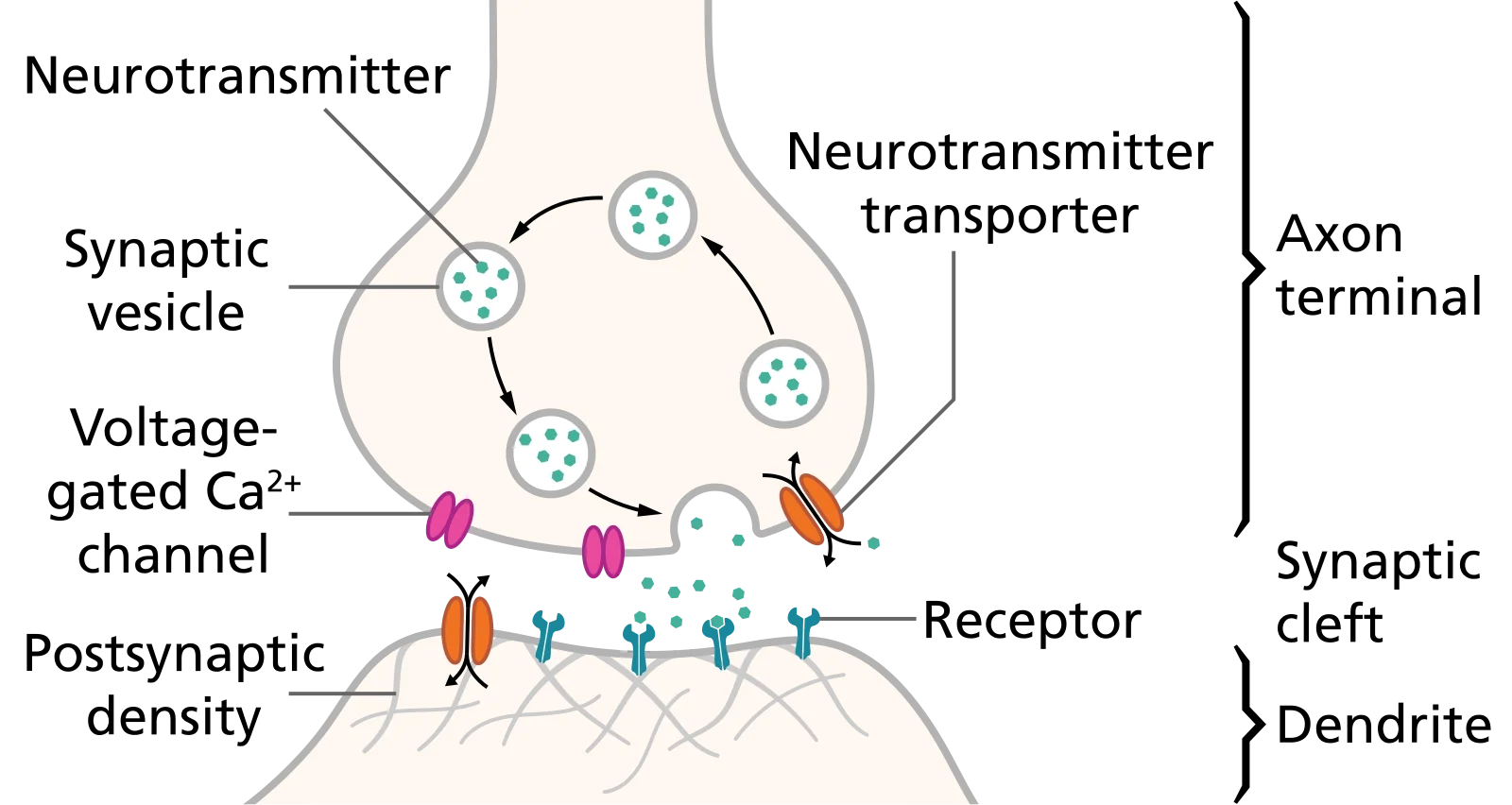
Action potential arrives at the presynaptic terminal.
Voltage-gated Ca2+ channels open. Depolarization of the terminal opens these channels, and Ca2+ floods into the presynaptic cell.
Ca2+ triggers vesicle fusion. The rise in intracellular Ca2+ causes synaptic vesicles to fuse with the presynaptic membrane, releasing neurotransmitter into the cleft by exocytosis.
Neurotransmitters diffuse across the synaptic cleft (~20 nm).
Neurotransmitters bind postsynaptic receptors, opening or closing ion channels and changing the postsynaptic membrane potential.
Signal termination occurs by one of three mechanisms: enzymatic degradation (e.g., acetylcholinesterase breaks down ACh), reuptake into the presynaptic terminal (e.g., serotonin transporter), or simple diffusion away from the cleft.
Neurotransmitter release at a chemical synapse. Vesicles fuse with the presynaptic membrane and release their contents into the cleft, where they bind postsynaptic receptors. Credit: Wikimedia Commons, CC BY-SA 4.0
Electrical Synapses
Not all synapses use chemicals. Electrical synapses use gap junctions - protein channels (connexons) that directly connect the cytoplasm of two neurons. Ions flow directly from one cell to the next.
Electrical synapses are bidirectional and extremely fast because there is no chemical intermediary. However, they are less modulatable - you cannot easily fine-tune or inhibit them the way you can with chemical synapses.
Electrical synapses are found in cardiac muscle (allowing synchronized contraction), smooth muscle, and certain brain circuits where speed is critical.
EPSPs and IPSPs
When neurotransmitters bind postsynaptic receptors, the effect depends on which ion channels are opened or closed:
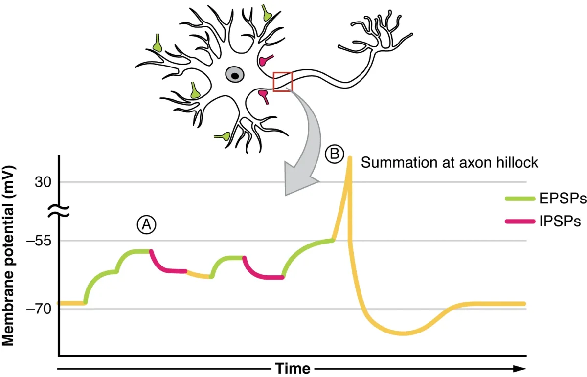
Excitatory postsynaptic potentials (EPSPs) result when the postsynaptic membrane is depolarized, bringing it closer to threshold. This typically happens when Na+ channels open and Na+ flows into the cell. EPSPs increase the probability that the postsynaptic neuron will fire an action potential.
Inhibitory postsynaptic potentials (IPSPs) result when the postsynaptic membrane is hyperpolarized, pushing it further from threshold. This happens when Cl- channels open (Cl- flows in) or K+ channels open (K+ flows out). IPSPs decrease the probability of firing.
A single EPSP is usually too small to reach threshold on its own. For the postsynaptic neuron to fire, multiple EPSPs must add together - a process called summation.
Summation: Adding Signals at the Axon Hillock
The axon hillock is the integration zone of the neuron. It receives all the EPSPs and IPSPs from the dendrites and cell body and determines whether their sum reaches threshold.
Temporal summation occurs when a single presynaptic neuron fires rapidly in succession. Each EPSP arrives before the previous one fades, and they stack on top of each other. Think of it as one person repeatedly pushing a swing - if they push fast enough, the swing goes higher each time.
Spatial summation occurs when multiple presynaptic neurons fire simultaneously, and their EPSPs (arriving at different locations on the dendrites) combine at the axon hillock. Think of it as several people pushing the same swing at the same time.
In reality, both temporal and spatial summation happen simultaneously. The axon hillock sums all excitatory and inhibitory inputs. If the net result reaches threshold, the neuron fires. If not, it stays quiet.
Temporal summation (rapid firing from one neuron) and spatial summation (simultaneous input from multiple neurons) both contribute to reaching threshold at the axon hillock. Credit: OpenStax Anatomy & Physiology 2e, CC BY 4.0
Ionotropic vs. Metabotropic Receptors
Postsynaptic receptors fall into two broad categories:
Ionotropic receptors (ligand-gated ion channels) are fast-acting. When the neurotransmitter binds, the receptor itself is the ion channel - it opens immediately, allowing ions to flow within milliseconds. Examples include nicotinic acetylcholine receptors and GABA-A receptors.
Metabotropic receptors (G-protein coupled receptors, or GPCRs) are slower but longer-lasting. Neurotransmitter binding activates an intracellular G-protein, which triggers a second messenger cascade (e.g., cAMP, IP3). This can open or close ion channels indirectly, modify gene expression, or produce other cellular effects. Examples include muscarinic acetylcholine receptors, dopamine receptors, and GABA-B receptors.
Neurotransmitter signaling must be turned off quickly to allow precise control. Three mechanisms accomplish this:
Enzymatic degradation: Enzymes in the synaptic cleft break down the neurotransmitter. The classic example is acetylcholinesterase (AChE), which rapidly hydrolyzes ACh into acetate and choline.
Reuptake: Transporter proteins on the presynaptic membrane pump the neurotransmitter back into the presynaptic terminal for recycling. This is the primary termination mechanism for dopamine, serotonin, and norepinephrine.
Diffusion: The neurotransmitter simply drifts away from the synaptic cleft and is absorbed by surrounding glial cells or degraded elsewhere.
What is the role of Ca2+ at the presynaptic terminal during chemical synaptic transmission?
Click to reveal answer
When the action potential arrives at the presynaptic terminal, voltage-gated Ca2+ channels open and Ca2+ enters the cell. This Ca2+ influx triggers synaptic vesicles to fuse with the presynaptic membrane, releasing neurotransmitter into the synaptic cleft by exocytosis. Without Ca2+, no neurotransmitter is released.
What is the difference between temporal summation and spatial summation?
Click to reveal answer
Temporal summation occurs when a single presynaptic neuron fires rapidly, stacking EPSPs before previous ones fade. Spatial summation occurs when multiple presynaptic neurons fire simultaneously, combining their EPSPs at the axon hillock. Both increase the chance of reaching threshold.
What are the main inhibitory and excitatory neurotransmitters in the CNS?
Click to reveal answer
GABA is the main inhibitory neurotransmitter in the CNS (glycine is the main inhibitory NT in the spinal cord). Glutamate is the main excitatory neurotransmitter in the CNS. Remember: GABA = "Going Away" (inhibition), Glutamate = "Go" (excitation).
The brain is the most complex organ in the human body, containing roughly 86 billion neurons connected by trillions of synapses. For the MCAT, you do not need to memorize every gyrus and sulcus, but you absolutely need to know the major regions, what they do, and what happens when they are damaged. Brain anatomy shows up in biology, psychology, and even biochemistry passages.
This section covers the cerebrum, diencephalon, brainstem, cerebellum, limbic system, basal ganglia, meninges, and cerebrospinal fluid. It is one of the longest sections in this chapter for a reason - every one of these structures is fair game on test day.
The Cerebrum: Four Lobes
The cerebrum is the largest part of the brain, accounting for about 85% of total brain mass. Its surface is covered by the cerebral cortex, a thin layer of gray matter (neuron cell bodies) folded into ridges (gyri) and grooves (sulci) to maximize surface area. Beneath the cortex lies white matter (myelinated axons connecting different cortical regions).
The cerebrum is divided into two hemispheres (left and right) connected by the corpus callosum, a massive bundle of axons that allows the hemispheres to communicate. Each hemisphere is divided into four lobes.
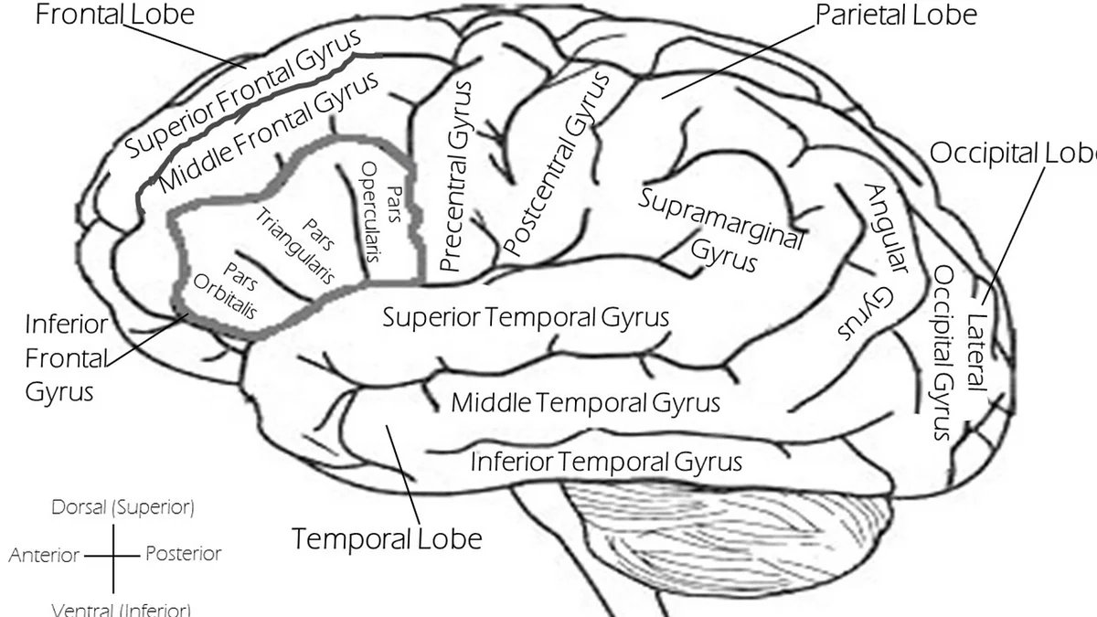
The four lobes of the cerebral cortex. Each lobe has specialized functions, though they work together constantly. Credit: Pixabay, free to use
Interactive 3D Brain. Rotate to see how the four lobes, cerebellum, and brainstem fit together spatially. Click the annotation pins to identify each region.Credit: AbdulMuhaymin via Sketchfab, CC BY
Frontal Lobe
The frontal lobe is the largest lobe, occupying the front third of each hemisphere. It handles some of the most “human” functions:
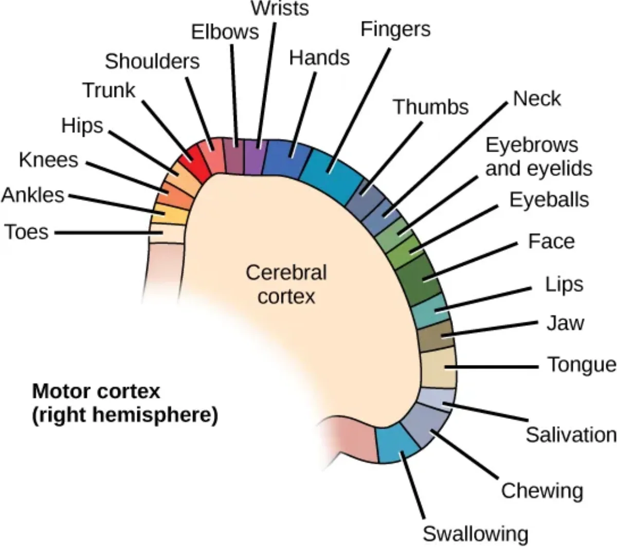
Primary motor cortex (precentral gyrus): Initiates voluntary movement. It is organized as a motor homunculus - a map where different body parts are represented in proportion to how finely they are controlled (hands and face get disproportionately large areas).
Prefrontal cortex: Executive functions - planning, judgment, personality, impulse control, working memory, and social behavior. Damage here produces dramatic personality changes.
- **Broca's area** (usually left hemisphere): Controls speech production - the motor planning of speaking. Damage causes **Broca's aphasia** - the patient understands language but cannot produce fluent speech. Speech is halting and effortful ("Broca's is Broken speech").
Parietal Lobe
The parietal lobe sits behind the frontal lobe, separated by the central sulcus:
Primary somatosensory cortex (postcentral gyrus): Processes touch, pressure, temperature, and pain from the body. It is also organized as a homunculus, with the most sensitive body parts (lips, fingertips) getting the most cortical real estate.
Spatial awareness and integration: The parietal lobe integrates sensory information to create a sense of body position and spatial relationships. Damage to the right parietal lobe can cause hemispatial neglect - patients ignore the entire left side of their world.
Temporal Lobe
The temporal lobe sits below the lateral sulcus on each side of the brain:
Primary auditory cortex: Processes sound.
Wernicke’s area (usually left hemisphere): Language comprehension. Damage causes Wernicke’s aphasia - the patient speaks fluently but the words are nonsensical. They cannot understand spoken or written language (“Wernicke’s is Wordy but Wrong”).
Hippocampus (medial temporal lobe): Critical for forming new long-term memories. Bilateral hippocampal damage causes anterograde amnesia - the inability to form new declarative memories (the case of patient H.M.).
Occipital Lobe
The occipital lobe occupies the back of the brain:
Primary visual cortex: Processes visual information received from the eyes via the optic nerve and lateral geniculate nucleus of the thalamus.
Damage to the occipital lobe causes cortical blindness - the eyes work fine, but the brain cannot process what they see.
The Diencephalon: Thalamus and Hypothalamus
The diencephalon sits deep within the brain, between the cerebrum and the brainstem.
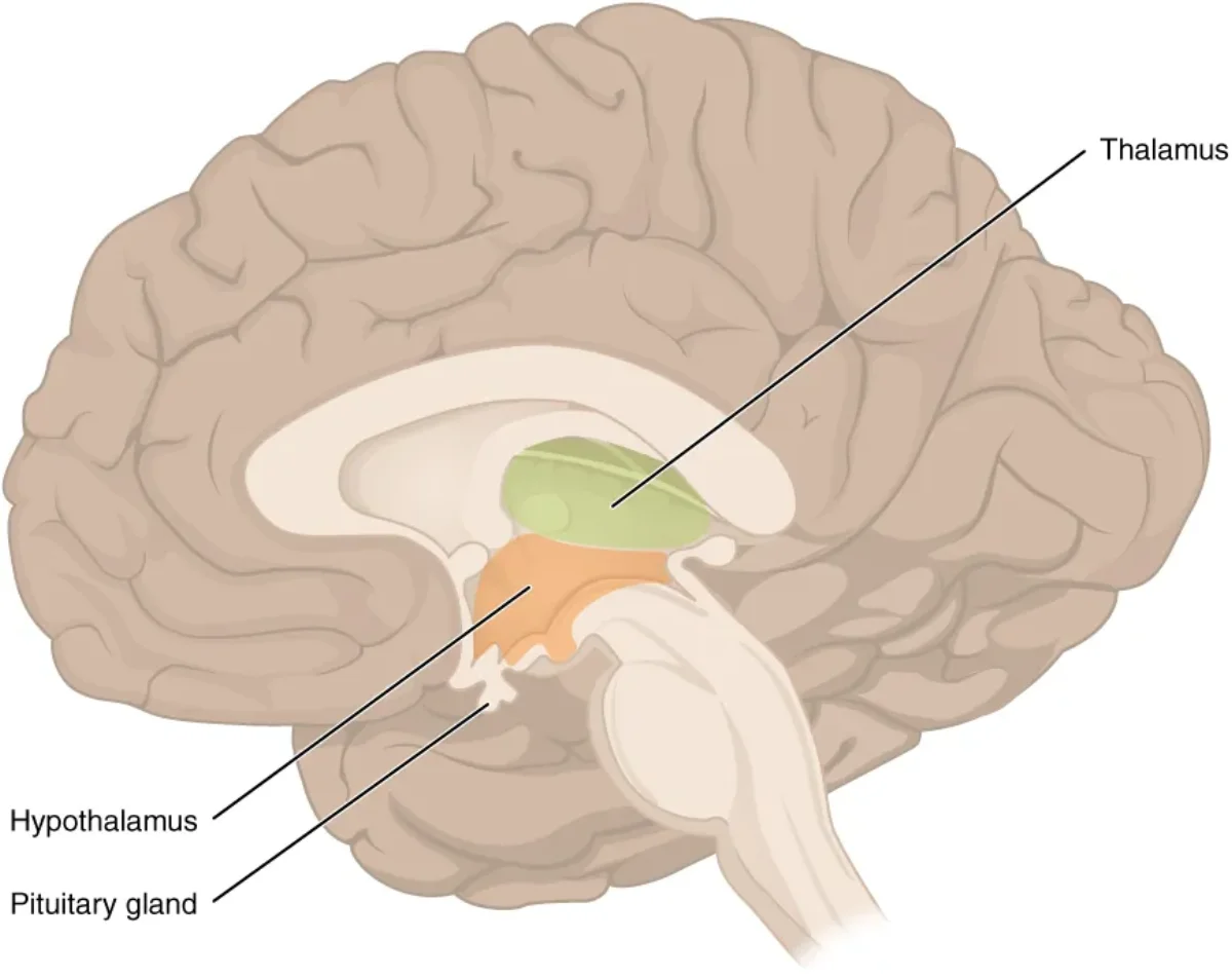
The diencephalon, showing the thalamus (sensory relay) and hypothalamus (homeostatic control center). Credit: OpenStax Anatomy & Physiology 2e, CC BY 4.0
The thalamus is the brain’s sensory relay station. Nearly all sensory information passes through the thalamus before reaching the cerebral cortex. Vision goes through the lateral geniculate nucleus, hearing through the medial geniculate nucleus, and somatosensory information through the ventral posterior nucleus.
The one major exception: smell (olfaction) bypasses the thalamus entirely and projects directly to the olfactory cortex. This is a classic MCAT fact.
The hypothalamus is the master regulator of homeostasis. Despite being only about the size of an almond, it controls:
Autonomic nervous system output (sympathetic and parasympathetic tone)
Endocrine function - the hypothalamus controls the pituitary gland (the “master gland”) through releasing and inhibiting hormones. This hypothalamic-pituitary axis is one of the most tested endocrine concepts on the MCAT.
Emotional responses (works with the limbic system)
The Brainstem: Midbrain, Pons, and Medulla Oblongata
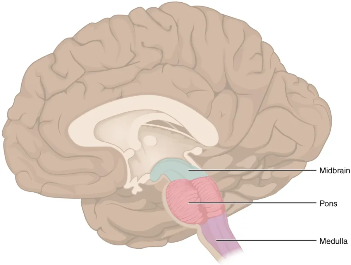
The brainstem connects the cerebrum to the spinal cord and controls the most fundamental life-sustaining functions. It consists of three regions, from top to bottom:
The three regions of the brainstem: midbrain, pons, and medulla oblongata. Credit: OpenStax Anatomy & Physiology 2e, CC BY 4.0
Midbrain (mesencephalon): Processes visual and auditory reflexes. Contains the superior colliculus (visual reflexes - e.g., turning your head toward a flash of light) and the inferior colliculus (auditory reflexes - e.g., orienting toward a sudden sound). Also contains the substantia nigra, which produces dopamine for the basal ganglia (degeneration here causes Parkinson’s disease).
Pons: Serves as a bridge (Latin pons = bridge) between the cerebellum and the cerebral cortex. It relays information between these structures and also plays a role in regulating breathing (works with the medulla’s respiratory centers). Several cranial nerve nuclei are located here.
Medulla oblongata: The most vital part of the brainstem. It controls autonomic functions that keep you alive:
Cardiovascular center: Regulates heart rate and blood pressure
Respiratory center: Controls the basic rhythm of breathing
Other reflexes: Vomiting, coughing, sneezing, swallowing, hiccupping
Damage to the medulla is often fatal because these are the functions you cannot live without. The medulla is also where most descending motor tracts and ascending sensory tracts decussate (cross over to the opposite side), which is why the left brain controls the right body and vice versa.
Reticular formation: A network of neurons running through the entire length of the brainstem. It regulates arousal, consciousness, and the sleep-wake cycle. The reticular activating system (RAS) is the ascending portion that keeps the cortex alert. Damage to the RAS can cause coma.
The Cerebellum
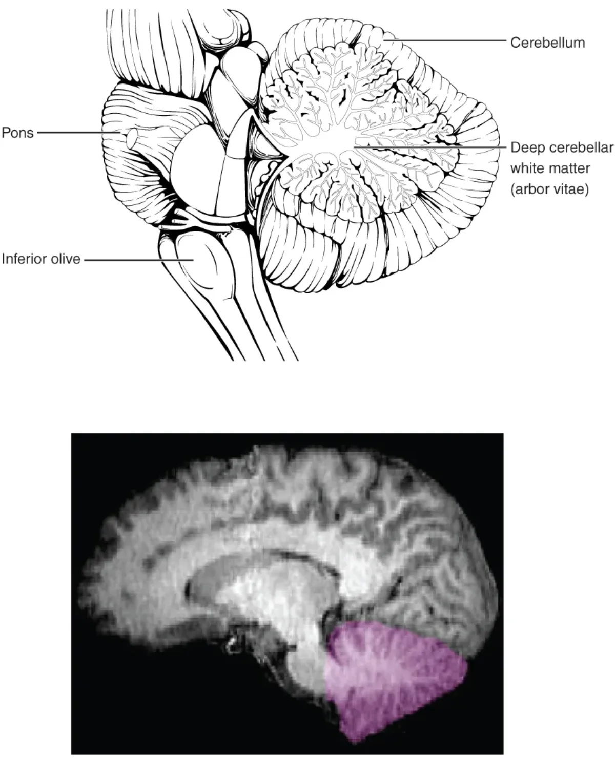
The cerebellum (“little brain”) sits behind the brainstem at the back of the skull. It contains more neurons than the rest of the brain combined, despite being only about 10% of brain volume.
The cerebellum coordinates movement, balance, and motor learning but does not initiate voluntary movement. Credit: OpenStax Anatomy & Physiology 2e, CC BY 4.0
Key functions of the cerebellum:
Motor coordination: Smooths and fine-tunes movements initiated by the motor cortex. Damage causes ataxia - jerky, uncoordinated movement.
Balance and posture: Integrates input from the vestibular system to maintain equilibrium.
Motor learning: Stores procedural memories like riding a bike, playing piano, or typing. Once learned, these tasks become “automatic” and run through cerebellar circuits.
A critical distinction: the cerebellum does not initiate movement. That is the job of the primary motor cortex (frontal lobe). The cerebellum compares intended movement (from the cortex) with actual movement (from proprioceptive feedback) and sends corrections in real time. Think of it as a GPS that constantly recalculates your route - it does not decide where you are going, but it makes sure you get there smoothly.
The Limbic System
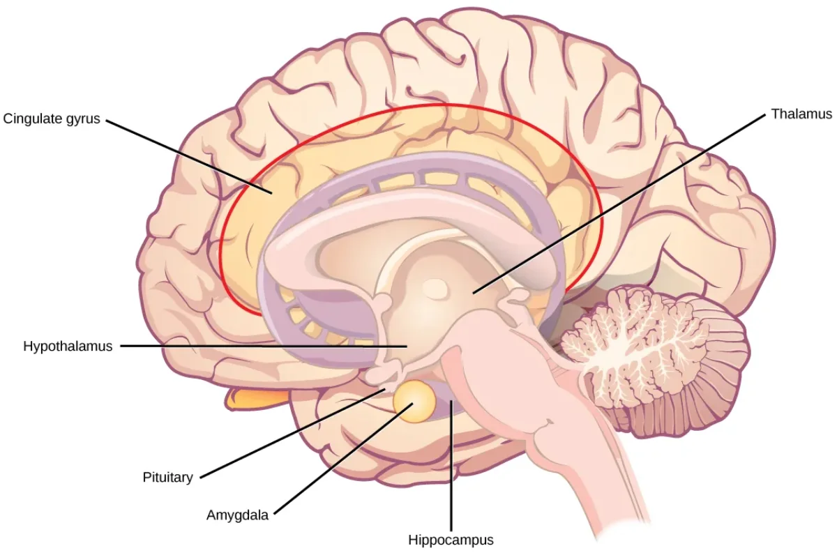
The limbic system is a group of interconnected structures that sit deep within the brain, forming a ring around the top of the brainstem. It handles emotions, motivation, and memory formation.
Major components of the limbic system. These structures work together to process emotion and form memories. Credit: OpenStax Anatomy & Physiology 2e, CC BY 4.0
Key limbic structures:
Hippocampus: Essential for converting short-term memories into long-term declarative memories (facts and events). It does not store memories permanently - it transfers them to the cortex over time. Bilateral damage causes anterograde amnesia.
Amygdala: Processes fear, aggression, and emotional significance. It attaches emotional weight to memories (which is why emotionally charged events are remembered more vividly). Damage to the amygdala reduces fear responses.
Cingulate gyrus: Links emotions to behavior and plays a role in attention and pain perception.
The limbic system works closely with the hypothalamus (emotional responses trigger autonomic and endocrine changes) and the prefrontal cortex (which modulates limbic output - this is how you can suppress an emotional impulse).
Basal Ganglia (Basal Nuclei)
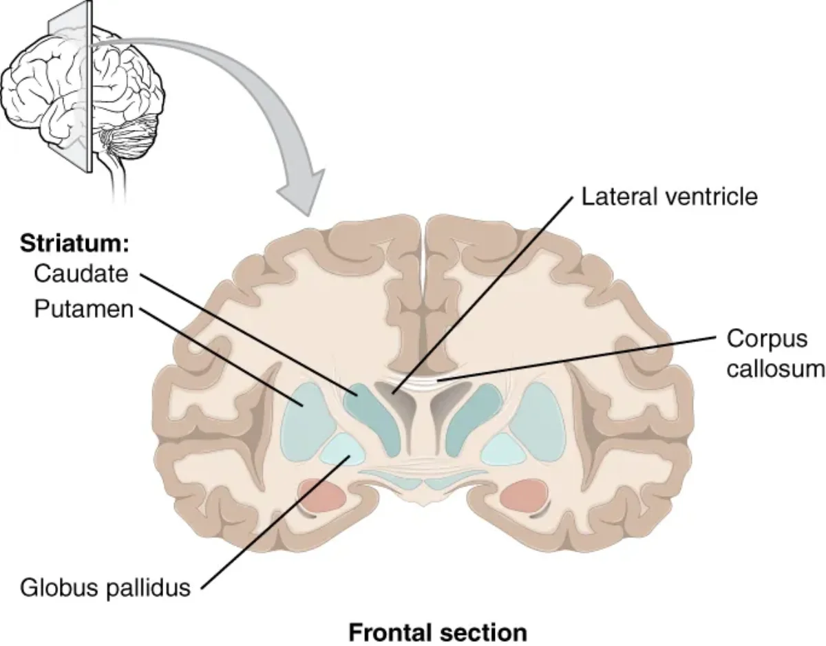
The basal ganglia are a group of subcortical nuclei (caudate nucleus, putamen, globus pallidus, and associated structures) involved in motor planning and initiation.
The basal nuclei (basal ganglia) play a critical role in regulating voluntary movement. Credit: OpenStax Anatomy & Physiology 2e, CC BY 4.0
The basal ganglia form a loop with the motor cortex and thalamus. They help select which movements to perform and suppress unwanted movements. Disruption of this circuit - whether from loss of dopaminergic input or degeneration of the basal ganglia neurons themselves - produces characteristic movement disorders.
The Meninges
The brain and spinal cord are wrapped in three protective membranes called meninges, from outermost to innermost:
Dura mater (“tough mother”): The thickest, outermost layer. It is a dense, fibrous membrane that adheres to the inner surface of the skull. Epidural bleeding (between the skull and dura) is typically caused by arterial rupture and is a neurosurgical emergency.
Arachnoid mater (“spider mother”): A web-like middle layer. The space beneath it - the subarachnoid space - is filled with cerebrospinal fluid (CSF). Subarachnoid hemorrhage causes “the worst headache of my life.”
Pia mater (“gentle mother”): The thinnest, innermost layer. It clings directly to the surface of the brain, following every gyrus and sulcus.
Cerebrospinal Fluid (CSF)
CSF is a clear fluid that bathes the brain and spinal cord. It serves three functions: cushioning (shock absorption), buoyancy (reduces the effective weight of the brain from ~1,400 g to ~50 g), and waste removal.
CSF is produced by the choroid plexus, a network of capillaries and ependymal cells lining the ventricles. It circulates through the four ventricles (two lateral ventricles, third ventricle, fourth ventricle), exits into the subarachnoid space, and is eventually reabsorbed into the venous blood via arachnoid granulations.
Blockage of CSF flow causes a dangerous buildup of fluid that increases intracranial pressure.
Putting It All Together: Brain Region Summary
Structure
Key Functions
Clinical Connection
Frontal lobe
Motor initiation, planning, personality, Broca’s area (speech production)
Broca’s aphasia, personality changes
Parietal lobe
Somatosensory processing, spatial awareness
Hemispatial neglect
Temporal lobe
Auditory processing, Wernicke’s area (language comprehension), hippocampus
Wernicke’s aphasia, amnesia
Occipital lobe
Visual processing
Cortical blindness
Thalamus
Sensory relay (all senses except smell)
Sensory processing disruption
Hypothalamus
Homeostasis, pituitary control, ANS regulation
Endocrine dysfunction
Midbrain
Visual/auditory reflexes, substantia nigra
Parkinson’s disease
Pons
Bridge to cerebellum, breathing regulation
Breathing dysfunction
Medulla
Heart rate, breathing, blood pressure, vomiting
Fatal if damaged
Cerebellum
Motor coordination, balance, motor learning
Ataxia
Hippocampus
Memory formation (short-term to long-term)
Anterograde amnesia
Amygdala
Fear, emotion, emotional memory
Reduced fear response
Basal ganglia
Motor planning, suppressing unwanted movement
Parkinson’s, Huntington’s
Which sense is the only one that does NOT relay through the thalamus before reaching the cortex?
Click to reveal answer
Smell (olfaction). Olfactory signals project directly to the olfactory cortex, bypassing the thalamus entirely. All other senses - vision, hearing, touch, taste - are relayed through specific thalamic nuclei before reaching the cerebral cortex.
A patient can understand language perfectly but speaks in short, effortful phrases. Which brain area is damaged, and what is this condition called?
Click to reveal answer
Broca's area (in the frontal lobe, usually left hemisphere) is damaged. This condition is Broca's aphasia - characterized by non-fluent, effortful speech with intact comprehension. Remember: "Broca's is Broken speech."
What is the key functional difference between the cerebellum and the primary motor cortex?
Click to reveal answer
The primary motor cortex (frontal lobe) initiates voluntary movement. The cerebellum coordinates and fine-tunes that movement but does NOT initiate it. Cerebellar damage causes ataxia (clumsy, uncoordinated movement), while motor cortex damage causes paralysis or weakness.
Name the three meninges from outermost to innermost, and state where CSF circulates.
Click to reveal answer
From outermost to innermost: Dura mater, Arachnoid mater, Pia mater (remember "DAP"). CSF is produced by the choroid plexus in the ventricles, circulates through the four ventricles (two lateral, third, and fourth), exits into the subarachnoid space (between the arachnoid and pia mater), and is reabsorbed via arachnoid granulations.
The spinal cord is far more than a simple relay cable between the brain and body. It is an integration center in its own right, capable of processing sensory information and generating motor commands without any input from the brain. Every time you jerk your hand away from a hot surface before you consciously feel pain, your spinal cord has already handled the situation.
Understanding spinal cord anatomy and reflex arcs is a high-yield MCAT topic that connects neuron structure, signal transmission, and nervous system organization into a single testable pathway.
Spinal Cord Gross Anatomy
The spinal cord is a cylindrical structure roughly 45 cm long in adults. It begins at the foramen magnum, where it is continuous with the brainstem (medulla oblongata), and extends inferiorly to approximately the L1-L2 vertebral level. Below this point, the spinal cord tapers into the conus medullaris, and only a bundle of nerve roots called the cauda equina (“horse’s tail”) continues through the lower vertebral canal.
The spinal cord is protected by the vertebral column, three layers of meninges (dura mater, arachnoid mater, pia mater), and cerebrospinal fluid circulating in the subarachnoid space. Two notable enlargements exist: the cervical enlargement (C4-T1), which serves the upper limbs, and the lumbar enlargement (L1-S3), which serves the lower limbs.
A total of 31 pairs of spinal nerves emerge from the spinal cord: 8 cervical, 12 thoracic, 5 lumbar, 5 sacral, and 1 coccygeal. Each spinal nerve is a mixed nerve containing both sensory (afferent) and motor (efferent) fibers.
Cross-Sectional Anatomy: Grey and White Matter
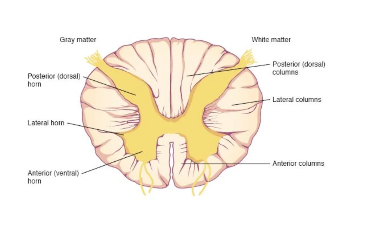
When you look at a cross-section of the spinal cord, a distinctive butterfly-shaped (or “H”-shaped) region of grey matter sits in the center, surrounded by white matter on the outside. This arrangement is the opposite of the brain, where grey matter forms the outer cortex and white matter is internal.
Spinal cord cross-section. Grey matter (yellow) contains cell bodies; white matter contains myelinated axon tracts. Dorsal (posterior) horns handle sensory input, ventral (anterior) horns handle motor output. Credit: Lumen Learning / OpenStax, CC BY 4.0
Grey matter contains neuron cell bodies, dendrites, unmyelinated axons, and interneurons. It is the site of synaptic integration. The grey matter is organized into horns:
Dorsal horns (posterior): receive incoming sensory information from afferent neurons. Think “D” for Dorsal, “D” for Data coming in.
Ventral horns (anterior): contain motor neuron cell bodies that send commands out to skeletal muscles. Think “V” for Ventral, “V” for Vacate (signals leaving).
Lateral horns (present only at thoracic and upper lumbar levels, T1-L2): contain cell bodies of preganglionic sympathetic neurons.
White matter surrounds the grey matter and is composed of myelinated axon tracts. The myelin gives it its white appearance. These tracts are organized into three columns (funiculi) on each side: dorsal, lateral, and ventral columns.
Dorsal and Ventral Roots
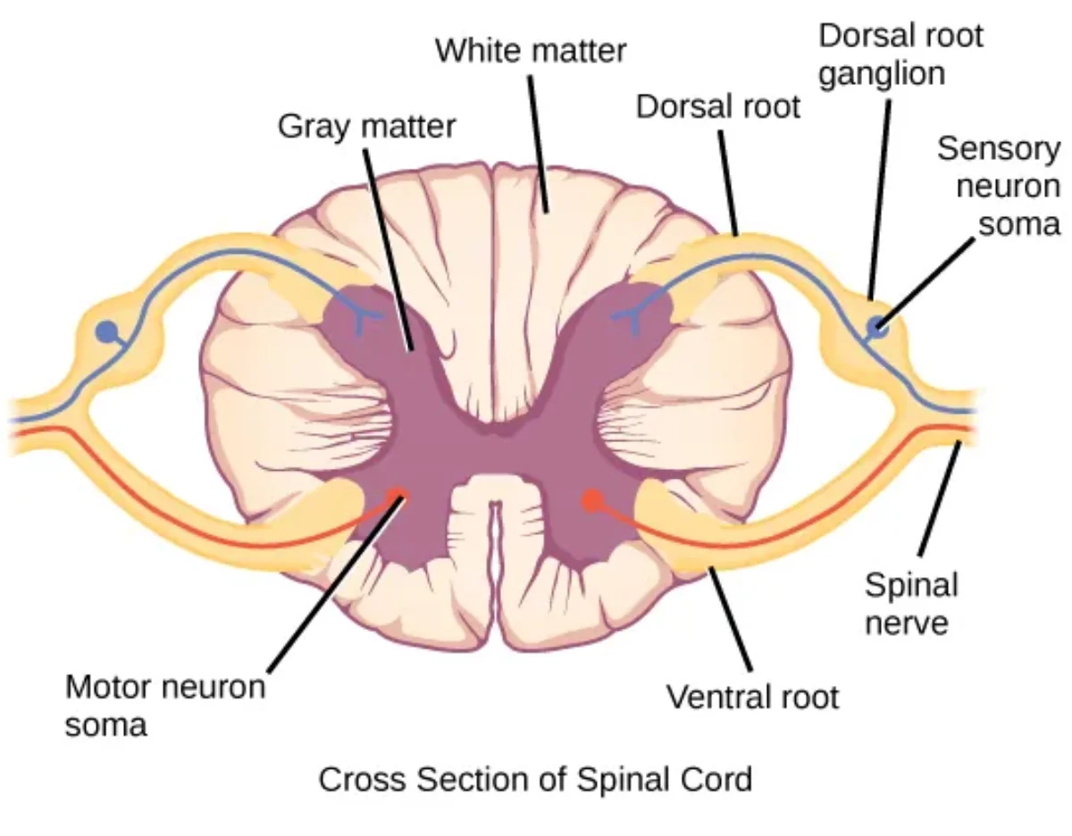
Each spinal nerve forms from the union of a dorsal root and a ventral root.
The dorsal root carries sensory (afferent) fibers into the spinal cord. Just before the dorsal root enters the cord, it swells into the dorsal root ganglion (DRG), which houses the cell bodies of sensory neurons. These are pseudounipolar neurons - their single process splits into a peripheral branch (extending to the receptor) and a central branch (entering the spinal cord).
The ventral root carries motor (efferent) fibers out of the spinal cord. The cell bodies of these motor neurons reside in the ventral horn of the grey matter.
The dorsal and ventral roots merge just lateral to the spinal cord to form a single mixed spinal nerve. This is why spinal nerves carry both sensory and motor information.
Ascending and Descending Tracts
The white matter tracts of the spinal cord serve as information highways connecting the spinal cord to the brain.
Ascending tracts carry sensory information upward from the body to the brain. Two major ascending pathways to know:
Dorsal column-medial lemniscus pathway: transmits fine touch, vibration, and proprioception. Fibers ascend ipsilaterally (same side) in the dorsal columns and cross over in the medulla.
Spinothalamic tract: transmits pain, temperature, and crude touch. Fibers cross over in the spinal cord itself and ascend contralaterally (opposite side).
Descending tracts carry motor commands downward from the brain to the body. The most important is:
Corticospinal (pyramidal) tract: transmits voluntary motor commands from the motor cortex. Most fibers cross over in the medullary pyramids (decussation of pyramids), which is why the left brain controls the right body and vice versa.
Tract
Direction
Information Carried
Where It Crosses
Dorsal column-medial lemniscus
Ascending
Fine touch, vibration, proprioception
Medulla
Spinothalamic
Ascending
Pain, temperature, crude touch
Spinal cord
Corticospinal
Descending
Voluntary motor commands
Medullary pyramids
The Reflex Arc
A reflex is a rapid, involuntary, predictable motor response to a stimulus. Reflexes are mediated by a neural circuit called the reflex arc, which is the simplest functional unit of the nervous system.
Every reflex arc has five components, always in this order:
Receptor - detects the stimulus (e.g., stretch receptor in muscle, pain receptor in skin)
Sensory (afferent) neuron - transmits the signal from the receptor to the CNS via the dorsal root
Integration center - processes the signal within the CNS (spinal cord for spinal reflexes); this may be a single synapse or involve interneurons
Motor (efferent) neuron - carries the response command from the CNS to the effector via the ventral root
Effector - the muscle or gland that carries out the response
The five components of a reflex arc: receptor, sensory neuron, integration center (spinal cord), motor neuron, and effector. Credit: OpenStax Anatomy & Physiology 2e, CC BY 4.0
Monosynaptic vs. Polysynaptic Reflexes
Monosynaptic reflexes involve only one synapse - a direct connection between the sensory neuron and the motor neuron with no interneuron in between. The classic example is the patellar (knee-jerk) reflex. When the patellar tendon is tapped, muscle spindles in the quadriceps detect the stretch. The sensory neuron synapses directly onto a motor neuron in the ventral horn, which fires and contracts the quadriceps, causing the leg to kick forward.
Polysynaptic reflexes involve one or more interneurons between the sensory and motor neurons. The withdrawal (flexor) reflex is the classic example. When you step on a sharp object, sensory neurons activate interneurons in the spinal cord, which then activate motor neurons to flex the injured limb away from the stimulus. At the same time, a crossed-extensor reflex activates extensors in the opposite leg to maintain balance.
Supraspinal Modulation of Reflexes
Spinal reflexes do not operate in isolation. Descending pathways from the brain continuously modulate the local reflex arc, either suppressing it (most of the time) or enhancing it when needed. This is why you can consciously override reflex urges, why reflex strength changes with attention or stress, and why upper motor neuron damage produces hyperreflexia (the “brakes” from above are lost, leaving the intact local arc unchecked).
The take-home: a reflex arc is the fastest possible response, but it is always running inside a larger loop that includes supraspinal feedback from the motor cortex, brainstem, and cerebellum.
Clinical Relevance: Upper vs. Lower Motor Neuron Lesions
Understanding the reflex arc helps explain clinical findings that appear on the MCAT. The key concept is that the reflex arc itself is a local spinal cord circuit. Whether a lesion is “above” or “below” this circuit determines the pattern of symptoms.
What are the five components of a reflex arc, in order?
Click to reveal answer
1) Receptor - detects the stimulus. 2) Sensory (afferent) neuron - transmits signal to the CNS. 3) Integration center - processes the signal in the spinal cord. 4) Motor (efferent) neuron - carries the command to the effector. 5) Effector - the muscle or gland that responds.
What is the difference between a monosynaptic and a polysynaptic reflex? Give an example of each.
Click to reveal answer
A monosynaptic reflex has only one synapse (sensory neuron directly to motor neuron) - e.g., the patellar (knee-jerk) reflex. A polysynaptic reflex includes interneurons between the sensory and motor neurons - e.g., the withdrawal (flexor) reflex when stepping on a sharp object.
In a spinal cord cross-section, where is grey matter located vs. white matter? What does each contain?
Click to reveal answer
Grey matter is the butterfly-shaped region in the center, containing neuron cell bodies, interneurons, and synapses. White matter surrounds the grey matter on the outside, containing myelinated axon tracts (ascending sensory and descending motor pathways). This arrangement is the opposite of the brain.
Your heart rate, pupil diameter, digestive activity, and bronchiole diameter are all adjusted continuously without a single conscious thought. The autonomic nervous system (ANS) manages these vital functions automatically, keeping your internal environment stable whether you are sprinting from danger or digesting a meal. For the MCAT, you need to know exactly how the ANS is organized, which neurotransmitters it uses at each synapse, and what each division does to every major organ system.
Overview: Two-Neuron Chain
Both divisions of the ANS use a two-neuron chain to connect the CNS to the target organ. The first neuron (preganglionic neuron) has its cell body in the CNS and synapses at an autonomic ganglion. The second neuron (postganglionic neuron) has its cell body in the ganglion and sends its axon to the effector organ.
This two-neuron design differs from the somatic nervous system, which uses a single motor neuron to reach skeletal muscle. The location and length of these neurons differ between the sympathetic and parasympathetic divisions, and this is a commonly tested distinction.
The Sympathetic Division: “Fight or Flight”
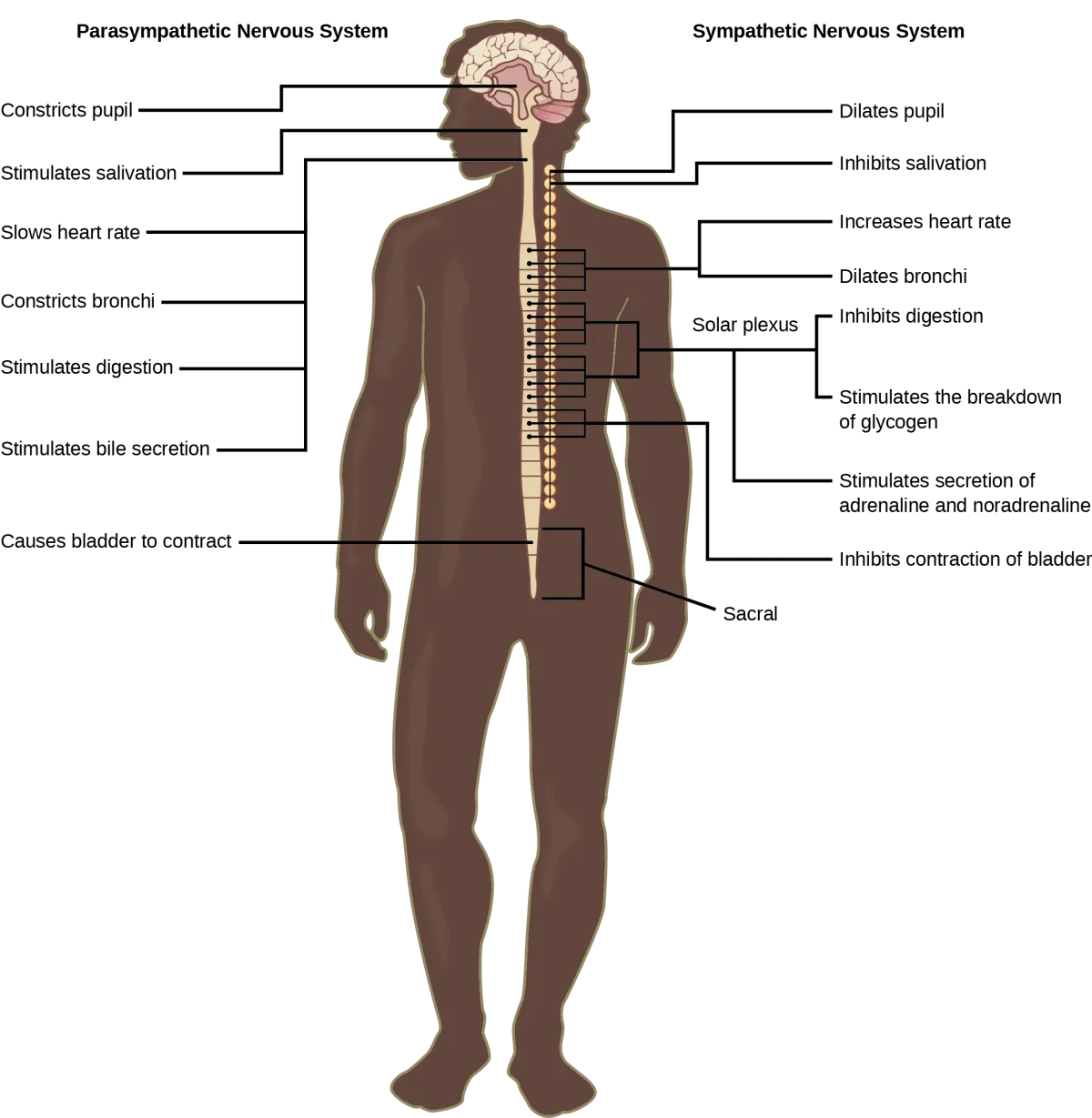
The sympathetic nervous system prepares the body for intense physical activity. Think of every change it makes as an answer to the question: “What does the body need to fight or run away?”
Origin: Preganglionic neurons emerge from the thoracolumbar spinal cord (T1-L2). Their cell bodies reside in the lateral horns of the spinal cord grey matter.
Neuron lengths: Preganglionic fibers are short (they synapse in ganglia close to the spinal cord). Postganglionic fibers are long (they must travel from the paravertebral or prevertebral ganglia to the distant target organs).
Neurotransmitters:
Preganglionic neurons release acetylcholine (ACh) at the ganglion (all preganglionic neurons in both divisions use ACh).
Postganglionic neurons release norepinephrine (NE) at the target organ. The one major exception: sympathetic postganglionic fibers to sweat glands release ACh, not NE.
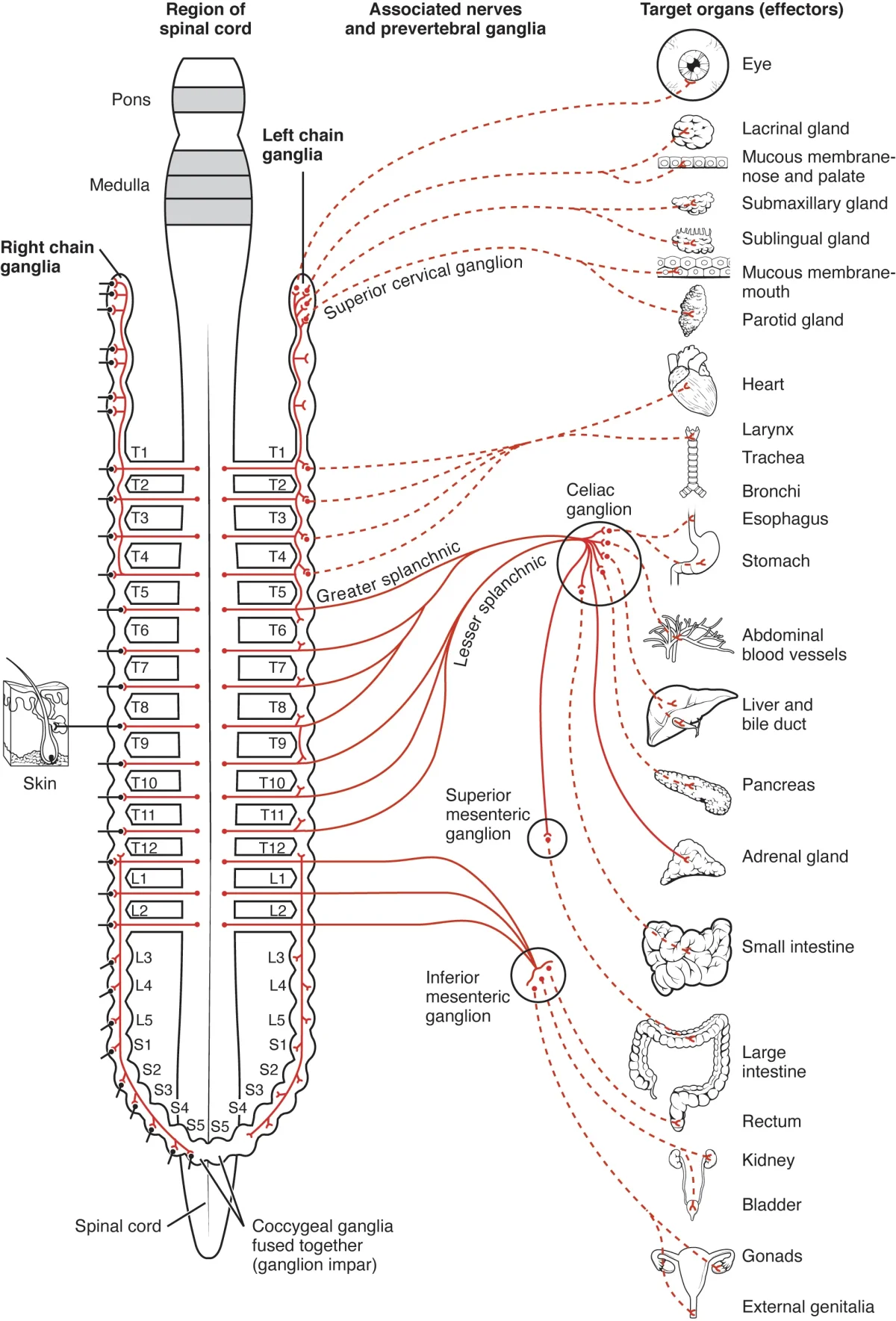
The sympathetic division. Focus on: thoracolumbar origin, short preganglionic/long postganglionic fibers, and the target organ effects (heart rate up, bronchodilation, pupils dilate). Ignore the specific ganglia names. Credit: OpenStax Anatomy & Physiology 2e, CC BY 4.0
The adrenal medulla deserves special attention. It is a modified sympathetic ganglion. Preganglionic sympathetic fibers synapse directly on chromaffin cells within the adrenal medulla. Instead of sending a postganglionic neuron to a target, these cells release epinephrine (~80%) and norepinephrine (~20%) directly into the bloodstream. This allows the sympathetic response to reach the entire body at once, amplifying the fight-or-flight effect.
The Parasympathetic Division: “Rest and Digest”
The parasympathetic nervous system promotes calming, energy-conserving, and maintenance activities. It dominates during rest, digestion, and recovery.
Origin: Preganglionic neurons emerge from the craniosacral regions - specifically cranial nerves III, VII, IX, and X, plus sacral spinal segments S2-S4.
Neuron lengths: Preganglionic fibers are long (they travel from the brainstem or sacral cord all the way to ganglia near or within the target organ). Postganglionic fibers are short (the ganglion is right next to or embedded in the organ wall).
Neurotransmitters: Both preganglionic and postganglionic neurons release acetylcholine (ACh). This is a key distinction from the sympathetic division, where the postganglionic neurotransmitter is norepinephrine.
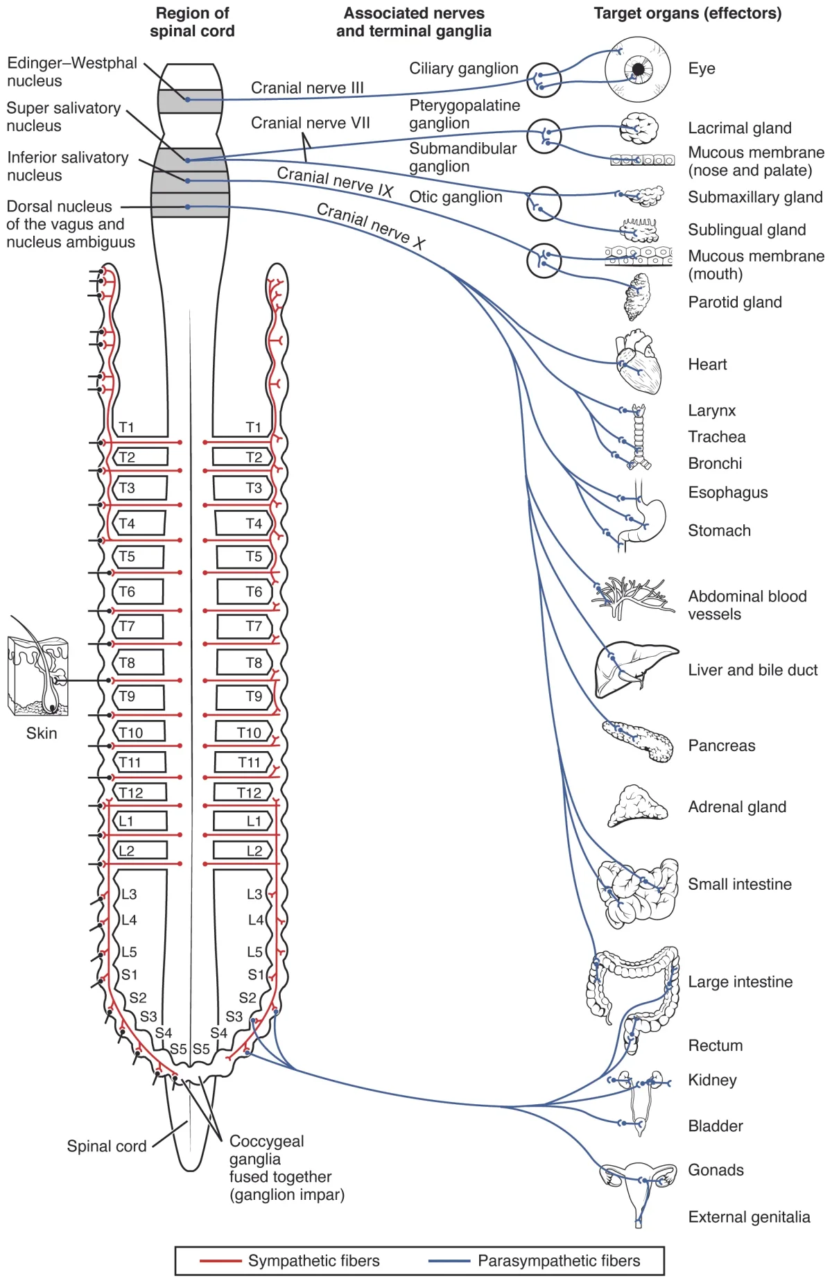
The parasympathetic division originates from craniosacral regions. Long preganglionic fibers synapse at ganglia near or within the target organ. Credit: OpenStax Anatomy & Physiology 2e, CC BY 4.0
The vagus nerve (cranial nerve X) is by far the most important parasympathetic nerve. It carries approximately 75% of all parasympathetic fibers and innervates the heart, lungs, and virtually the entire gastrointestinal tract. When the MCAT mentions parasympathetic effects on thoracic or abdominal organs, the vagus nerve is almost always the pathway involved.
Dual Innervation and Organ-Specific Effects
Most organs receive input from both sympathetic and parasympathetic divisions. This dual innervation allows precise control through the balance of opposing signals. The table below summarizes the effects on each organ system:
Little direct effect (most vessels lack parasympathetic innervation)
Lungs (bronchioles)
Dilation
Constriction
Pupils
Dilation (mydriasis)
Constriction (miosis)
GI tract (motility)
Decreases motility and secretion
Increases motility and secretion
GI sphincters
Contracts (closes)
Relaxes (opens)
Bladder (detrusor)
Relaxes (filling)
Contracts (voiding)
Bladder sphincter
Contracts (retention)
Relaxes (voiding)
Liver
Glycogenolysis (glucose release)
Glycogen synthesis
Sweat glands
Stimulates secretion (via ACh)
No innervation
Adrenal medulla
Stimulates epinephrine/NE release
No innervation
Notable exceptions to dual innervation: blood vessels (primarily sympathetic only), sweat glands (sympathetic only via ACh), and the adrenal medulla (sympathetic only).
Comparison of sympathetic vs. parasympathetic effects on target organs. Note the opposing actions that allow fine-tuned control. Credit: OpenStax Anatomy & Physiology 2e, CC BY 4.0
Receptor Types
The neurotransmitters of the ANS act on specific receptor types. The MCAT expects you to know the major categories.
Cholinergic receptors bind acetylcholine (ACh):
Nicotinic receptors: found at all autonomic ganglia (both sympathetic and parasympathetic) and at the neuromuscular junction. They are ionotropic (ligand-gated ion channels) and always excitatory.
Muscarinic receptors: found on target organs innervated by parasympathetic postganglionic fibers (and on sweat glands innervated by sympathetic postganglionic fibers). They are metabotropic (G-protein coupled) and can be excitatory or inhibitory depending on the tissue.
Adrenergic receptors bind norepinephrine and epinephrine:
What neurotransmitter does each type of ANS neuron release? What is the major exception?
Click to reveal answer
All preganglionic neurons (both divisions) release ACh. Parasympathetic postganglionic neurons release ACh. Sympathetic postganglionic neurons release norepinephrine (NE). The major exception: sympathetic postganglionic fibers to sweat glands release ACh instead of NE.
What is the adrenal medulla, and why is it considered a modified sympathetic ganglion?
Click to reveal answer
The adrenal medulla is the inner portion of the adrenal gland. It is a modified sympathetic ganglion because preganglionic sympathetic fibers synapse directly onto its chromaffin cells (which are embryologically derived from neural crest cells, like postganglionic neurons). Instead of sending a postganglionic neuron to a target, the chromaffin cells release epinephrine (~80%) and norepinephrine (~20%) directly into the blood.
Compare the preganglionic and postganglionic neuron lengths in the sympathetic vs. parasympathetic divisions.
Click to reveal answer
Sympathetic: short preganglionic, long postganglionic (ganglia are near the spinal cord, far from target organs). Parasympathetic: long preganglionic, short postganglionic (ganglia are near or within the target organ). Both divisions always use a two-neuron chain from CNS to effector.
Every sensation you experience - the pressure of a handshake, the burn of a hot pan, the position of your limbs in space - begins with a sensory receptor converting a stimulus into an electrical signal. That signal must then travel along specific pathways to reach the brain, where it is consciously perceived. Likewise, every voluntary movement originates in the motor cortex and travels along descending pathways to reach the muscles. The MCAT tests both the receptor types and the major pathways, along with the twelve cranial nerves that serve as direct lines between the brain and specific structures.
Sensory Receptor Types by Stimulus
Sensory receptors are classified by the type of stimulus they detect. Each receptor type responds best to one specific form of energy, known as its adequate stimulus.
Mechanoreceptors respond to mechanical forces - pressure, vibration, stretch, and touch. Examples include Meissner’s corpuscles (light touch), Pacinian corpuscles (deep pressure and vibration), Merkel’s discs (sustained pressure), and muscle spindles (stretch).
Thermoreceptors respond to changes in temperature. Separate populations of free nerve endings detect warming versus cooling.
Nociceptors respond to potentially damaging stimuli and generate the sensation of pain. They are free nerve endings activated by extreme heat, extreme cold, intense mechanical force, or inflammatory chemicals.
Chemoreceptors respond to specific chemical molecules. Examples include olfactory receptors (smell), taste receptors (gustation), and peripheral chemoreceptors that monitor blood oxygen and pH.
Photoreceptors respond to light. Rods and cones in the retina are the primary examples.
Sensory receptors are classified by the type of stimulus they detect. Each receptor is optimally tuned to its adequate stimulus. Credit: OpenStax Anatomy & Physiology 2e, CC BY 4.0
Sensory Receptor Classification by Location
Receptors can also be grouped by where in the body they are found:
Exteroceptors are located at or near the body surface and detect external stimuli - touch, pressure, temperature, pain, and light. They keep you aware of the outside environment.
Interoceptors (visceroceptors) are located within internal organs and blood vessels. They monitor conditions such as blood pressure, blood oxygen levels, and organ stretch. You are usually not consciously aware of these signals.
Proprioceptors are located in muscles, tendons, joints, and the inner ear. They provide information about body position, movement, and equilibrium. Muscle spindles detect stretch, Golgi tendon organs detect tension, and joint kinesthetic receptors detect joint position.
Ascending (Sensory) Pathways
Sensory information from the body travels to the brain through two major ascending pathways. Both ultimately reach the somatosensory cortex in the parietal lobe, but they carry different types of information and cross the midline at different levels.
Dorsal column-medial lemniscus (DCML) pathway: Carries fine touch, vibration, two-point discrimination, and proprioception. First-order neurons ascend ipsilaterally in the dorsal columns of the spinal cord, synapse in the medulla, cross the midline there, and then ascend to the thalamus. Third-order neurons relay the signal from the thalamus to the somatosensory cortex.
Spinothalamic tract: Carries pain, temperature, and crude (non-discriminative) touch. First-order neurons synapse soon after entering the spinal cord. Second-order neurons cross the midline within the spinal cord and then ascend contralaterally to the thalamus. Third-order neurons relay to the somatosensory cortex.
Feature
DCML Pathway
Spinothalamic Tract
Carries
Fine touch, vibration, proprioception
Pain, temperature, crude touch
Crosses midline at
Medulla
Spinal cord
Ascends on
Ipsilateral side (until medulla)
Contralateral side
Descending (Motor) Pathways
Motor commands from the brain travel down to the spinal cord and out to muscles through descending pathways.
Corticospinal (pyramidal) tract: This is the primary pathway for voluntary movement. Upper motor neurons originate in the primary motor cortex (precentral gyrus) and descend through the brainstem. Approximately 90% of fibers cross the midline at the medullary pyramids (pyramidal decussation) and descend in the lateral corticospinal tract. They synapse on lower motor neurons in the ventral horn of the spinal cord, which then innervate skeletal muscles.
This crossing explains why the left motor cortex controls the right side of the body and vice versa.
Upper motor neurons (UMNs) reside entirely within the CNS - their cell bodies are in the motor cortex and their axons descend through the brainstem and spinal cord. Lower motor neurons (LMNs) have cell bodies in the ventral horn of the spinal cord (or brainstem motor nuclei for cranial nerves) and their axons exit the CNS to directly innervate skeletal muscles.
The motor cortex homunculus illustrates how different body regions are represented in the motor cortex. Areas requiring fine motor control (hands, face) occupy disproportionately large cortical areas. Credit: OpenStax Anatomy & Physiology 2e, CC BY 4.0
The Twelve Cranial Nerves
Unlike spinal nerves, which emerge from the spinal cord, cranial nerves emerge directly from the brain (mostly the brainstem). There are 12 pairs, numbered I through XII in order from anterior to posterior. Some are purely sensory, some are purely motor, and some carry both types of fibers.
The twelve cranial nerves. Focus on: the vagus nerve (CN X - parasympathetic to viscera), optic nerve (CN II - vision), and facial nerve (CN VII). Know all 12 by name and number, but prioritize function over anatomical origin. Credit: OpenStax Anatomy & Physiology 2e, CC BY 4.0
Number
Name
Type
Key Functions
I
Olfactory
Sensory
Smell
II
Optic
Sensory
Vision
III
Oculomotor
Motor
Most eye movements; pupil constriction; lens accommodation
IV
Trochlear
Motor
Superior oblique muscle (eye movement - looking down and inward)
Taste (posterior 31 of tongue); swallowing; monitors carotid body/sinus
X
Vagus
Both
Parasympathetic to thoracic and abdominal viscera; voice; swallowing; taste (epiglottis)
XI
Accessory (Spinal Accessory)
Motor
Trapezius and sternocleidomastoid muscles (head/shoulder movement)
XII
Hypoglossal
Motor
Tongue movement
Somatotopic Organization
Both the somatosensory cortex (in the postcentral gyrus) and the motor cortex (in the precentral gyrus) are organized somatotopically, meaning that specific cortical regions correspond to specific body parts. This creates the homunculus - a distorted “little human” map where the size of each body region reflects the density of its innervation, not its physical size.
The hands, lips, and tongue occupy disproportionately large areas in both the sensory and motor homunculi because these regions require extremely fine sensory discrimination and precise motor control. In contrast, the trunk and legs occupy relatively small cortical areas.
What are the five major types of sensory receptors classified by stimulus type?
Which ascending pathway carries pain and temperature, and where does it cross the midline?
Click to reveal answer
The spinothalamic tract carries pain, temperature, and crude touch. It crosses the midline within the spinal cord (shortly after the first-order neuron enters). This early crossing means the signal ascends on the contralateral side, which is important for understanding spinal cord injury patterns.
Name the cranial nerve that carries approximately 75% of parasympathetic fibers. What organs does it innervate?
Click to reveal answer
The vagus nerve (CN X). It provides parasympathetic innervation to the heart (slows heart rate), lungs (bronchoconstriction), and nearly the entire gastrointestinal tract (increases motility and secretion). It does not innervate pelvic organs - those receive parasympathetic input from sacral nerves S2-S4.
The special senses - vision, hearing, equilibrium, taste, and smell - are served by complex receptor organs concentrated in the head. Unlike general senses (touch, temperature, pain) that use relatively simple receptors distributed throughout the body, the special senses rely on highly specialized structures that convert very specific forms of energy into neural signals. The MCAT tests the anatomy of these organs, the mechanism of signal transduction in each, and the neural pathways that carry sensory information to the brain.
Vision: Eye Anatomy
The eye is a fluid-filled sphere designed to focus light onto the retina, a thin layer of neural tissue at the back of the eye that contains photoreceptors.
Outer layer (fibrous tunic):
Sclera: the white, protective outer coat of the eye. It maintains eye shape and provides attachment points for extraocular muscles.
Cornea: the transparent anterior portion of the outer layer. It is the primary refractive structure, bending incoming light rays to begin the focusing process.
Middle layer (vascular tunic/uvea):
Choroid: a pigmented, highly vascular layer that nourishes the retina and absorbs stray light.
Ciliary body: contains the ciliary muscle, which controls the shape of the lens for focusing (accommodation). It also produces aqueous humor.
Iris: the colored muscular ring that controls pupil diameter, regulating the amount of light entering the eye.
Inner layer (neural tunic):
Retina: the innermost layer containing the photoreceptors (rods and cones), bipolar cells, and ganglion cells. It is where light is converted into neural signals.
The human eye. Focus on: the light path (cornea to pupil to lens to retina), the lens and ciliary muscle (accommodation), and the retina (rods, cones, fovea). You do not need every layer name - know cornea, lens, retina, optic nerve, and vitreous/aqueous humor. Credit: Wikimedia Commons, CC BY-SA 3.0
Interactive 3D Eye. Rotate to see how the cornea, lens, and retina are arranged. Trace the path light takes from the cornea through the vitreous to the retina.Credit: MotionCow via Sketchfab, CC BY
The lens sits behind the iris and fine-tunes the focusing of light onto the retina. It is held in place by suspensory ligaments (zonular fibers) attached to the ciliary body. When the ciliary muscle contracts, the suspensory ligaments loosen, and the elastic lens rounds up to focus on near objects. When the ciliary muscle relaxes, the ligaments pull taut and flatten the lens for distance vision. This process is called accommodation.
Vision
Lens Rx
Aqueous humor fills the anterior and posterior chambers (in front of the lens) and is continuously produced and drained. If drainage is blocked, intraocular pressure rises, which can damage the optic nerve. Vitreous humor is the gel-like substance filling the large posterior cavity behind the lens.
Vision: Rods, Cones, and Phototransduction
The retina contains two types of photoreceptors, each optimized for different conditions.
Rods (~120 million per eye):
Extremely sensitive to light - responsible for scotopic (dim-light) vision
Produce images in shades of grey only (no color discrimination)
Concentrated in the peripheral retina
Contain the photopigment rhodopsin (retinal + opsin)
Many rods converge onto a single ganglion cell, increasing sensitivity but decreasing acuity
Cones (~6 million per eye):
Require brighter light - responsible for photopic (daylight) vision and color vision
Three subtypes based on their peak sensitivity: red (long wavelength), green (medium wavelength), and blue (short wavelength)
Concentrated in the fovea centralis, a small pit in the center of the retina that produces the sharpest image
Fewer cones converge per ganglion cell, giving high acuity but lower sensitivity
Rods and cones differ in shape, photopigment, distribution, and function. Rods dominate the periphery and handle dim-light vision; cones are concentrated at the fovea and provide color vision. Credit: OpenStax Anatomy & Physiology 2e, CC BY 4.0
Phototransduction is the process by which light energy is converted to a neural signal. In the dark, photoreceptors are partially depolarized and continuously release glutamate. When light strikes rhodopsin (in rods) or photopsin (in cones), retinal changes from the cis to trans configuration, activating a G-protein signaling cascade that closes cGMP-gated sodium channels, causing the photoreceptor to hyperpolarize and reduce glutamate release. This change in neurotransmitter release is detected by bipolar cells and ultimately transmitted to ganglion cells.
Vision: The Visual Pathway
The visual pathway from retina to cortex involves several processing steps:
Photoreceptors (rods/cones) synapse on bipolar cells
Bipolar cells synapse on ganglion cells (whose axons form the optic nerve)
The optic nerve (CN II) exits the eye at the optic disc (the blind spot - no photoreceptors here)
At the optic chiasm, fibers from the nasal (medial) half of each retina cross to the opposite side, while fibers from the temporal (lateral) half remain ipsilateral
The optic tracts carry the reorganized fibers to the lateral geniculate nucleus (LGN) of the thalamus
From the LGN, fibers project to the primary visual cortex in the occipital lobe
Because of the partial crossing at the optic chiasm, each hemisphere receives visual information from the contralateral visual field. The left visual cortex processes the right visual field, and vice versa.
Refractive errors:
Myopia (nearsightedness): the eyeball is too long or the lens is too curved, causing the focal point to fall in front of the retina. Corrected with concave (diverging) lenses.
Hyperopia (farsightedness): the eyeball is too short or the lens is too flat, causing the focal point to fall behind the retina. Corrected with convex (converging) lenses.
Hearing: Ear Anatomy
The ear is divided into three anatomical regions, each playing a distinct role in converting sound waves into neural signals.
Outer ear: The pinna (auricle) funnels sound waves into the external auditory canal, which directs them toward the tympanic membrane.
Middle ear: The tympanic membrane (eardrum) vibrates in response to sound waves. These vibrations are transmitted through three tiny bones called ossicles - the malleus (hammer), incus (anvil), and stapes (stirrup) - which amplify the sound energy by approximately 20-fold. The stapes footplate pushes against the oval window, transferring vibrations into the fluid-filled inner ear.
The middle ear also connects to the pharynx via the Eustachian (auditory) tube, which equalizes air pressure on both sides of the tympanic membrane.
The inner ear contains both the hearing organ (cochlea) and the balance organs (semicircular canals). Credit: Blausen Medical Communications, CC BY 3.0
Interactive 3D Ear. Rotate to see the outer, middle, and inner ear. Identify the ossicles, cochlea, and semicircular canals.Credit: zames1992 via Sketchfab, CC BY
Inner ear: The inner ear contains both the hearing organ (cochlea) and the balance organs (semicircular canals, utricle, and saccule). It is filled with fluid and embedded within the temporal bone.
Hearing: Sound Transduction in the Cochlea
The cochlea is a snail-shaped, fluid-filled structure divided into three chambers: the scala vestibuli, scala media (cochlear duct), and scala tympani. The organ of Corti sits on the basilar membrane within the scala media and contains the hair cells - the mechanoreceptor cells responsible for hearing.
Cochlea cross-section. Focus on: the basilar membrane (where hair cells sit), the organ of Corti (hair cells + tectorial membrane), and that different positions along the basilar membrane respond to different frequencies. You do not need to memorize the three scala names. Credit: OpenStax Anatomy & Physiology 2e, CC BY 4.0
The transduction process follows a precise sequence:
Sound waves vibrate the tympanic membrane
Ossicles amplify the vibration and transmit it to the oval window
Pressure waves travel through the cochlear fluid (perilymph)
The basilar membrane vibrates in response
Hair cells on the basilar membrane are displaced, and their stereocilia bend against the tectorial membrane
Bending of stereocilia opens mechanically gated ion channels, depolarizing the hair cell
The hair cell releases neurotransmitter onto the cochlear branch of CN VIII (vestibulocochlear nerve)
The signal travels to the auditory cortex in the temporal lobe
The Vestibular System: Balance and Equilibrium
The vestibular apparatus is located in the inner ear, adjacent to the cochlea. It detects head position and movement, allowing you to maintain balance and coordinate eye movements.
Semicircular canals (three per ear): These three fluid-filled rings are oriented in three perpendicular planes (anterior, posterior, and lateral). They detect rotational (angular) acceleration. When the head rotates, the fluid (endolymph) lags behind due to inertia. This deflects a gelatinous structure called the cupula within the ampulla at the base of each canal, bending the hair cells embedded within it.
The three semicircular canals detect rotational acceleration in three planes. Each canal contains an ampulla with a cupula that bends in response to fluid movement. Credit: OpenStax Anatomy & Physiology 2e, CC BY 4.0
Utricle and saccule: These two chambers detect linear acceleration and the static position of the head relative to gravity. They contain a gelatinous layer topped with calcium carbonate crystals called otoliths (literally “ear stones”). When the head tilts or undergoes linear acceleration, gravity or inertial force shifts the otoliths, bending the underlying hair cells.
All vestibular signals travel via the vestibular branch of CN VIII to the brainstem and cerebellum.
Taste (Gustation)
Taste receptors are located within taste buds, which are found on papillae (raised bumps) on the tongue surface, as well as on the soft palate, epiglottis, and pharynx. Each taste bud contains 50-100 taste receptor cells.
Taste buds are found within papillae on the tongue. Each taste bud contains receptor cells that detect one or more of the five basic tastes. Credit: OpenStax Anatomy & Physiology 2e, CC BY 4.0
Taste signals are carried by three cranial nerves: CN VII (facial) for the anterior two-thirds of the tongue, CN IX (glossopharyngeal) for the posterior one-third, and CN X (vagus) for the epiglottis region. All taste information synapses in the solitary nucleus of the medulla, passes through the thalamus, and reaches the gustatory cortex in the insula.
Smell (Olfaction)
Olfactory receptor neurons are located in the olfactory epithelium high in the nasal cavity. Each receptor neuron expresses a single type of odorant receptor protein on its cilia. Humans have approximately 400 different odorant receptor types, and each odorant activates a specific combination of receptors, allowing us to distinguish thousands of different smells.
Olfactory receptor neurons in the nasal epithelium send axons through the cribriform plate to the olfactory bulb. From there, signals project to the olfactory cortex without first passing through the thalamus. Credit: OpenStax Anatomy & Physiology 2e, CC BY 4.0
The axons of olfactory receptor neurons pass through tiny holes in the cribriform plate of the ethmoid bone and synapse in the olfactory bulb. From the olfactory bulb, the signal travels via the olfactory tract (CN I) to the olfactory cortex (piriform cortex) and the limbic system (amygdala, hippocampus).
How do rods and cones differ in their sensitivity, distribution, and function?
Click to reveal answer
Rods are highly sensitive to light (dim-light/scotopic vision), located primarily in the peripheral retina, and produce only black-and-white images. Cones require brighter light (daylight/photopic vision), are concentrated in the fovea, and provide color vision via three subtypes (red, green, blue). Rods have high convergence (high sensitivity, low acuity); cones have low convergence (low sensitivity, high acuity).
What is unique about the olfactory pathway compared to all other sensory pathways?
Click to reveal answer
Olfaction is the only special sense that does not relay through the thalamus before reaching the cerebral cortex. Olfactory signals travel from the olfactory epithelium to the olfactory bulb and then directly to the olfactory cortex and limbic system. This direct limbic connection explains why smells are particularly effective at triggering emotional memories.
Describe the pathway of sound from the external environment to the auditory cortex.
Click to reveal answer
Sound waves enter the external auditory canal, vibrate the tympanic membrane, are amplified by the ossicles (malleus, incus, stapes), and transmitted through the oval window into cochlear fluid. Fluid waves vibrate the basilar membrane, bending hair cell stereocilia against the tectorial membrane. This opens mechanically gated ion channels, depolarizing the hair cells, which release neurotransmitter onto CN VIII. The signal travels to the auditory cortex in the temporal lobe.