
Neuron Structure
Neurons are the functional units of the nervous system - specialized cells that generate and transmit electrical signals across long distances at remarkable speed. Although they come in many shapes and sizes, all neurons share the same basic architecture. Understanding each structural component and its role is essential for making sense of signal transmission, synaptic communication, and the clinical conditions the MCAT tests.
Dendrites: The Receivers
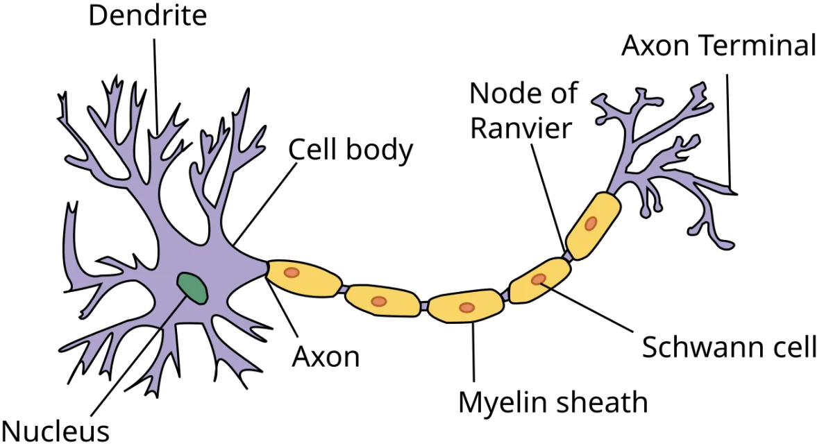
Dendrites are highly branched extensions that project from the neuron’s cell body. Their job is to receive incoming signals from other neurons, sensory receptors, or the environment. The more branches a dendrite has, the more connections it can make - some neurons in the brain have thousands of dendritic branches.
Dendrites contain receptors and ligand-gated ion channels on their surface that bind neurotransmitters released by neighboring neurons. When a neurotransmitter binds, it may produce a small local change in voltage called a graded potential. These graded potentials are not all-or-nothing; they vary in size and can be either excitatory or inhibitory.
Soma (Cell Body): The Integration Center
The soma is the metabolic heart of the neuron. It contains the nucleus, rough endoplasmic reticulum (called Nissl bodies in neurons), Golgi apparatus, and mitochondria. All the proteins and organelles the neuron needs are manufactured here and transported outward.
Critically, the soma integrates all the graded potentials arriving from the dendrites. Excitatory signals (EPSPs) and inhibitory signals (IPSPs) are summed together. If the net signal is strong enough when it reaches the next structure - the axon hillock - an action potential fires.
Axon Hillock: The Decision Point
The axon hillock is the cone-shaped region where the soma transitions into the axon. It has the highest density of voltage-gated sodium channels anywhere on the neuron, giving it the lowest threshold for firing an action potential.
This is where the all-or-nothing decision is made. If the summed graded potentials depolarize the membrane at the axon hillock to threshold (approximately -55 mV), an action potential is generated. If they do not reach threshold, nothing happens. There is no “partial” action potential.
Axon: The Transmission Line
The axon is a long, slender projection that carries the action potential away from the cell body toward the target cell. Axons can be extremely short (a fraction of a millimeter for interneurons in the brain) or remarkably long (over one meter for motor neurons extending from the spinal cord to the toes).
The cytoplasm of the axon is called axoplasm, and its membrane is called the axolemma. Axons do not contain rough ER or Golgi apparatus, so they depend on the soma for protein synthesis. Materials are moved along the axon by motor proteins: kinesin transports cargo toward the synaptic terminal (anterograde transport), while dynein transports cargo back toward the soma (retrograde transport).
Myelin Sheath and Nodes of Ranvier
Many axons are wrapped in a fatty insulating layer called the myelin sheath. Myelin is composed of lipid-rich cell membrane wound tightly around the axon in concentric layers. It serves two critical functions: it insulates the axon to prevent current leakage, and it dramatically increases the speed of signal conduction.
The cells that produce myelin differ by location:
- In the PNS, Schwann cells each wrap around a single segment of one axon.
- In the CNS, oligodendrocytes extend multiple processes, each myelinating a segment of a different axon. A single oligodendrocyte can myelinate portions of up to 50 axons.
Between each myelinated segment are small gaps called nodes of Ranvier. These gaps are packed with voltage-gated sodium channels. Because the action potential effectively “jumps” from node to node rather than traveling continuously along the axon, myelinated neurons conduct signals much faster. This jumping conduction is called saltatory conduction (from the Latin “saltare,” meaning to jump).
Synaptic Terminals: The Output Zone
At its end, the axon branches into many fine extensions that terminate in synaptic terminals (also called terminal boutons or axon terminals). Each terminal contains synaptic vesicles loaded with neurotransmitter molecules.
When an action potential arrives at the synaptic terminal, voltage-gated calcium channels open. The influx of Ca2+ triggers the vesicles to fuse with the presynaptic membrane and release their neurotransmitter into the synaptic cleft - the tiny gap between the sending and receiving cells. This process is called exocytosis, and it converts the electrical signal back into a chemical one.
Structural Classification of Neurons
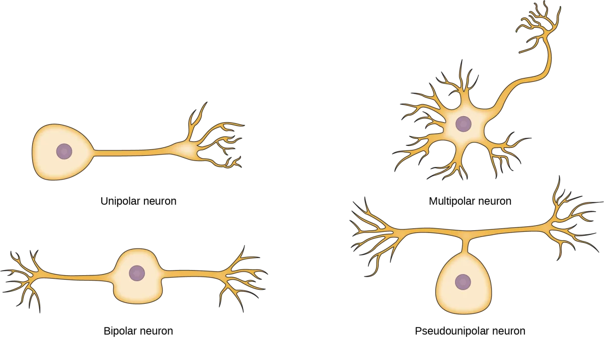
Neurons are classified by how many processes (extensions) emerge from the cell body.
Multipolar neurons have one axon and many dendrites. They are the most common type in the CNS and include most motor neurons and interneurons.
Bipolar neurons have one axon and one dendrite on opposite sides of the cell body. They are found in special sensory organs - the retina, olfactory epithelium, and inner ear.
Unipolar (pseudounipolar) neurons have a single process that splits into two branches - one heading toward the periphery and one toward the CNS. Most sensory neurons in the PNS are pseudounipolar. Their cell bodies sit in the dorsal root ganglia.
Functional Classification of Neurons
Neurons can also be classified by the direction they carry signals:
- Sensory (afferent) neurons carry information from receptors toward the CNS. Most are pseudounipolar.
- Motor (efferent) neurons carry commands from the CNS to muscles and glands. Most are multipolar.
- Interneurons connect neurons within the CNS. They are exclusively multipolar and represent the vast majority of all neurons in the body.