
Glial Cells
Neurons get all the attention, but they could not function for a second without glial cells. Glia (from the Greek word for “glue”) outnumber neurons by roughly 10:1 in certain regions and perform every essential support function the nervous system requires - from insulation and nutrition to immune defense and waste removal. Six major types of glial cells appear on the MCAT, four in the CNS and two in the PNS. Knowing each one’s location, function, and clinical relevance is non-negotiable for test day.
CNS Glial Cells
Astrocytes: The Multitaskers
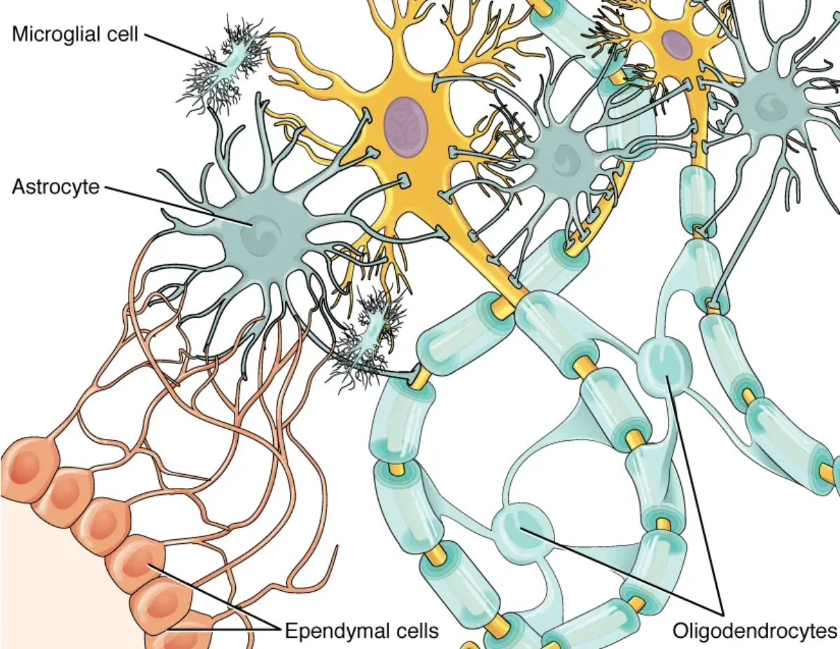
Astrocytes are the most abundant glial cells in the CNS and arguably the most versatile. They are star-shaped cells with numerous processes that contact both neurons and blood vessels. Their functions include:
- Blood-brain barrier (BBB) maintenance: Astrocyte foot processes (end-feet) wrap around CNS capillaries and help maintain the tight junctions that form the BBB. This barrier prevents most pathogens and large molecules in the blood from entering brain tissue.
- Nutrient transfer: Astrocytes shuttle glucose from blood vessels to neurons and can convert glucose to lactate for neuronal fuel.
- Neurotransmitter recycling: They take up excess neurotransmitters (especially glutamate) from the synaptic cleft, preventing excitotoxicity.
- Ion homeostasis: Astrocytes buffer extracellular K+ concentrations, which is critical because even small changes in K+ levels alter neuronal excitability.
- Structural support and repair: After CNS injury, astrocytes proliferate and form a glial scar (a process called reactive gliosis).
Oligodendrocytes: The CNS Insulators
Oligodendrocytes produce the myelin sheath in the CNS. Unlike Schwann cells, a single oligodendrocyte extends multiple flat, paddle-like processes that each wrap around a segment of a different axon. One oligodendrocyte can myelinate portions of up to 50 axons simultaneously.
This efficiency comes with a clinical downside: if a single oligodendrocyte is damaged, multiple axons lose their myelin at once.
Microglia: The Immune Defense
Microglia are the resident immune cells of the CNS. Unlike other glial cells, which derive from neural ectoderm, microglia originate from mesoderm (specifically, from yolk sac macrophage precursors that migrate into the brain during development). They function as the brain’s macrophages.
In their resting state, microglia extend long, thin processes that continuously survey the local environment. When they detect pathogens, debris, or damaged neurons, they retract their processes, become amoeboid, and phagocytose the threat. They also release pro-inflammatory cytokines to recruit additional immune responses.
Ependymal Cells: The Fluid Managers
Ependymal cells are ciliated epithelial cells that line the ventricles of the brain and the central canal of the spinal cord. Their beating cilia help circulate cerebrospinal fluid (CSF). Specialized ependymal cells in the choroid plexus actively produce CSF by filtering blood plasma.
CSF serves as a shock absorber for the brain, a medium for nutrient and waste exchange, and a source of buoyancy that reduces the effective weight of the brain from about 1,400 g to roughly 50 g.
PNS Glial Cells
Schwann Cells: The PNS Insulators
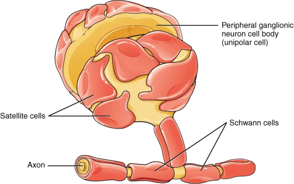
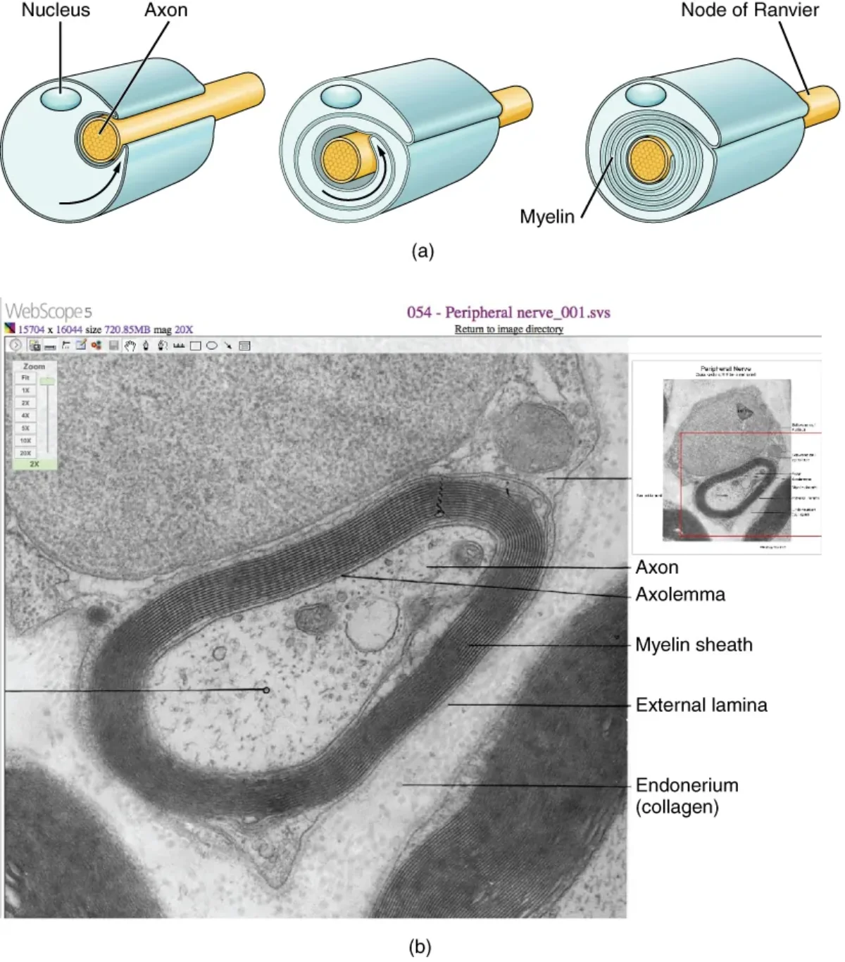
Schwann cells are the myelinating glia of the PNS. Each Schwann cell wraps around a single segment of a single axon, forming one internode of the myelin sheath. The gaps between adjacent Schwann cells are the nodes of Ranvier.
Schwann cells also play a crucial role in nerve regeneration. After PNS axon damage, Schwann cells form a regeneration tube that guides the regrowing axon back to its target. This is a major reason PNS nerves can regenerate while CNS axons generally cannot - the CNS lacks this guidance mechanism.
Not all Schwann cells produce myelin. Non-myelinating Schwann cells loosely envelop small-diameter axons without wrapping them in myelin layers. These unmyelinated fibers conduct signals more slowly.
Satellite Cells: The Ganglia Supporters
Satellite cells surround neuron cell bodies in PNS ganglia (such as dorsal root ganglia and autonomic ganglia). They provide structural support, regulate the chemical environment around the neuron, and may play a role in chronic pain signaling.
Think of satellite cells as the PNS counterpart to astrocytes - they nurture and protect neuronal cell bodies, just as astrocytes do in the CNS.
The Myelination Process
Whether performed by oligodendrocytes or Schwann cells, myelination follows the same basic principle: the glial cell membrane wraps concentrically around the axon, squeezing out cytoplasm to form a dense, lipid-rich sheath. The high lipid content (roughly 80% lipid, 20% protein) is what makes myelin such an effective electrical insulator.
Summary Table: All Six Glial Cell Types
| Glial Cell | Location | Key Function(s) | Origin |
|---|---|---|---|
| Astrocytes | CNS | BBB maintenance, nutrient transfer, neurotransmitter recycling, K+ buffering | Neural ectoderm |
| Oligodendrocytes | CNS | Myelinate CNS axons (one cell, many axon segments) | Neural ectoderm |
| Microglia | CNS | Immune defense, phagocytosis | Mesoderm |
| Ependymal cells | CNS | Line ventricles, produce and circulate CSF | Neural ectoderm |
| Schwann cells | PNS | Myelinate PNS axons (one cell, one axon segment), aid regeneration | Neural crest |
| Satellite cells | PNS | Support neuron cell bodies in ganglia | Neural crest |