
Bone Growth and Remodeling
You were not born with the same skeleton you have now. Most of your bones started as cartilage models that were gradually replaced by bone tissue - a process that began in the womb and was not fully complete until your mid-twenties. Understanding how bone forms and grows is a reliable MCAT topic, and there are two fundamentally different pathways.
Two Pathways of Bone Formation
Intramembranous ossification and endochondral ossification are the two mechanisms the body uses to create bone. They differ in their starting material:
| Feature | Intramembranous | Endochondral |
|---|---|---|
| Starting material | Mesenchymal connective tissue (membrane) | Hyaline cartilage model |
| Bones formed | Flat bones of the skull, clavicle, mandible | Most other bones (long bones, vertebrae, pelvis, ribs) |
| Cartilage intermediate? | No | Yes |
| Growth plate involvement? | No | Yes (epiphyseal plate) |
Intramembranous Ossification
This is the simpler pathway. It produces the flat bones of the skull (frontal, parietal, occipital), the mandible, and the clavicle.
Steps:
- Mesenchymal cells cluster together and differentiate directly into osteoblasts
- Osteoblasts secrete osteoid (unmineralized bone matrix), which then mineralizes
- The mineralized matrix forms small islands called trabeculae, which grow and merge
- Blood vessels are incorporated between the trabeculae
- The outer surface condenses into a layer of compact bone; the interior remains spongy bone
- The periosteum forms from the surrounding mesenchyme
The key point: there is no cartilage intermediate. Mesenchyme transforms directly into bone.
Endochondral Ossification
This is the pathway used by most bones in the body, including all long bones. It starts with a cartilage “blueprint” that is systematically replaced by bone.
Steps of endochondral ossification:
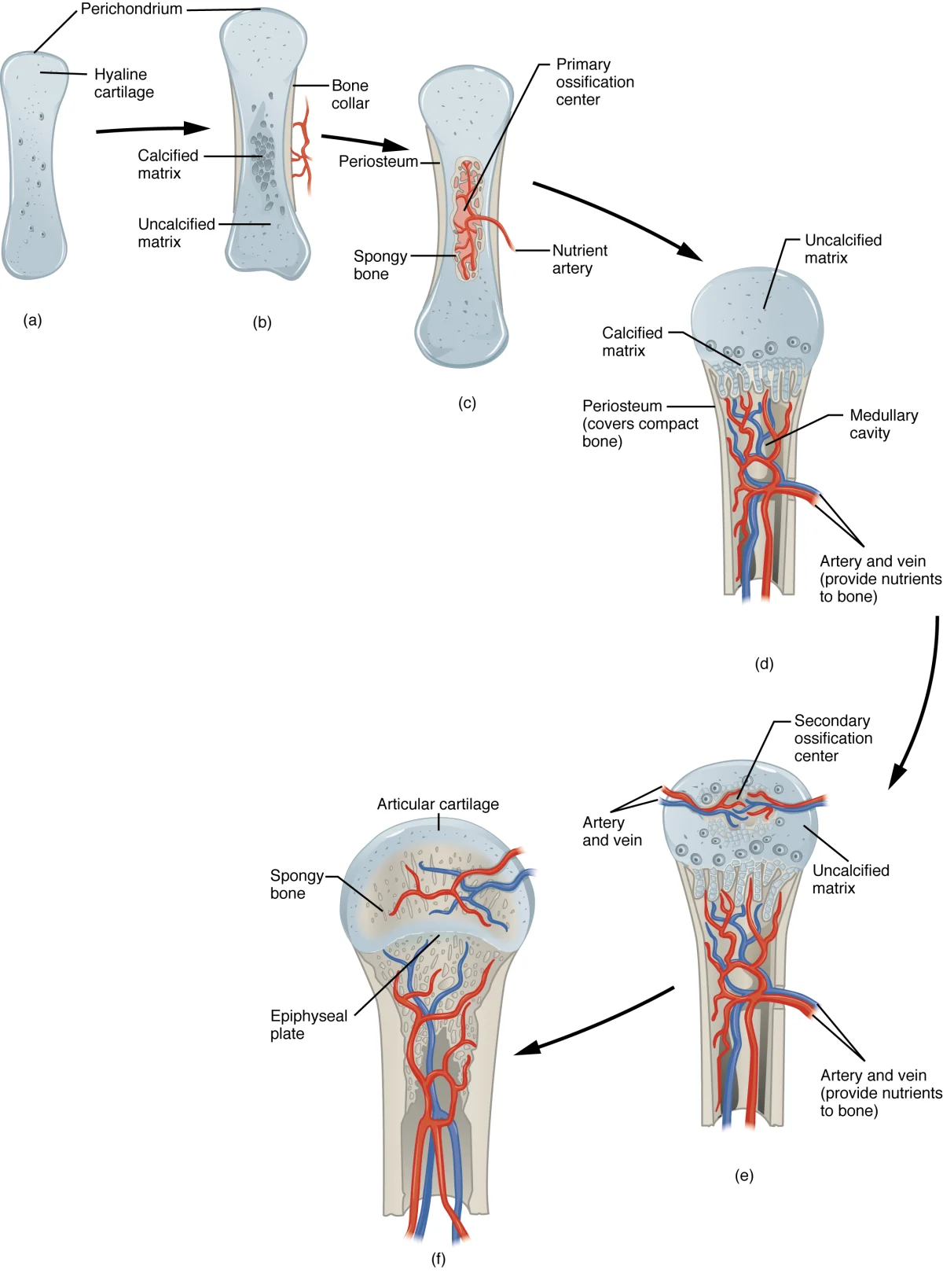
- Cartilage model forms - Mesenchymal cells differentiate into chondrocytes, which secrete a hyaline cartilage model in the shape of the future bone
- Primary ossification center - In the center of the diaphysis (shaft), chondrocytes enlarge (hypertrophy), calcify the surrounding cartilage, and then die. Blood vessels invade the area, bringing osteoblasts that begin depositing true bone on the calcified cartilage scaffolding
- Bone collar forms - Osteoblasts in the perichondrium (the membrane surrounding the cartilage) lay down a collar of compact bone around the diaphysis, converting the perichondrium into the periosteum
- Medullary cavity forms - Osteoclasts break down the newly formed spongy bone in the center of the diaphysis, creating the medullary cavity
- Secondary ossification centers - Blood vessels invade the epiphyses (ends), and the same process of cartilage replacement occurs there. However, spongy bone remains in the epiphyses (it is not hollowed out like the diaphysis)
- Epiphyseal plate remains - A band of cartilage persists between the diaphysis and each epiphysis - this is the growth plate
The Epiphyseal (Growth) Plate
The epiphyseal plate is the engine of longitudinal bone growth. It is a disc of hyaline cartilage located between the epiphysis and diaphysis. The basic process is straightforward:
- On the epiphyseal side, chondrocytes actively divide and produce new cartilage (pushing the plate outward)
- On the diaphyseal side, older cartilage calcifies, the chondrocytes die, and osteoblasts replace the cartilage with bone
This means new cartilage is constantly being added on one side and replaced by bone on the other. The net result: the bone gets longer as the epiphysis is pushed farther from the diaphysis.
Bone Remodeling
Even after growth is complete, bone is continuously remodeled throughout life. Remodeling is the coordinated process of bone resorption by osteoclasts followed by new bone deposition by osteoblasts. About 10% of the adult skeleton is replaced each year.
Wolff’s law states that bone remodels in response to mechanical stress placed upon it. Bone is deposited where stress is high and resorbed where stress is low. This explains why:
- Weight-bearing exercise increases bone density
- Astronauts lose bone mass in microgravity (no mechanical loading)
- A bedridden patient develops osteoporosis in immobilized limbs
- The dominant arm of a tennis player has denser bones than the non-dominant arm
The Calcium Axis: Bone Meets Endocrine
Remodeling is the skeleton’s role in calcium homeostasis. Three hormones work together to keep blood Ca²⁺ in range:
- PTH (parathyroid) rises when blood Ca²⁺ is low. It stimulates osteoclast-driven resorption (via osteoblasts), boosts Ca²⁺ reabsorption in the kidney, and activates vitamin D.
- Calcitriol (active vitamin D, made in the kidney) increases gut Ca²⁺ absorption and supports mineralization.
- Calcitonin (thyroid C cells) opposes PTH: it inhibits osteoclasts, lowering blood Ca²⁺.
Fracture Repair
When a bone breaks, the repair process recapitulates many of the steps of endochondral ossification:
- Hematoma formation - Blood from ruptured vessels clots at the fracture site
- Fibrocartilaginous callus - Fibroblasts and chondrocytes invade the clot and produce a soft callus of fibrocartilage that bridges the gap
- Bony (hard) callus - Osteoblasts replace the fibrocartilage with spongy bone (endochondral ossification in miniature)
- Remodeling - Osteoclasts reshape the bony callus into compact bone, restoring the original bone structure over months to years