
Bone Structure
Pick up a chicken drumstick next time you eat one. Snap it in half and look inside. You will see a hard, dense outer shell surrounding a spongy, honeycomb-like interior. That is not a design flaw - it is brilliant engineering. The dense outer layer resists bending and compression, while the spongy interior saves weight and houses bone marrow. Your bones use the exact same blueprint.
The Two Types of Bone Tissue
All bones contain two types of tissue working together:
Compact (cortical) bone forms the dense outer shell. Under a microscope, compact bone is organized into cylindrical units called osteons (Haversian systems). Each osteon is built like a tree trunk: concentric rings of bone matrix (called lamellae) surround a central canal (the Haversian canal) that carries blood vessels and nerves. Tiny channels called canaliculi radiate outward from each lacuna (the small pocket where a bone cell sits), connecting neighboring bone cells like a network of underground tunnels.
Spongy (cancellous/trabecular) bone fills the interior. It looks like a honeycomb or scaffold - an open lattice of bony struts called trabeculae. This structure is not random. The trabeculae align along lines of mechanical stress, reinforcing the bone exactly where it needs it most. Spongy bone is lighter than compact bone and contains red bone marrow, where blood cells are produced (hematopoiesis).
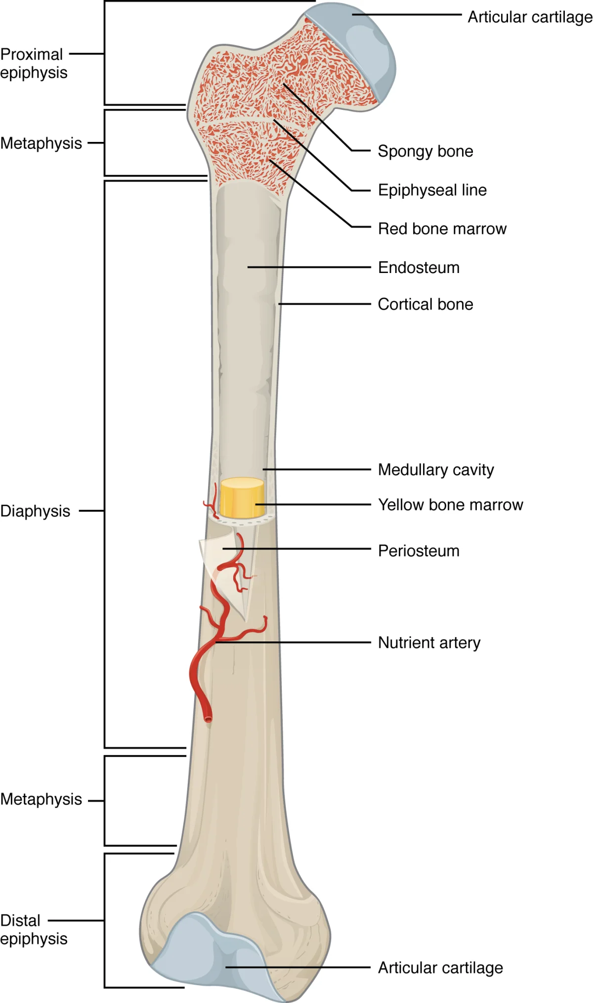
Anatomy of a Long Bone
Long bones (like the femur, humerus, and tibia) are the classic model for learning bone anatomy. Here are the key structures:
| Structure | Location | Function |
|---|---|---|
| Diaphysis | Shaft (middle) | Thick tube of compact bone; encloses the medullary cavity |
| Epiphysis | Ends (proximal and distal) | Spongy bone covered by thin compact bone; articulates with other bones |
| Metaphysis | Between diaphysis and epiphysis | Contains the epiphyseal plate (growth plate) in growing bones |
| Epiphyseal plate | Between metaphysis and epiphysis | Cartilage growth zone; becomes the epiphyseal line when growth stops |
| Medullary cavity | Interior of diaphysis | Contains yellow bone marrow (fat) in adults |
| Periosteum | Outer surface (except at joints) | Dense connective tissue; anchors tendons and ligaments; contains osteoblasts for bone repair |
| Endosteum | Inner surface (lines medullary cavity) | Thin membrane containing osteoblasts and osteoclasts |
| Articular cartilage | Covers epiphyses at joints | Smooth hyaline cartilage; reduces friction during movement |
The Bone Matrix
Bone is not solid mineral. It is a composite material made of two components:
-
Organic component (~35%) - primarily Type I collagen fibers, plus proteoglycans and glycoproteins. Collagen gives bone its tensile strength and flexibility. Without collagen, bone would be brittle like chalk and shatter on impact.
-
Inorganic component (~65%) - primarily hydroxyapatite crystals (Ca₁₀(PO₄)₆(OH)₂), a calcium phosphate mineral. Hydroxyapatite gives bone its hardness and compressive strength. Without minerals, bone would be rubbery and bend like cartilage.
This is why bone is both a mineral reservoir and a structural material. When blood calcium drops, the body can dissolve hydroxyapatite to release calcium. When calcium is abundant, it gets deposited back into bone.
The Osteon (Haversian System)
The osteon is the structural and functional unit of compact bone. Understanding its anatomy is essential:
- Central (Haversian) canal - runs longitudinally through the center of each osteon; contains blood vessels and nerves
- Lamellae - concentric rings of calcified bone matrix surrounding the central canal
- Lacunae - small pockets between lamellae where osteocytes (mature bone cells) reside
- Canaliculi - tiny channels radiating from each lacuna, forming a network that connects neighboring osteocytes; these allow nutrient exchange and cell-to-cell communication
The Two Skeletal Divisions
The human skeleton is divided into two parts:
-
Axial skeleton (80 bones) - the central axis of the body: skull, vertebral column (spine), rib cage, and hyoid bone. Provides protection for the brain, spinal cord, and thoracic organs.
-
Appendicular skeleton (126 bones) - the limbs and the girdles that attach them to the axial skeleton: pectoral girdle (clavicle + scapula), upper limbs, pelvic girdle (hip bones), and lower limbs. Provides the framework for movement.
Embryological Origin
Bones, cartilage, and connective tissues of the musculoskeletal system are derived from the mesoderm, one of the three primary germ layers.