
Cartilage Types
Bend your ear forward. It springs back. Now press on the tip of your nose - it squishes and returns to shape. That flexibility comes from cartilage, a specialized connective tissue that is softer and more flexible than bone but firmer than most other tissues. There are three types of cartilage in your body, and they each have a different job based on their specific combination of collagen and elastic fibers.
General Properties of Cartilage
Before diving into the three types, know these features that ALL cartilage shares:
- Avascular - cartilage has no blood supply. Nutrients reach chondrocytes by diffusing through the matrix from surrounding tissues. This is why cartilage heals slowly (or not at all) after injury.
- No nerves - cartilage is not innervated (aneural), which is why you can press on your ear without pain
- Chondrocytes - the only cell type in cartilage; they sit in small cavities called lacunae and produce the surrounding matrix
- Perichondrium - a dense connective tissue membrane covering most cartilage (except articular cartilage and fibrocartilage). It provides blood supply to the outer edge and contains stem cells for cartilage growth
- High water content - the matrix is heavily hydrated, giving cartilage its ability to resist compression
Hyaline Cartilage
Hyaline cartilage is the most common type. Its name comes from the Greek word “hyalos” (glass) because it appears glassy and translucent when fresh.
Matrix composition: Fine Type II collagen fibers in a gel-like ground substance rich in proteoglycans (especially aggrecan). The collagen fibers are too thin to see individually, giving the matrix its smooth, glassy appearance.
Key locations:
- Articular surfaces of joints (covering bone ends - no perichondrium here)
- Tracheal rings and bronchi (keeps airways open)
- Tip of the nose
- Costal cartilage (connects ribs to sternum)
- Epiphyseal plates (growth plates in growing bones)
- Fetal skeleton (the template for endochondral ossification)
Function: Provides smooth, low-friction surfaces at joints; provides flexible support for airways; serves as the template for bone growth.
Elastic Cartilage
Elastic cartilage is similar to hyaline cartilage but contains a dense network of elastic fibers in addition to Type II collagen. This makes it extremely flexible - it can be bent repeatedly without breaking.
Key locations:
- External ear (pinna/auricle)
- Epiglottis (the flap that covers the larynx during swallowing)
- Auditory (Eustachian) tube
Function: Provides flexible support that can spring back to its original shape after being deformed.
Fibrocartilage
Fibrocartilage is the toughest type. It contains dense bundles of Type I collagen fibers (the same collagen found in bone, tendons, and ligaments), making it extremely resistant to compression and tension.
Key locations:
- Intervertebral discs (between vertebrae)
- Menisci of the knee
- Pubic symphysis
- Labrum of the hip and shoulder joints
- At the insertion points of tendons and ligaments into bone
Function: Absorbs compressive shock and resists tearing at sites that experience heavy mechanical stress.
Important: Fibrocartilage has no perichondrium (unlike hyaline and elastic cartilage). Its chondrocytes are scattered among thick collagen bundles rather than enclosed in a smooth matrix.
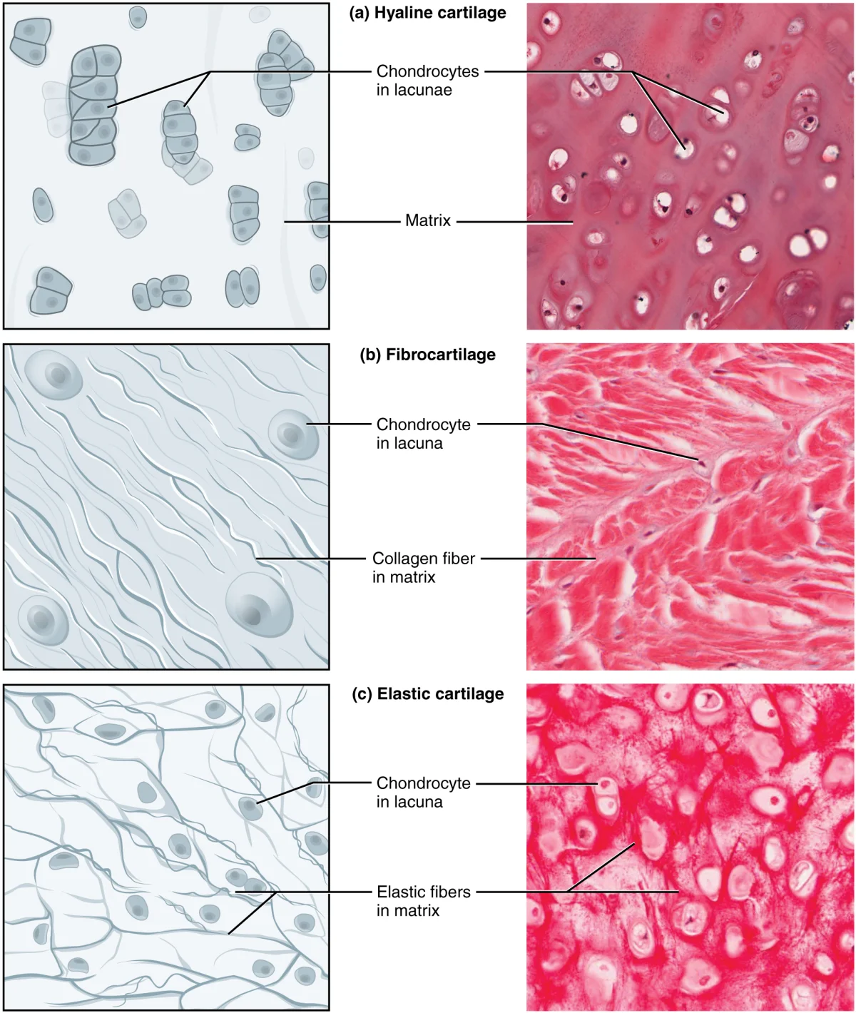
Comparison Table
| Feature | Hyaline | Elastic | Fibrocartilage |
|---|---|---|---|
| Collagen type | Type II | Type II + elastic fibers | Type I (thick bundles) |
| Flexibility | Moderate | Very high | Low |
| Strength | Moderate | Moderate | Very high |
| Perichondrium? | Yes (except articular) | Yes | No |
| Key locations | Trachea, joints, nose, growth plates | Ear, epiglottis | Intervertebral discs, meniscus, pubic symphysis |
| Appearance | Glassy, translucent | Yellow, flexible | Dense, fibrous |
Cartilage Growth
Cartilage can grow in two ways:
-
Interstitial growth - chondrocytes within the cartilage divide and produce new matrix from the inside. This occurs mainly during childhood and at the epiphyseal plate.
-
Appositional growth - new cartilage is added to the surface by cells in the perichondrium that differentiate into chondrocytes. This can occur throughout life.