
Joints and Connective Tissue
Bones are rigid. If your skeleton were a single solid piece, you could not move at all. Joints are the points where two or more bones meet, and they range from completely immovable (like the sutures in your skull) to highly mobile (like your shoulder). The type of joint determines how much movement is possible.
Joint Classification by Structure
Joints are classified into three structural categories based on the type of tissue connecting the bones:
| Structural Type | Connecting Tissue | Movability | Examples |
|---|---|---|---|
| Fibrous | Dense connective tissue (collagen fibers) | Mostly immovable | Skull sutures |
| Cartilaginous | Cartilage (hyaline or fibrocartilage) | Slightly movable (amphiarthrosis) | Pubic symphysis, intervertebral discs, epiphyseal plates |
| Synovial | Synovial capsule with fluid-filled cavity | Freely movable (diarthrosis) | Knee, shoulder, hip, elbow, wrist |
Joint Classification by Movability
Joints can also be classified by how much movement they allow:
- Synarthrosis - immovable (e.g., skull sutures)
- Amphiarthrosis - slightly movable (e.g., pubic symphysis, intervertebral discs)
- Diarthrosis - freely movable (e.g., all synovial joints)
The Synovial Joint
Synovial joints are the most common and most tested type on the MCAT. They share a characteristic set of structures:
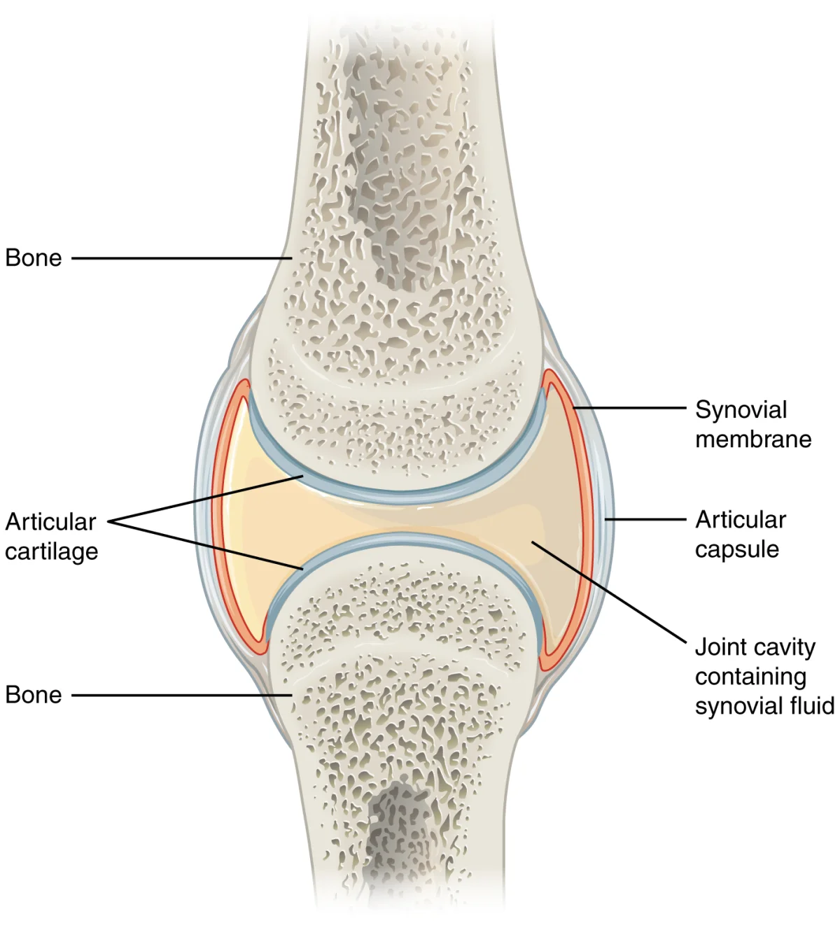
Key components of a synovial joint:
-
Articular (hyaline) cartilage - smooth cartilage covering the bone ends; reduces friction and absorbs shock. Has no perichondrium and is avascular.
-
Joint (synovial) cavity - the space between the bones, filled with synovial fluid
-
Synovial membrane - lines the inner surface of the joint capsule (but NOT the articular cartilage). Secretes synovial fluid.
-
Synovial fluid - viscous fluid that lubricates the joint surfaces and nourishes the avascular articular cartilage. Contains hyaluronic acid (for viscosity) and filtered plasma (for nutrients).
-
Fibrous capsule - tough outer layer of the joint capsule made of dense connective tissue. Reinforced by ligaments.
-
Ligaments - bands of dense connective tissue connecting bone to bone. They stabilize the joint and limit excessive movement.
-
Bursae - small, fluid-filled sacs that reduce friction between tendons, ligaments, and bones at points of high mechanical stress
Types of Synovial Joints
The three synovial joint types worth knowing for the MCAT:
| Type | Movement | Example |
|---|---|---|
| Hinge | Flexion/extension (1 axis) | Elbow, knee |
| Ball-and-socket | Movement in all directions (3 axes) | Shoulder, hip |
| Pivot | Rotation (1 axis) | Atlas/axis (turning the head) |
Tendons and Ligaments
Tendons and ligaments are both made of dense regular connective tissue (primarily Type I collagen fibers arranged in parallel bundles), but they connect different structures:
| Feature | Tendons | Ligaments |
|---|---|---|
| Connects | Muscle to bone | Bone to bone |
| Function | Transmits muscle force to bone to produce movement | Stabilizes joints, limits excessive movement |
| Vascularity | Relatively avascular | Relatively avascular |
| Healing | Slow (poor blood supply) | Slow (poor blood supply) |
| Elasticity | Low (stiff - efficient force transfer) | Slightly more elastic than tendons |
| Example | Achilles tendon, rotator cuff tendons | ACL (anterior cruciate ligament), MCL |
Movement Terminology
The MCAT expects you to recognize the basic types of joint movements:
| Movement | Description | Example |
|---|---|---|
| Flexion | Decreasing the angle between bones | Bending the elbow, bending the knee |
| Extension | Increasing the angle between bones | Straightening the elbow, straightening the knee |
| Abduction | Moving a limb away from the body’s midline | Raising your arm out to the side |
| Adduction | Moving a limb toward the body’s midline | Lowering a raised arm back to your side |
| Rotation | Turning a bone around its own axis | Turning your head left or right |
| Circumduction | Circular movement combining flexion, extension, abduction, and adduction | Swinging your arm in a circle |
Antagonistic and Synergistic Muscles
Muscles work in coordinated pairs and groups:
-
Antagonistic muscles produce movement in opposite directions. When one contracts, the other relaxes. Example: the biceps (flexor) and triceps (extensor) at the elbow. When the biceps contracts and flexes the elbow, the triceps relaxes. When the triceps contracts and extends the elbow, the biceps relaxes.
-
Synergistic muscles assist each other to produce the same movement. Example: the hamstring muscles (biceps femoris, semitendinosus, semimembranosus) all work together to flex the knee.
-
Agonist (prime mover) - the muscle primarily responsible for a movement
-
Antagonist - the muscle that opposes the agonist
-
Synergist - a muscle that assists the agonist
Other Connective Tissues
A few additional structures are worth knowing:
- Aponeurosis - a flat, sheet-like tendon (e.g., the aponeurosis connecting the abdominal muscles)
- Fascia - dense connective tissue that surrounds muscles, groups of muscles, blood vessels, and nerves. It compartmentalizes structures and reduces friction.
- Meniscus - a crescent-shaped pad of fibrocartilage in certain joints (knee, temporomandibular joint) that acts as a shock absorber and improves joint fit